

Color.Atlas.of.Endodontics-ublog.tk
.pdf26 |
Color Atlas o f Endodontics |
Maxillary Molars
Treatment of maxillary molars is never routine (Figure 2-22). In a recent study of maxillary first and second molars an MB2 canal was found in 96% of the mesiobuccal roots of maxillary first molars and 94% of the maxillary second molars. Approximately 54% were located in the traditional access opening, 31% were found with the use of a bur, and 10% were found with the aid of a microscope. The MBZ canal orifice was found on average 1.82 mm lingual to the main MB canal orifice. 9 In another study of the maxillary first molar using microscopy, the MB Z canal was located in
93% of first molars and 60% of second molars 4 (Figures 2-23 and 2-24). The difficulty in access, high percentage of fourth and even fifth canals, and root curvatures put even the "routine" maxillary molar in a high-risk category.3 Complicating factors such as limited opening, crowns, changes in tooth angulation, tooth position, and calcification make predictable treatment of these teeth challenging for even the most experienced clinician trained in microscopy, ultrasonics, and rotary instrumentation.
The clinician wishing to treat these highto extremerisk cases should perform a 6-month chart review to de-
FIGURE 2-22 Buccal view of the access for maxillary molars. |
FIGURE 2-23 An occlusal view of the access for maxillary |
|
molars. |
FIGURE 2-24 The location of the MB 2 canal is mesial to the line connecting the MB and palatal orifice. In the maxillary first molar the MB 2 canal is generally located within the range shown. In the maxillary second molar the location of the MB 2 is highly variable and can be located from the MB orifice to the palatal orifice (see Figs. 2-46 and 2-47).

Chapter Two Endodontic Access |
27 |
termine the percentage of cases with at least four canals. If the percentage is less than 45% for first molars or less than 35% for second molars, these cases should be carefully screened for referral because the MBZ canal is being missed and untreated about half of the time.
Guidelines for canal location in the maxillary first molar (Figure 2-25) differ from that in the maxillary second molar. In the maxillary first molar the MB canal is located under the mesial buccal cusp (see Figure 2-25, D). The MBZ canal is located mesial to a line from the
MB canal toward the palatal canal (see Figures 2-25, E, and 2-26). The DB canal is located distal to the MB canal in the buccal groove area, slightly lingual to the MB canal (see Figure 2-25, G). The palatal canal is generally the largest canal and is located under the mesiolingual (ML) cusp (see Figure 2-25, F). These general locations remain the same as the pulp calcifies with age ( Figure 2-27). Although these general principles apply to the maxillary second molar, the chamber may be narrower, resembling a straight line (see Fig. 2-55).
FIGURE 2-25 A, Preoperative photograph of a maxillary molar. The pronounced cervical bulge over the MB is highly suggestive of a large root and two canals. B, Preoperative radiograph of this necrotic maxillary first molar. Note the constricted pulp chamber. The angle of entry to the mesial canals is from the distal. C, The canals have been prepared to help illustrate their locations and angles. Note that in the following illustrations the access form may need to be extended or modified on the side opposite the canal to clear the rotary instruments and avoid cervical flexure of the instruments. D, The angle of entry into the MB canal is markedly from the distal and palatal. The access may need to be extended distally and palatally to allow clean placement of instruments.
Continued

28 |
Color Atlas o f Endodontics |
FIGURE 2-25, cont'd E, The entrance into the prepared MB 2 is from the distal and slightly from the palatal. The access may need to be extended distally to allow clean placement of rotary instruments into the MB2. F, The angle of entry into the palatal is from the buccal and mesial. Occasionally the access may need to be extended to the MB to allow clean placement of rotary instruments into the palatal canal. G, The angle of entry into the DB canal is from the mesial and palatal. A diamond bur (see Figure 2-26) can be used to relieve the impeding restorative material or tooth structure. H, The completed case. Note that the access is not in the center of the tooth. Adequate access to locate, negotiate, prepare, and obturate can be obtained without violating the transverse ridge. Note that the access extends almost to the MB cusp tip. I, The HFetched silanated composite crown repair of the endodontic access is outlined in black. The presence of this type of radicular anatomy is usually unconfirmed until the case is complete. Missing the MB 2 canal here would doom the case to failure because of the presence of separate foramina.

Cbapter Two Endodontic Access |
29 |
FIGURE 2-26 A, On entering the pulp chamber in this maxillary second molar, the clinician can readily locate the MB2 orifice along a line connecting the MB and palatal orifices.This "false orifice" is a source of frustration for many dentists because although it can be probed, it is often resistant to negotiation.The reason for this is that the MB 2 canal proceeds mesially (horizontally) before making a 90-degree turn down the root. B, The prepared and obturated MB 2 canal is considerably more mesial than the original orifice. The red dot to the distal of the obturated MB2 is the location of the false original orifice. A technique routinely employed is to notch this area with a tapered roundtipped diamond, Mueller bur, or Gates Glidden bur to gain straight line access to the MB 2 canal.This technique is detailed in later figures.
FIGURE 2-27 A, Original view of canal shapes in a 9-year-old's maxillary first molar. Note that the very broad MB "canal" is full of debris. B, Computer-enhanced view of canal spaces at 9 years.
Continued

30 |
Color Atlas of Endodontics |
C D
FIGURE 2-27, cont'd C, Computer simulation of typical calcific metamorphosis (calcification) in an adult tooth. D, Computer simulation of significant calcific metamorphosis.
A B
FIGURE 2-28 A, Access for a mandibular incisor as viewed from the lingual. The access is quite high on the lingual surface of the tooth. This gives the clinician a much straighter shot down the canal and minimizes the chance of perforating out the facial surface. B, This mesial view shows the access extending nearly to the incisal edge.
Mandibular Incisors
As with the other anterior teeth the traditional access to the mandibular incisor was more cervically placed than necessary because of esthetic constraints. The optimal access for the mandibular central and lateral incisor is actually through the incisal edge, but this is balanced with the desire to maintain an intact incisal edge where
possible (Figure 2-28). In the mature adult tooth, attrition has generally caused the access to extend through the incisal edge'° (Figure 2-29).
Because two canals are present in about 40% of all mandibular incisors,11 these teeth should be assumed to have two canals until substantial evidence to the contrary is discovered.

Chapter Two Endodontic Access |
31 |
FIGURE 2-29 In this attrited and rotated incisor with two canals, an incisal access greatly facilitated location of the lingual canal.
FIGURE 2-30 The typical error in access on a mandibular incisor is to perforate toward the facial (white dot). The clinician had already "located" the canal but bypassed it while continuing to drill down and to the facial. Mandibular incisors are rarely perforated to the lingual.
FIGURE 2-31 While searching for a calcified canal, clinicians tend to drill in an apical direction but neglect to take into account the natural angulation of the mandibular incisor, resulting in buccal perforation.
Because of the facial inclination of the tooth, perforation of the facial aspect of the root is a common procedural error in accessing mandibular incisors (Figures 2-30 and 2-31). In cases of rotation or crowding a facial approach to access should be considered. 12
ally has two canals and therefore requires a broad faciallingual access. The access opening is ovoid and located on the lingual portion of the crown (see Figures 2-32 and 2-33). As wear occurs, the access may involve the incisal edge (Figure 2-34).
Mandibular Canines |
Mandibular Premolars |
The mandibular canine has a very broad facial-lingual dimension to its root (Figure 2-32). This root occasion-
The broad buccolingual dimension of the mandibular premolar dictates an access form that is about twice as

32 |
Color Atlas o f Endodontics |
FIGURE 2-32 Access openings for the mandibular canine.
A
FIGURE 2-33 An ideal access opening in a mandibular canine. Viewed from the incisal surface, the access is slightly to the lingual and can be seen extending to nearly the incisal edge.
B
FIGURE 2-34 A, Preoperative view of a mandibular canine with incisal attrition. B, Occasionally the mandibular canine has two canals. A more incisally and facially positioned access facilitates location of the lingual canal.
broad in the buccolingual dimension than it is mesiodistally (Figures 2-35 through 2-37). Although most mandibular premolars have a single canal, two canals occur about 25% of the time in mandibular first premolars 13; rarely, three canals are present. When numerous
canals are present, the preoperative radiograph often indicates a "fast break." This appears as a relatively patent canal space in the coronal portion of the tooth that suddenly disappears (Figure 2-38). Locating the two canals requires an appropriate access (Figure 2-39).

Chapter Two Endodontic Access |
33 |
FIGURE 2-35 Viewed from the occlusal, the access is relatively well centered in the buccolingual and mesiodistal dimensions. It is about twice as broad buccolingually as it is mesiodistally.
FIGURE 2-36 A, Viewed from the buccal surface, the access is conservative mesiodistally. B, Viewed from the mesial, the crowns of the mandibular premolars have a slightly lingual inclination relative to the root.
FIGURE 2-37 Occlusal view of access through a PFM crown.

34 |
Color Atlas o f Endodontics |
FIGURE 2-38 Radiographic appearance of a fast break in the mandibular right first premolar. The coronal extent of the canal is readily visible but abruptly disappears in the middle of the root, indicating at least two canals. Note that the second premolar has three roots.
FIGURE 2-39 A, The mandibular first premolar has two canals about one quarter of the time. This crown of the mandibular first premolar can have a marked lingual inclination that may misdirect the clinician's bur too far to the buccal. This increases the likelihood of perforation and greatly decreases the chances of locating a lingual canal. In this retreatment clinical case, the access error manifests as a small round access too far to the lingual. B, Extending the access to the buccal allows the clinician to locate the missed lingual canal (top) and obturate both canals. C, Obturation.
Chapter Two Endodontic Access |
35 |
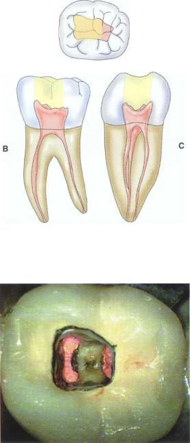
Mandibular Molars
The access to the mandibular molars has been presented by many as triangular in shape. This access shape greatly hinders the clinician's ability to locate the DB canal when one is present and to treat the full buccolingual extent of the broad distal canal when a single distal canal is present. The naturally present slight mesial and lingual inclination of the tooth, coupled with the clinical access to the tooth, dictates an access that is placed more toward the mesial half of the tooth and may extend buc-
cally to nearly the MB cusp tip (Figure 2-40). The access may occasionally cross the central pit (Figure 2-41).
In mandibular molars the MB canal lies under the mesiobuccal cusp tip. The ML canal often appears in line with the central groove crossing the mesial marginal ridge. The lingual inclination of the tooth in the arch, coupled with the lingual constriction of the crown, accounts for this anatomic relationship (see Figure 2-40, C). The distal canal is generally at the intersection of the buccal, lingual, and central grooves as viewed from the
A
FIGURE 2-40 A, Viewed from the occlusal, the access can be seen to just cross the central pit area and extend to nearly the MB cusp tip. B, Viewed from the buccal, the access is slightly mesially inclined. C, Viewed from the mesial, the buccal extent of the access can extend to nearly the MB cusp tip.
FIGURE 2-41 The ideal access is demonstrated on this mandibular first molar through a PFM crown.