

39
Figure 4: X-ray examination data, Case N3
Case 4 – a digital X-ray of a patient with a ground-glass nodule (via CT scan) in C1+2 of the left lung, max. size of 22 mm – adenocarcinoma via histological examination (Figure 5) [6].
Figure 5: X-ray examination data, Case N4
Case 5 – a digital X-ray of a patient with a solid nodule in C3 of the right lung, max. size of 19 mm – squamous cell cancer via histological examination (Figure 6) [6].

40
Figure 6: X-ray examination data, Case N5
Case 6 – a digital X-ray of a patient with a solid nodule in the subpleural area C1+2 of the left lung, max. size of 13 mm – TB via histological examination (Figure 7) [6].
Figure 7: X-ray examination data, Case N6
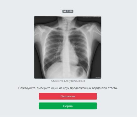
The next stage of researching the interpretation quality of digital X-rays by radiologists was the online platform testing (Figure 8); 1,387 specialists have taken part, with 651 results subject to consistent assessment, where 516 of such results were done by radiologists with work experience from less than 1 year to 20 years and more. Many of the specialists worked from one to five years, what makes 62.4% (Table 4) [6].
Also, there were two categories of radiologists: radiologists exposed to thoracic radiology (N=130) and radiologists with no such exposure (N=386).
Рекомендовано к изучению разделом по лучевой диагностике сайта https://meduniver.com/
41
Figure 8: Testing of radiologists through online platform
Table 4: Ranked data of radiologists from online testing
Work Experience |
Quantity |
Share |
0 |
56 |
10.8% |
1-2 |
179 |
34.7% |
3-5 |
143 |
27.7% |
6-10 |
87 |
16.9% |
>10 |
51 |
9.9% |
During the online testing, a slightly smaller number of the specialists with more than six years of experience were identified among the tested radiologists. The share amounted to 26.8%, whereas the value of the previous research was 39%. The share of radiologists with work experience of up to one year was also smaller, reaching 10.8% against 31% from the previous in-person testing; at the same time, the share of radiologists with work experience from one year to five years was significantly higher and amounted to 62.4% against 31% of the in-person testing. The difference in the shares of radiologists under the in-person and online testing is probably caused by the

42
social differences between the Internet audience and the people taking traditional fulltime educational courses [137].
A sampling package was used to evaluate the results; it consists of depersonalized digital PA chest X-rays of 100 persons with health status confirmed by histology and CT scans, with the 94:6 norm/pathology ratio – six persons with confirmed pulmonary pathology and 94 persons with no significant pathology [137].
X-rays of six persons with pulmonary nodules and masses were used as pathology, same as for the in-person testing [137].
Case 1 – a digital X-ray of a patient with a part-solid nodule in C1 of the right lung, max. size of 18 mm – adenocarcinoma via histological examination (Figure 9).
Figure 9: X-ray examination data, Case N1
Case 2 – a digital X-ray of a patient with a solid mass in C1 of the right lung, max. size of 67 mm – adenocarcinoma via histological examination (Figure 10).
Рекомендовано к изучению разделом по лучевой диагностике сайта https://meduniver.com/

43
Figure 10: X-ray examination data, Case N2
Case 3 – a digital X-ray of a patient with a solid round mass in C4 of the left lung, max. size of 8 mm, adenocarcinoma via histological examination (Figure 11).
Figure 11: X-ray examination data, Case N3
Case 4 – a digital X-ray of a patient with a solid round mass in C1+2 of the left lung, max. size of 34 mm, sarcoma via histological examination (Figure 12).

44
Figure 12: X-ray examination data, Case N4
Case 5 – a digital X-ray of a patient with a solid nodule in C3 of the left lung, max. size of 35 mm – small-cell cancer via histological examination (Figure 13).
Figure 13: X-ray examination data, Case N5
Case 6 – a digital X-ray of a patient with a solid mass in C2 of the right lung, max. size of 19 mm – tuberculoma via histological examination (Figure 14).
Рекомендовано к изучению разделом по лучевой диагностике сайта https://meduniver.com/

45
Figure 14: X-ray examination data, Case N6
2.3 Testing of automated analysis systems
We have chosen four software programs based on convolutional neural networks pitched as systems of automated analysis of digital chest X-rays [44].
Below are given the testing selection criteria:
1.availability of a computer program registration certificate/patent;
2.availability of a test online access;
3.availability to detect pulmonary nodules and masses described in the
software’s details.
Total of four software products have been chosen based on these criteria (three domestic systems and one foreign system) [44].
Since the research was aimed at the general assessment of diagnostic indicators of currently available systems and not at the assessment of a particular product, all programs in the research were disguised as A, B, C, D [44].
Following the recommendations of developers, Program A (Registration No.2019665266) can be used in medical institutions as a medical decision support system providing radiologists with a ‘second opinion’ [9], or as an addition to archives of medical images with a follow-up analysis of incoming images aimed at detecting pathologies to be checked by radiologists. A module for analyzing chest X-rays can detect 14 types of pathologies: opacity, infiltrations, pneumonia, tumors, pulmonary nodules, atelectasis, cardiomegaly, edema, emphysema, fibrosis, hernia, infiltrate,

46
tumor, nodule, pleural thickening, pneumonia, and pneumothorax [29]. Based on the analysis results, the program provides users with the information on the pathology detected and the image with the pathology localization shown in red. The program has a web interface allowing users to view the results of image analysis, to change the brightness and contrast of images (Figure 15). The program uses PyTorch library to work with neural networks and Flask as a webserver [29].
Figure 15: Program A interface and chest X-ray results
Developers bill Program B as a system for analysis of possible pathology on digital PA chest X-rays and their description (Medical Product Registration Certificate
No.РЗН 2020/11137). Options for the automated analysis system: Webshow, API is designed to analyze PA chest X-rays and then generate a conclusion in a cloud infrastructure [98]. Program B is a software with a set of artificial neural networks. Artificial neural networks are designed to carry out analysis of diagnostic examinations and categorization: ‘with visible pathology’ and ‘without pathology’ [98]. Based on the results of the diagnostics analysis, Program B provides users with a research protocol with the X-rays classified as ‘with visible pathology’ and ‘without pathology’ following
Рекомендовано к изучению разделом по лучевой диагностике сайта https://meduniver.com/

47
the algorithms of trained neural networks [98]. The set of artificial neural networks is a basic element of a medical product; this set is completed with a specific environment allowing users to interact with the set of trained networks. Modifications of the following neural networks were used in the development: InceptionVS, Inception ResNet-2, ResNet-50. Webshow WEB (presentation layer) is a web application that provides users with an opportunity to upload X-rays in real time and obtain results of their analysis from a neural network (Figure 16). The product is designed to analyze depersonalized diagnostic images in PNG, JPEG or DICOM formats.
Program B determines the presence/absence of a pathology, defines its areas and type using a classifier of radiographic signs containing 12 classes of pathology in organs (classification provided by the developer): atelectasis, cardiomegaly, pneumothorax, hydrothorax, nodule, cavity, infiltrate, dissemination, pulmonary opacity, pulmonary emphysema, pulmonary edema, local infiltrate syndrome [25].
Figure 16: Program B interface and chest X-ray results
48
Based on the data of the developers, Program C (ТУ 62.01.29-001-96876180-
2019, Medical Product Registration Certificate No.РЗН 2022/17406) is aimed at the automated analysis of chest images. Program C processes images from users and develops conclusions on suspected pathologies or their absence. Such analysis is done to automatically detect X-rays with suspected conditions of chest organs [28]. The pathology classifier of Program C can distinguish and categorize nine pathology types (pleural effusion, pneumothorax, atelectasis, nodule, infiltrate or consolidation, a dissemination, a cavity, a calcification / a calcified shadow, violation of cortex continuity). [97]. Moreover, the program provides numerical values of a possible pathology [97]. Images are downloaded for the further processing with the help of a special software module (Figure 17). The images are processed one by one by a convolutional neural network. Based on the training results, each of the three convolutional neural networks provides its own opinion, highlights a suspicious area, if any. The next program module collects results of all three networks, puts them together and layers them over the initial image. An image output module shows the processed image with a pathology highlighted (if any). Suspected pathology is shown with various color schemes where different colors correspond with possible pathology classes as per the automated analysis of X-rays. The color scheme comes with a sign about suspicions of possible pathology class [28, 27, 97]. The color intensity depends on the number of network replies and is directly proportional to the possible pathology as evidenced by the program [28].
Рекомендовано к изучению разделом по лучевой диагностике сайта https://meduniver.com/