

ESC cardiology guideline 2012 valvular disease
.pdfESC/EACTS Guidelines |
2481 |
|
|
durability has been demonstrated so far.193 Sutureless bioprostheses are an incoming technology, allowing quick placement of a bioprosthesis without a sewing cuff and also having larger effective orifice areas.
The two transcatheter-implantable prostheses which are most widely used are made of pericardial tissue inserted into a baremetal balloon-expanding stent or a nitinol self-expanding stent.
All mechanical valves require lifelong anticoagulation. In biological valves, long-term anticoagulation is not required unless AF or other indications are present, but they are subject to structural valve deterioration (SVD) over time.
Homografts and pulmonary autografts are mainly used in the aortic position in adults, although they account for ,1% of AVRs in large databases. Homografts are subject to SVD. A propensity-matched analysis did not find the durability of homografts to be better than that of pericardial bioprostheses and a randomized trial showed superior durability of stentless bioprostheses over homografts.194,195 Median time to reoperation for SVD of homografts is age-dependent and varies from an average of 11 years in a 20-year-old patient to 25 years in a 65-year-old patient.194,195 Technical concerns, limited availability, and increased complexity of reoperation restrict the use of homografts.196 Although under debate, the main indication for homografts is acute infective endocarditis with perivalvular lesions.10,197
The transfer of the pulmonary autograft in the aortic position (Ross procedure) provides excellent haemodynamics but requires expertise and has several disadvantages: the risk of early stenosis of the pulmonary homograft, the risk of recurrence of AR due to subsequent dilatation of the native aortic root or the pulmonary autograft itself when used as a mini-root repair, and the risk of rheumatic involvement.198 Although the Ross operation is occasionally carried out in adults (professional athletes or women contemplating pregnancy), its main advantage is in children, as the valve and new aortic annulus appear to grow with the child, which is not the case with homografts. Potential candidates for a Ross procedure should be referred to centres that are experienced and successful in performing this operation.11
In practice, the choice is between a mechanical and a stented biological prosthesis in the majority of patients.
The heterogeneity of VHD and the variability of outcomes following these procedures make the design and execution of prospective randomized comparisons difficult. Two randomized trials comparing older models of mechanical and biological valves found no significant difference in rates of valve thrombosis and thromboembolism, in accordance with numerous individual valve series. Long-term survival was very similar.199,200 A more recent trial randomized 310 patients aged 55–70 years to mechanical or biological prostheses.201 No differences were found in survival, thromboembolism or bleeding rates, but a higher rate of valve failure and reoperation was observed following implantation of bioprostheses. Meta-analyses of observational series do not find differences in survival when patient characteristics are taken into account. Microsimulation models may assist in making individual patient choices by enabling valve-related event-free survival to be assessed according to patient age and type of prosthesis.202
Apart from haemodynamic considerations, the choice between a mechanicaland a biological valve in adults is mainly determined
by estimating the risk of anticoagulant-related bleeding and thromboembolism with a mechanical valve, as compared with the risk of SVD with a bioprosthesis, and by considering the patient’s goals, values, and life and healthcare preferences.46,203 – 205 The former is determined mainly by the target INR, the quality of anticoagulation control, the concomitant use of aspirin, and the patient’s risk factors for bleeding. The risk linked to SVD must take into account the rate of SVD—which decreases with age and is higher in the mitral than the aortic position—and the risk of reoperation, which is only slightly higher than for a first operation.203
Rather than setting arbitrary age limits, prosthesis choice should be individualized and discussed in detail between the informed patient, cardiologists and surgeons, taking into account the factors detailed in Tables 17 and 18. In patients aged 60–65 years, who are to receive an aortic prosthesis, and those 65–70 years in the case of mitral prosthesis, both valves are acceptable
Table 17 Choice of the aortic/mitral prosthesis. In favour of a mechanical prosthesis.
|
|
|
|
|
|
|
|
|
|
|
|
Class a |
Level b |
|
|
A mechanical prosthesis is recommended |
|
|
|
|
according to the desire of the informed |
I |
C |
|
|
patient and if there are no contraindications |
|
||
|
|
|
|
|
|
for long-term anticoagulation.c |
|
|
|
|
A mechanical prosthesis is recommended in |
|
|
|
|
patients at risk of accelerated structural valve |
I |
C |
|
|
deterioration.d |
|
|
|
|
A mechanical prosthesis is recommended |
|
|
|
|
in patients already on anticoagulation as a |
I |
C |
|
|
result of having a mechanical prosthesis in |
|
||
|
|
|
|
|
|
another valve position. |
|
|
|
|
|
|
|
|
|
A mechanical prosthesis should be |
|
|
|
|
considered in patients aged <60 years for |
|
|
|
|
prostheses in the aortic position and |
IIa |
C |
|
|
<65 years for prostheses in the mitral |
|
|
|
|
position.e |
|
|
|
|
A mechanical prosthesis should be |
|
|
|
|
considered in patients with a reasonable |
IIa |
C |
|
|
life expectancy,f for whom future redo valve |
|
||
|
|
|
|
|
|
surgery would be at high risk. |
|
|
|
|
|
|
|
|
|
A mechanical prosthesis may be considered in |
|
|
|
|
patients already on long-term |
IIb |
C |
|
|
anticoagulation due to high risk of |
|
||
|
|
|
|
|
|
thromboembolism.g |
|
|
|
|
|
|
|
|
The decision is based on the integration of several of the following factors aClass of recommendation.
bLevel of evidence.
cIncreased bleeding risk because of comorbidities, compliance concerns, geographic, lifestyle and occupational conditions.
dYoung age (,40 years), hyperparathyroidism.
eIn patients aged 60–65 years who should receive an aortic prosthesis, and those between 65–70 years in the case of mitral prosthesis, both valves are acceptable and the choice requires careful analysis of other factors than age.
fLife expectancy should be estimated .10 years, according to age, gender, comorbidities, and country-specific life expectancy.
gRisk factors for thromboembolism are atrial fibrillation, previous thromboembolism, hypercoagulable state, severe left ventricular systolic dysfunction.
2015 18, October on guest by org/.oxfordjournals.http://eurheartj from Downloaded

2482 |
ESC/EACTS Guidelines |
|
|
Table 18 Choice of the aortic/mitral prosthesis. In favour of a bioprosthesis.
|
|
|
|
|
|
|
|
|
|
|
|
Class a |
Level b |
|
|
A bioprosthesis is recommended according |
I |
C |
|
|
to the desire of the informed patient |
|
||
|
|
|
|
|
|
|
|
|
|
|
A bioprosthesis is recommended when |
|
|
|
|
good quality anticoagulation is unlikely |
|
|
|
|
(compliance problems; not readily available) |
|
|
|
|
or contraindicated because of high bleeding |
I |
C |
|
|
risk (prior major bleed; comorbidities; |
|
|
|
|
unwillingness; compliance problems; lifestyle; |
|
|
|
|
occupation). |
|
|
|
|
|
|
|
|
|
A bioprosthesis is recommended for |
|
|
|
|
reoperation for mechanical valve thrombosis |
I |
C |
|
|
despite good long-term anticoagulant |
|
||
|
|
|
|
|
|
control. |
|
|
|
|
|
|
|
|
|
A bioprosthesis should be considered in |
|
|
|
|
patients for whom future redo valve surgery |
IIa |
C |
|
|
would be at low risk. |
|
|
|
|
|
|
|
|
|
A bioprosthesis should be considered in |
IIa |
C |
|
|
young women contemplating pregnancy. |
|
||
|
|
|
|
|
|
|
|
|
|
|
A bioprosthesis should be considered in |
|
|
|
|
patients aged >65 years for prosthesis in |
|
|
|
|
aortic position or >70 years in mitral position, |
IIa |
C |
|
|
or those with life expectancyc lower than the |
|
|
|
|
presumed durability of the bioprosthesis.d |
|
|
|
|
|
|
|
|
The decision is based on the integration of several of the following factors
aClass of recommendation. bLevel of evidence.
cLife expectancy should be estimated according to age, gender, comorbidities, and country-specific life expectancy.
dIn patients aged 60–65 years who should receive an aortic prosthesis and those
65–70 years in the case of mitral prosthesis, both valves are acceptable and the choice requires careful analysis of factors other than age.
and the choice requires careful analysis of additional factors. The following considerations should be taken into account:
†Bioprostheses should be considered in patients whose life expectancy is lower than the presumed durability of the bioprosthesis, particularly if comorbidities may necessitate further surgical procedures, and in those with increased bleeding risk. Although SVD is accelerated in chronic renal failure, poor longterm survival with either type of prosthesis and an increased risk
of complications with mechanical valves may favour the choice of a bioprosthesis in this situation.206
†In women who wish to become pregnant, the high risk of thromboembolic complications with a mechanical prosthesis during pregnancy—whatever the anticoagulant regimen used—and the low risk of elective reoperation are incentives
to consider a bioprosthesis, despite the rapid occurrence of SVD in this age group.207
†Quality of life issues and informed patient preferences must also be taken into account. The inconvenience of oral anticoagulation can be minimized by self-management of the therapy. Although bioprosthetic recipients can avoid long-term use of anticoagulation, they face the possibility of deterioration in functional status due to SVD and the prospect of reoperation if they live long enough.
†During mid-term follow-up, certain patients receiving a bioprosthetic valve may develop another condition requiring oral anticoagulation (AF, stroke, peripheral arterial disease and others).
The impact of valve prosthesis–patient mismatch in the aortic position supports the use of a prosthesis with the largest possible effective orifice area, although the use of in vitro data and the geometric orifice area lacks reliability.208 If the valve prosthesis– patient ratio is expected to be ,0.65 cm2/m2 BSA, enlargement of the annulus to allow placement of a larger prosthesis may be considered.209
11.2 Management after valve replacement
Thromboembolism and anticoagulant-related bleeding represent the majority of complications experienced by prosthetic valve recipients.12 Endocarditis prophylaxis and management of prosthetic valve endocarditis are detailed in separate ESC Guidelines.10
11.2.1Baseline assessment and modalities of follow-up
A complete baseline assessment should, ideally, be performed 6– 12 weeks after surgery. This includes clinical assessment, chest X-ray, ECG, TTE, and blood testing. This assessment is of the utmost importance in interpreting changes in murmur and prosthetic sounds, as well as ventricular function, transprosthetic gradients, and absence of paravalvular regurgitation. This postoperative visit is also useful to improve patient education on endocarditis prophylaxis and, if needed, on anticoagulant therapy and to emphasize that new symptoms should be reported as soon as they occur.
All patients who have undergone valve surgery require lifelong follow-up by a cardiologist, in order to detect early deterioration in prosthetic function or ventricular function, or progressive disease of another heart valve. Clinical assessment should be performed yearly—or as soon as possible if new cardiac symptoms occur. TTE should be performed if any new symptoms occur after valve replacement or if complications are suspected. Yearly echocardiographic examination is recommended after the fifth year in patients with a bioprosthesis and earlier in young patients. Trans-prosthetic gradients are best interpreted in comparison with the baseline values, rather than in comparison with theoretical values for a given prosthesis, which lack reliability. TOE should
be considered if TTE is of poor quality and in all cases of suspected prosthetic dysfunction or endocarditis.210 Cinefluoroscopy and MSCT provide useful additional information if valve thrombus or pannus are suspected.211
11.2.2Antithrombotic management
11.2.2.1 General management
Antithrombotic management should address effective control of modifiable risk factors for thromboembolism, in addition to the prescription of antithrombotic drugs.203,212,213
Indications for antithrombotic therapy after valve repair or replacement are summarized in Table 19.
The need for a three-month period of postoperative anticoagulant therapy has been challenged in patients with aortic bioprostheses, with the use of low-dose aspirin now favoured as an alternative.214,215
2015 18, October on guest by org/.oxfordjournals.http://eurheartj from Downloaded

ESC/EACTS Guidelines |
2483 |
|
|
Table 19 Indications for antithrombotic therapy after valvular surgery
|
|
|
|
|
|
|
|
|
|
|
|
|
|
Class a |
Level b |
Ref C |
|
|
Oral anticoagulation is |
|
|
|
|
|
recommended lifelong for all |
I |
B |
213 |
|
|
patients with a mechanical |
|
|||
|
|
|
|
|
|
|
prosthesis. |
|
|
|
|
|
Oral anticoagulation is |
|
|
|
|
|
recommended lifelong for |
|
|
|
|
|
patients with bioprostheses |
I |
C |
|
|
|
who have other indications for |
|
|
|
|
|
anticoagulation.d |
|
|
|
|
|
The addition of low-dose |
|
|
|
|
|
aspirin should be considered |
|
|
|
|
|
in patients with a mechanical |
IIa |
C |
|
|
|
prosthesis and concomitant |
|
|
|
|
|
atherosclerotic disease. |
|
|
|
|
|
|
|
|
|
|
|
The addition of low-dose |
|
|
|
|
|
aspirin should be considered |
|
|
|
|
|
in patients with a mechanical |
IIa |
C |
|
|
|
prosthesis after |
|
|
||
|
|
|
|
|
|
|
thromboembolism despite |
|
|
|
|
|
adequate INR. |
|
|
|
|
|
|
|
|
|
|
|
Oral anticoagulation should be |
|
|
|
|
|
considered for the first three |
|
|
|
|
|
months after implantation |
IIa |
C |
|
|
|
of a mitralor tricuspid |
|
|
|
|
|
bioprosthesis. |
|
|
|
|
|
|
|
|
|
|
|
Oral anticoagulation should be |
|
|
|
|
|
considered for the first three |
IIa |
C |
|
|
|
months after mitral valve repair. |
|
|
|
|
|
|
|
|
|
|
|
Low-dose aspirin should be |
|
|
|
|
|
considered for the first three |
IIa |
C |
|
|
|
months after implantation of |
|
|
||
|
|
|
|
|
|
|
an aortic bioprosthesis. |
|
|
|
|
|
|
|
|
|
|
|
Oral anticoagulation may be |
|
|
|
|
|
considered for the first three |
IIb |
C |
|
|
|
months after implantation of |
|
|
||
|
|
|
|
|
|
|
an aortic bioprosthesis. |
|
|
|
|
|
|
|
|
|
|
|
|
|
|
|
|
INR ¼ international normalized ratio. aClass of recommendation.
bLevel of evidence.
cReference(s) supporting class I (A + B) and IIa + IIb (A + B) recommendations. dAtrial fibrillation, venous thromboembolism, hypercoagulable state, or with a lesser degree of evidence, severely impaired left ventricular dysfunction (ejection fraction ,35%).
The substitution of vitamin K antagonists by direct oral inhibitors of factor IIa or Xa is not recommended in patients with a mechanical prosthesis, because specific clinical trials in such patients are not available at this time.
When postoperative anticoagulant therapy is indicated, oral anticoagulation should be started during the first postoperative days. Intravenous unfractionated heparin (UFH), monitored to an activated partial thromboplastin time (aPTT) of 1.5–2.0 times control value, enables rapid anticoagulation to be obtained before the INR rises. Low molecular weight heparin (LMWH) seems to offer effective and stable anticoagulation and has been used in small observational series.216 This is off-label use. The limiting factors for the use of LMWH early after mechanical valve
replacement are the lack of randomized controlled trials, concerns about pharmacokinetics in obese patients and target anti-Xa activity, contraindication in the presence of severe renal dysfunction, and our inability to neutralize it. If LMWH is used, anti-Xa monitoring is recommended.
The first postoperative month is a high-risk period for thromboembolism and anticoagulation should not be lower than the target value during this time, particularly in patients with mechanical mitral prostheses.217,218 In addition, during this period, anticoagulation is subject to increased variability and should be monitored more frequently.
Despite the lack of evidence, a combination of low-dose aspirin and a thienopyridine is used early after TAVI and percutaneous edge-to-edge repair, followed by aspirin or a thienopyridine alone. In patients in AF, a combination of vitamin K antagonist and aspirin or thienopyridine is generally used, but should be weighed against increased risk of bleeding.
11.2.2.2 Target INR
In choosing an optimum target INR, one should consider patient risk factors and the thrombogenicity of the prosthesis, as determined by reported valve thrombosis rates for that prosthesis in relation to specific INR levels (Table 20).203,219 Currently available randomized trials comparing different INR values cannot be used to determine target INR in all situations and varied methodologies make them unsuitable for meta-analysis.220 – 222
Certain caveats apply in selecting the optimum INR:
†Prostheses cannot be conveniently categorized by basic design (e.g. bileaflet, tilting disc, etc.) or date of introduction for the purpose of determining thrombogenicity.
†For many currently available prostheses—particularly newly introduced designs—there is insufficient data on valve thrombosis rates at different levels of INR, which would otherwise allow for categorisation. Until further data become available, they should be placed in the ‘medium thrombogenicity’ category.
Table 20 Target international normalized ratio (INR) for mechanical prostheses
|
|
|
|
|
|
|
|
|
|
|
Prosthesis |
Patient-related risk factorsb |
|
|
|
|
|
|
|
|
thrombogenicity a |
No risk factor |
Risk factor ≥1 |
|
|
|
|
|
|
|
Low |
2.5 |
3.0 |
|
|
|
|
|
|
|
Medium |
3.0 |
3.5 |
|
|
|
|
|
|
|
High |
3.5 |
4.0 |
|
|
|
|
|
|
|
|
|
|
|
aProsthesis thrombogenicity: Low ¼ Carbomedics, Medtronic Hall, St Jude Medical, ON-X; Medium ¼ other bileaflet valves; High ¼ Lillehei-Kaster, Omniscience, Starr-Edwards, Bjork-Shiley and other tilting-disc valves. bPatient-related risk factors: mitral or tricuspid valve replacement; previous thromboembolism; atrial fibrillation; mitral stenosis of any degree; left ventricular ejection fraction ,35%.
2015 18, October on guest by org/.oxfordjournals.http://eurheartj from Downloaded

2484 |
ESC/EACTS Guidelines |
|
|
†INR recommendations in individual patients may need to be revised downwards if recurrent bleeding occurs, or upwards in case of embolism, despite an acceptable INR level.
We recommend a median INR value, rather than a range, to avoid considering extreme values in the range as a valid target INR, since values at either end of a range are not as safe and effective as median values.
High variability of the INR is a strong independent predictor of reduced survival after valve replacement. Self-management of anticoagulation has been shown to reduce INR variability and clinical events, although appropriate training is required. Monitoring by an anticoagulant clinic should, however, be considered for patients with unstable INR or anticoagulant-related complications.
11.2.2.3 Management of overdose of vitamin K antagonists and bleeding
The risk of major bleeding increases considerably when the INR exceeds 4.5 and increases exponentially above an INR of 6.0. An INR ≥6.0 therefore requires rapid reversal of anticoagulation because of the risk of subsequent bleeding.
In the absence of bleeding, the management depends on the target INR, the actual INR, and the half-life of the vitamin K antagonist used. It is possible to stop oral anticoagulation and to allow the INR to fall gradually or to give oral vitamin K in increments of 1 or 2 mg.223 If the INR is .10, higher doses of oral vitamin K (5 mg) should be considered. The oral route should be favoured over the intravenous route, which may carry a higher risk of anaphylaxis.223
Immediate reversal of anticoagulation is required only for severe bleeding—defined as not amenable to local control, threatening life or important organ function (e.g. intracranial bleeding), causing haemodynamic instability, or requiring an emergency surgical procedure or transfusion. Intravenous prothrombin complex concentrate has a short half-life and, if used, should therefore be combined with oral vitamin K, whatever the INR.223 When available, the use of intravenous prothrombin complex concentrate is preferred over fresh frozen plasma. The use of recombinant activated factor VII cannot be recommended, due to insufficient data. There are no data suggesting that the risk of thromboembolism due to transient reversal of anticoagulation outweighs the consequences of severe bleeding in patients with mechanical prostheses. The optimal time to re-start anticoagulant therapy should be discussed in relation to the location of the bleeding event, its evolution, and interventions performed to stop bleeding and/or to treat an underlying cause. Bleeding while in the therapeutic INR range is often related to an underlying pathological cause and it is important that it be identified and treated.
11.2.2.4 Combination of oral anticoagulants with antiplatelet drugs
In determining whether an antiplatelet agent should be added to anticoagulation in patients with prosthetic valves, it is important to distinguish between the possible benefits in coronary and vascular disease and those specific to prosthetic valves. Trials showing a benefit from antiplatelet drugs in vascular disease and in patients with prosthetic valves and vascular disease should not be taken as evidence that patients with prosthetic valves and no vascular disease will also benefit.224 When added to anticoagulation,
antiplatelet agents increase the risk of major bleeding.225,226 They should, therefore, not be prescribed to all patients with prosthetic valves, but be reserved for specific indications, according to the analysis of benefit and increased risk of major bleeding. If used, the lower recommended dose should be prescribed (e.g. aspirin ≤100 mg daily).
Indications for the addition of an antiplatelet agent are detailed in Table 19. The addition of antiplatelet agents should be considered only after full investigation and treatment of identified risk factors and optimisation of anticoagulation management.
Addition of aspirin and a P2Y12 receptor blocker is necessary following intracoronary stenting, but increases the risk of bleeding. Bare-metal stents should be preferred over drug-eluting stents in patients with mechanical prostheses, to shorten the use of triple antithrombotic therapy to 1 month.20 Longer durations (3–6 months) of triple antithrombotic therapy should be considered in selected cases after acute coronary syndrome.47 During this period, close monitoring of INR is advised and any overanticoagulation should be avoided.20
Finally, there is no evidence to support the use of antiplatelet agents beyond 3 months in patients with bioprostheses who do not have an indication, other than the presence of the bioprosthesis itself.
11.2.2.5 Interruption of anticoagulant therapy
Anticoagulation during non-cardiac surgery requires very careful management, based on risk assessment.203,227 Besides prosthesis and patient-related prothrombotic factors (Table 20), surgery for malignant disease or an infective process carries a particular risk due to the hypercoagulability associated with these conditions.
It is recommended not to interrupt oral anticoagulation for most minor surgical procedures (including dental extraction, cataract removal) and those procedures where bleeding is easily controlled (recommendation class I, level of evidence C). Appropriate techniques of haemostasis should be used and the INR should be measured on the day of the procedure.228,229
Major surgical procedures require an INR ,1.5. In patients with a mechanical prosthesis, oral anticoagulant therapy should be stopped before surgery and bridging, using heparin, is recommended (recommendation class I, level of evidence C).227 – 229 UFH remains the only approved heparin treatment in patients with mechanical prostheses; intravenous administration should be favoured over the subcutaneous route (recommendation class IIa, level of evidence C). The use of subcutaneous LMWH should be considered as an alternative to UFH for bridging (recommendation class IIa, level of evidence C). However, despite their widespread use and the positive results of observational studies230,231 LMWHs are not approved in patients with mechanical prostheses, due to the lack of controlled comparative studies with UFH. When LMWHs are used, they should be administered twice a day using therapeutic doses, adapted to body weight, and, if possible, with monitoring of anti-Xa activity with a target of 0.5–1.0 U/ml.227 LMWHs are contraindicated in cases of severe renal failure. The last dose of LMWH should be administered .12 hours before the procedure, whereas UFH should be discontinued 4 hours before surgery. Effective anticoagulation should be resumed as soon as possible after the surgical procedure
2015 18, October on guest by org/.oxfordjournals.http://eurheartj from Downloaded

ESC/EACTS Guidelines |
2485 |
|
|
according to bleeding risk and maintained until the INR returns to the therapeutic range.227
If required, after a careful risk-benefit assessment, combined aspirin therapy should be discontinued 1 week before a noncardiac procedure.
Oral anticoagulation can be continued at modified doses in the majority of patients who undergo cardiac catheterisation, in particular using the radial approach. In patients who require transseptal catheterisation, direct LV puncture or pericardial drainage, oral anticoagulants should be stopped and bridging anticoagulation performed as described above.203
In patients who have a sub-therapeutic INR during routine monitoring, bridging with UFH—or preferably LMWH—in an outpatient setting is indicated as above until a therapeutic INR value is reached.
11.2.3 Management of valve thrombosis
Obstructive valve thrombosis should be suspected promptly in any patient with any type of prosthetic valve, who presents with recent dyspnoea or an embolic event. Suspicion should be higher after recent inadequate anticoagulation or a cause for increased coagulability (e.g. dehydration, infection, etc). The diagnosis should be confirmed by TTE and/or TOE or cinefluoroscopy.210,232
The management of prosthetic thrombosis is high-risk, whatever the option taken. Surgery is high-risk because it is most often performed under emergency conditions and is a reintervention. On the other hand, fibrinolysis carries risks of bleeding, systemic embolism and recurrent thrombosis.233
The analysis of the risks and benefits of fibrinolysis should be adapted to patient characteristics and local resources.
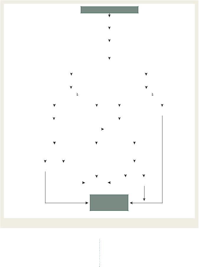
Urgent or emergency valve replacement is recommended for obstructive thrombosis in critically ill patients without serious comorbidity (recommendation class I, level of evidence C: Figure 5). If thrombogenicity of the prosthesis is an important factor, it should be replaced with a less thrombogenic prosthesis.
Fibrinolysis should be considered in:
†Critically ill patients unlikely to survive surgery because of comorbidities or severely impaired cardiac function before developing valve thrombosis.
†Situations in which surgery is not immediately available and the patient cannot be transferred.
†Thrombosis of tricuspid or pulmonary valve replacements, because of the higher success rate and low risk of systemic embolism.
In case of haemodynamic instability a short protocol is recommended, using either intravenous recombinant tissue plasminogen activator 10 mg bolus + 90 mg in 90 minutes with UFH, or streptokinase 1 500 000 U in 60 minutes without UFH. Longer durations of infusions can be used in stable patients.234
Fibrinolysis is less likely to be successful in mitral prostheses, in chronic thrombosis, or in the presence of pannus, which can be difficult to distinguish from thrombus.210,233
Non-obstructive prosthetic thrombosis is diagnosed using TOE, performed after an embolic event, or systematically following mitral valve replacement with a mechanical prosthesis. Management depends mainly on the occurrence of a thromboembolic
event and the size of the thrombus (Figure 6). Close monitoring by TOE is mandatory. The prognosis is favourable with medical therapy in most cases of small thrombus (,10 mm). A good response with gradual resolution of the thrombus obviates the need for surgery. Conversely, surgery should be considered for large (≥10 mm) non-obstructive prosthetic thrombus complicated by embolism (recommendation class IIa, level of evidence C) or which persists despite optimal anticoagulation.217 Fibrinolysis may be considered if surgery is at high risk. However, it should only be used where absolutely necessary because of the risks of bleeding and thromboembolism.
11.2.4Management of thromboembolism
Thromboembolism after valve surgery is multifactorial in origin.203 Although thromboembolic events frequently originate from the prosthesis, many others arise from other sources and are part of the background incidence of stroke and transient ischaemic attack in the general population.
Thorough investigation of each episode of thromboembolism is therefore essential (including cardiac and non-cardiac imaging: Figure 6), rather than simply increasing the target INR or adding an antiplatelet agent. Prevention of further thromboembolic events involves:
†Treatment or reversal of risk factors such as AF, hypertension, hypercholesterolaemia, diabetes, smoking, infection, and prothrombotic blood test abnormalities.
†Optimization of anticoagulation control, if possible with patient self-management, on the basis that better control is more effective than simply increasing the target INR. This should be discussed with the neurologist in case of recent stroke.
†Low-dose aspirin (≤100 mg daily) should be added, if it was not previously prescribed, after careful analysis of the risk-benefit ratio, avoiding excessive anticoagulation.
11.2.5Management of haemolysis and paravalvular leak
Blood tests for haemolysis should be part of routine follow-up after valve replacement. Haptoglobin measurement is too sensitive and lactate dehydrogenase, although non-specific, is better related to the severity of haemolysis. The diagnosis of haemolytic anaemia requires TOE to detect a paravalvular leak (PVL) if TTE is not contributive. Reoperation is recommended if PVL is related to endocarditis, or if PVL causes haemolysis requiring repeated blood transfusions or leading to severe symptoms (recommendation class I, level of evidence C). Medical therapy, including iron supplementation, beta-blockers and erythropoietin, is indicated in patients with severe haemolytic anaemia and PVL not related to endocarditis, where contraindications to surgery
are present, or in those patients unwilling to undergo reoperation.235 Transcatheter closure of PVL is feasible but experience
is limited and there is presently no conclusive evidence to show a consistent efficiency.236 It may be considered in selected patients in whom reintervention is deemed high-risk or is contraindicated.
11.2.6 Management of bioprosthetic failure
After the first 5 years following implantation—and earlier in young patients—yearly echocardiography is required indefinitely
2015 18, October on guest by org/.oxfordjournals.http://eurheartj from Downloaded

2486 |
ESC/EACTS Guidelines |
|
|
Suspicion of thrombosis
Echo (TTE + TOE/fluoroscopy)
Obstructive thrombus
Critically ill
|
|
|
|
|
Yes |
|
|
|
|
|
|
|
|
|
|
|
|
|
|
|
|
|
No |
|
|
|
|
|
|
|
|
|
|||
|
|
|
|
|
|
|
|
|
|
|
|
|
|
|
|
|
|
|
|
|
|
|
|
|
|
|
|
|
|
|
|
|
|
|
|
|
|
|
|
|
|
|
|
|
|
|
|
|
|
|
|
|
|
|
|
|
|
|
|
|
|
|
|
|
|
|
|
|
|
|
|
|
|
|
|
|
|
|
|
|
|
|
|||||||||||||||||||||||||
|
Surgery immediately available |
|
|
|
|
|
|
|
|
Recent inadequate anticoagulation |
|
|
|||||||||||||||||||||||
|
|
|
|
|
|
|
|
|
|
|
|
|
|
|
|
|
|
|
|
|
|
|
|
|
|
|
|
|
|
|
|
|
|
|
|
|
|
|
|
|
|
|
|
|
|
|
|
|
|
|
|
|
|
|
|
|
|
|
|
|
|
|
|
|
|
|
|
|
|
|
|
|
|
|
|
|
|
|
|
|
|
|
|
|
|
|
|
|
|
|
|
|
|
|
|
|
|
|
|
|
|
|
|
|
|
|
|
|
|
|
|
|
|
|
|
|
|
|
|
|
|
|
|
|
|
|
|
|
|
|
|
|
|
|
|
|
|
|
|
|
|||
|
Yes |
|
|
|
|
|
|
No |
|
|
|
|
|
|
|
Yes |
|
|
|
|
|
No |
|
|
|
|
|
||||||||
|
|
|
|
|
|
|
|
|
|
|
|
|
|
|
|
|
|
|
|
|
|
|
|
|
|
|
|
|
|
|
|
|
|
|
|
|
|
|
|
|
|
|
|
|
|
|
|
|
|
|
|
|
|
|
|
|
|
|
|
|
|
|
|
|
|
|
|
|
|
|
|
|
|
|
|
|
|
|
|
|
|
|
|
|
|
|
|
|
|
|
|
|
|
|
|
|
|
|
|
|
|
|
|||||
|
|
|
|
|
|
|
|
|
|
|
|
|
|
|
|
|
IV UFH ± aspirin |
|
|
|
|
|
|
|
|
|
|
|
|
|
|||||
|
|
|
|
|
|
|
|
|
|
|
|
|
|
|
|
|
|
|
|
|
|
|
|
|
|
|
|
|
|
|
|
|
|
||
|
|
|
|
|
|
|
|
|
|
|
|
|
|
|
|
|
|
|
|
|
|
|
|
|
|
|
|
|
|
|
|
|
|
|
|
|
|
|
|
|
|
|
|
|
|
|
|
|
|
|
|
|
|
|
|
|
|
|
|
|
|
|
|
|
|
|
|
|
|
||
|
|
|
|
|
|
|
|
|
|
|
|
|
|
|
|
|
|
|
|
|
|
|
|
|
|
|
|
|
|
||||||
|
|
|
|
|
|
|
|
|
|
|
|
|
|
|
Success |
|
|
|
|
Failure |
|
|
|
|
|
|
|
|
|||||||
|
|
|
|
|
|
|
|
|
|
|
|
|
|
|
|
|
|
|
|
|
|
|
|
|
|
|
|
|
|
|
|
|
|
|
|
|
|
|
|
|
|
|
|
|
|
|
|
|
|
|
|
|
|
|
|
|
|
|
|
|
|
|
|
|
|
|
|
|
|
|
|
|
|
|
|
|
|
|
|
|
|
|
|
|
|
|
|
|
|
|
|
|
|
|
|
|
|
|
|
||||||||
|
|
|
|
|
|
|
|
|
|
|
|
|
|
|
|
|
|
|
|
|
|
|
|
|
|
High risk for surgery |
|
||||||||
|
|
|
|
|
|
|
|
|
|
|
|
|
|
|
|
|
|
|
|
|
|
|
|
|
|
||||||||||
|
|
|
|
|
|
|
|
|
|
|
|
|
|
|
|
|
|
|
|
|
|
|
|
|
|
|
|
|
|
|
|
|
|
|
|
|
|
|
|
|
|
|
|
|
|
|
|
|
|
|
|
|
|
|
|
|
|
|
|
|
|
|
|
|
|
|
|
|
|
|
|
|
|
|
|
|
|
|
|
|
|
|
|
|
|
|
|
|
|
|
|
|
|
|
|
|
|
|
|
|
|
|
|
|
|
|
|
|
|
|
|
|
|
|
|
|
|
|
|
|
|
|
|
|
|
|
|
|
|
|
|
|
|
|
|
|
|
|
|
|
|
||
|
|
|
|
|
|
|
|
|
|
|
|
|
|
|
|
|
|
|
|
|
|
|
|
|
Yes |
|
|
|
No |
|
|
||||
|
|
|
|
|
|
|
|
|
|
|
|
|
|
|
|
|
|
|
|
|
|
|
|
|
|
|
|
|
|
|
|
|
|
|
|
|
|
|
|
|
|
|
|
|
|
|
|
|
|
|
|
|
|
|
|
|
|
|
|
|
|
|
|
|
|
|
|
|
|
|
|
|
|
|
|
|
|
|
|
|
|
|
|
|
|
|
|
|
|
|
|
|
|
|
|
|
|
|
|
|
|
|
|
|
|
|
|
Surgerya |
|
|
|
|
Fibrinolysisa |
|
|
Follow-up |
|
|
|
|
|
|
Fibrinolysisa |
|
|
Surgerya |
|||||||||||||||||
|
|
|
|
|
|
|
|
|
|
|
|
|
|
|
|
|
|
|
|
|
|
|
|
|
|
|
|
|
|
|
|
|
|
|
|
IV UFH = intravenous unfractionated heparin; TOE = transoesophageal echocardiography; TTE = transthoracic echocardiography. aRisk and benefits of both treatments should be individualized.The presence of a first-generation prosthesis is an incentive to surgery.
Figure 5 Management of left-sided obstructive prosthetic thrombosis.
to detect early signs of SVD, leaflet stiffening, calcification, reduced effective orifice area, and/or regurgitation. Auscultatory and echocardiographic findings should be carefully compared with previous examinations in the same patient. Reoperation is recommended in symptomatic patients with a significant increase in trans-prosthetic gradient or severe regurgitation (recommendation class I, level of evidence C). Reoperation should be considered in asymptomatic patients with any significant prosthetic dysfunction, provided they are at low risk for reoperation (recommendation class IIa, level of evidence C). Prophylactic replacement of a bioprosthesis implanted .10 years ago, without structural deterioration, may be considered during an intervention
on another valve or on the coronary arteries (recommendation class IIb, level of evidence C).
The decision to reoperate should take into account the risk of reoperation and the emergency situation. This underlines the need for careful follow-up to allow for timely reoperation.237
Percutaneous balloon interventions should be avoided in the treatment of stenotic left-sided bioprostheses.
Treating bioprosthetic failure by transcatheter valve-in-valve implantation has been shown to be feasible.238,239 Current evidence is limited, therefore it cannot be considered as a valid alternative to surgery except in inoperable or high-risk patients as assessed by a ‘heart team’.
2015 18, October on guest by org/.oxfordjournals.http://eurheartj from Downloaded
ESC/EACTS Guidelines |
2487 |
|
|
Suspicion of thrombosis
|
|
|
|
|
|
|
|
|
|
|
|
|
|
|
|
Echo (TTE + TOE/fluoroscopy) |
|
|
|
|
|
|
|
|
|
|
|
|
||||||||||||||||
|
|
|
|
|
|
|
|
|
|
|
|
|
|
|
|
|
|
|
|
|
|
|
|
|
|
|
|
|
|
|
|
|
|
|
|
|
|
|
|
|
|
|
|
|
|
|
|
|
|
|
|
|
|
|
|
|
|
|
|
|
|
|
|
|
|
|
|
|
|
|
|
|
|
|
|
|
|
|
|
|
|
|
|
|
|
|
|
|
|
|
|
|
|
|
|
|
|
|
|
|
|
|
|
|
|
|
|
|
|
|
|
|
|
|
|
|
|
|
|
|
|
|||||||||||||
|
|
|
|
|
|
|
|
|
|
|
|
|
|
|
|
|
|
Non-obstructive thrombus |
|
|
|
|
|
|
|
|
|
|
|
|
|
|
|
|||||||||||
|
|
|
|
|
|
|
|
|
|
|
|
|
|
|
|
|
|
|
|
|
|
|
|
|
|
|
|
|
|
|
|
|||||||||||||
|
|
|
|
|
|
|
|
|
|
|
|
|
|
|
|
|
|
|
|
|
|
|
|
|
|
|
|
|
|
|
|
|
|
|
|
|
|
|
|
|
|
|
|
|
|
|
|
|
|
|
|
|
|
|
|
|
|
|
|
|
|
|
|
|
|
|
|
|
|
|
|
|
|
|
|
||||||||||||||
|
|
|
|
|
|
|
|
|
|
|
|
|
|
|
|
|
|
Optimize anticoagulation. |
|
|
|
|
|
|
|
|
|
|
|
|
|
|
|
|||||||||||
|
|
|
|
|
|
|
|
|
|
|
|
|
|
|
|
|
|
Follow-up (clinical + echo) |
|
|
|
|
|
|
|
|
|
|
|
|
||||||||||||||
|
|
|
|
|
|
|
|
|
|
|
|
|
|
|
|
|
|
|
|
|
|
|
|
|
|
|
|
|||||||||||||||||
|
|
|
|
|
|
|
|
|
|
|
|
|
|
|
|
|
|
|
|
|
|
|
|
|
|
|
|
|
|
|
|
|
|
|
|
|
|
|
|
|
|
|
|
|
|
|
|
|
|
|
|
|
|
|
|
|
|
|
|
|
|
|
|
|
|
|
|
|
|
|
|||||||||||||||||||
|
|
|
|
|
|
|
|
|
|
|
|
|
Thromboembolism (clinical/cerebral imaging) |
|
|
|
|
|
|
|
|
|
|
|
||||||||||||||||||||
|
|
|
|
|
|
|
|
|
|
|
|
|
|
|
|
|
|
|
|
|
|
|
|
|
|
|
|
|
|
|
|
|
|
|
|
|
|
|
|
|
|
|
|
|
|
|
|
|
|
|
|
|
|
|
|
|
|
|
|
|
|
|
|
|
|
|
|
|
|
|
|
|
|
|
|
|
|
|
|
|
|
|
|
|
|
|
|
|
|
|
|
|
|
|
|
|
|
|
|
|
|
|
|
|
|
|
|
|
|
|
|
|
|
|
|
|
|
|
|
|
|
|
|
|
|
|
|
|
|
|
|
|
|
|
|
|
|
|
|
|
|
|
|
|
|
|
|
|
|
|
|
|
|
|
|
|
|
|
|
|
|
|
|
|
|
|
|
|
|
|
|
|
|
|
|
|
|
|
|
|
|
|
|
|
|
|
|
|
No |
|
|
|
|
|
|
|
|
|
|
|
|
|
|
|
|
|
|
|
|
|
|
|
|
|
Yes |
|
|
|
|
|
||||
|
|
|
|
|
|
|
|
|
|
|
|
|
|
|
|
|
|
|
|
|
|
|
|
|
|
|
|
|
|
|
|
|
|
|
|
|
|
|
|
|
|
|
|
|
|
|
|
|
|
|
|
|
|
|
|
|
|
|
|
|
|
|
|
|
|
|
|
|
|
|
|
|
|
|
|
|
|
|
|
|
|
|
|
|
|
|
|
|
|
|
|
|
|
|
|
|
|
|
|
|
|
|
|
|
|
|
|
|
|
|
|
|
|
|
|
|
|
|||||||||||||||||
|
|
|
|
|
|
Large thrombus ( |
10 mm) |
|
|
|
|
|
|
|
|
|
|
|
|
|
Large thrombus ( |
10 mm) |
|
|||||||||||||||||||||
|
|
|
|
|
|
|
|
|
|
|
|
|
|
|
|
|
|
|
|
|
|
|
|
|
|
|
|
|
|
|
|
|
|
|
|
|
|
|
|
|
|
|
|
|
|
|
|
|
|
|
|
|
|
|
|
|
|
|
|
|
|
|
|
|
|
|
|
|
|
|
|
|
|
|
|
|
|
|
|
|
|
|
|
|
|
|
|
|
|
|
|
|
|
|
|
|
|
|
|
|
|
|
|
|
|
|
|
|
|
|
|
|
|
|
|
|
|
|
|
|
|
|
|
|
|
|
|
|
|
|
|
|
|
|
|
|
|
|
|
|
|
|
|
|
|
|
|
|
|
|
|
|
|
|
|
|
|
|
|
|
|
|
|
|
|
|
|
|
|
|
|
|
|
|
|
|
|||
|
|
|
|
|
Yes |
|
|
|
|
|
|
|
|
No |
|
|
|
|
No |
|
|
|
|
|
|
|
|
|
|
|
|
|
|
Yes |
|
|||||||||
|
|
|
|
|
|
|
|
|
|
|
|
|
|
|
|
|
|
|
|
|
|
|
|
|
|
|
|
|
|
|
|
|
|
|
|
|
|
|
|
|
|
|
||
|
|
|
|
|
|
|
|
|
|
|
|
|
|
|
|
|
|
|
|
|
|
|
|
|
|
|
|
|
|
|
|
|
|
|
|
|
|
|
|
|
|
|
|
|
|
|
|
|
|
|
|
|
|
|
|
|
|
|
|
|
|
|
|
|
|
|
|
|
|
|
|
|
|
|
|
|
|||||||||||||
|
|
|
Optimize |
|
|
|
|
|
|
|
|
|
|
|
|
|
Optimize |
|
|
|
|
|
|
|
|
|
|
|
|
|||||||||||||||
|
anticoagulation. |
|
|
|
|
|
|
|
|
|
|
|
|
|
anticoagulation. |
|
|
|
|
|
|
|
|
|
|
|
|
|||||||||||||||||
|
|
|
|
|
|
|
|
|
|
|
|
|
|
|
|
|
|
|
|
|
|
|
||||||||||||||||||||||
|
|
|
Follow-up |
|
|
|
|
|
|
|
|
|
|
|
|
|
Follow-up |
|
|
|
|
|
|
|
|
|
|
|
|
|||||||||||||||
|
|
|
|
|
|
|
|
|
|
|
|
|
|
|
|
|
|
|
|
|
|
|
|
|
|
|
|
|
|
|
|
|
|
|
|
|||||||||
|
|
|
|
|
|
|
|
|
|
|
|
|
|
|
|
|
|
|
|
|
|
|
|
|
|
|
|
|
|
|
|
|
|
|
|
|
|
|
|
|
|
|
|
|
|
|
|
|
|
|
|
|
|
|
|
|
|
|
|
|
|
|
|
|
|
|
|
|
|
|
|
|
|
|
|
|
|
|
|
|
|||||||||
|
|
|
|
|
|
|
|
|
|
|
|
|
|
|
|
|
|
|
|
|
|
|
|
|
|
|
|
|
|
|
|
|
|
|
||||||||||
|
|
Persistence of |
|
|
|
|
|
Disappearance |
|
|
|
|
|
|
Persistence |
|
|
|
|
|
|
|
|
|
|
|||||||||||||||||||
|
thrombus or TE |
|
|
|
|
|
or decrease |
|
|
|
|
|
|
of thrombus |
|
|
|
|
|
|||||||||||||||||||||||||
|
|
|
|
|
|
|
|
|
|
|
|
|
|
|
|
of thrombus |
|
|
|
|
|
|
|
|
|
|
|
|
|
|
|
|
|
|
|
|
|
|
||||||
|
|
|
|
|
|
|
|
|
|
|
|
|
|
|
|
|
|
|
|
|
|
|
|
|
|
|
|
|
|
|
|
|
|
|
|
|
|
|||||||
|
|
|
|
|
|
|
|
|
|
|
|
|
|
|
|
|
|
|
|
|
|
|
|
|
|
|
|
|
|
|
|
|
|
|
|
|
|
|
|
|||||
|
|
|
|
|
|
|
|
|
|
|
|
|
|
|
|
|
|
|
|
|
|
|
|
|
|
|
|
|
|
|
|
|
|
|
|
|
|
|
|
|||||
|
Yes |
|
|
|
|
No |
|
|
|
|
|
|
|
|
|
|
|
|
|
|
|
|
|
|
|
|
Recurrent TE |
|
|
|
|
|
||||||||||||
|
|
|
|
|
|
|
|
|
|
|
|
|
|
|
|
|
|
|
|
|
|
|
|
|
|
|
|
|
|
|
|
|
|
|
|
|
|
|
||||||
|
|
|
|
|
|
|
|
|
|
|
|
|
|
|
|
|
|
|
|
|
|
|
|
|
|
|
|
|
|
|
|
|
|
|
|
|
|
|
|
|
|
|
|
|
|
|
|
|
|
|
|
|
|
|
|
|
|
|
|
|
|
|
|
|
|
|
|
|
|
|
|
|
|
|
|
|
|
|
|
|
|
|
|
|
|
|
|
|
|
|
|
|
|
|
|
|
|
|
|
|
|
|
|
|
|
|
|
|
|
|
|
|
|
|
|
|
|
|
|
|
|
|
|
|
|
|
|
|
|
|
|
|
|
|
|
|
|
|
|
|
|
|
|
|
|
|
|
|
|
|
|
Follow-up |
|
|
|
|
|
|
No |
|
|
|
|
Yes |
|
|
|
|
|
||||||||||
|
|
|
|
|
|
|
|
|
|
|
|
|
|
|
|
|
|
|
|
|
|
|
|
|
|
|
|
|
|
|
||||||||||||||
|
|
|
|
|
|
|
|
|
|
|
|
|
|
|
|
|
|
|
|
|
|
|
|
|
|
|
|
|
|
|
|
|
|
|
|
|
|
|
|
|
|
|
|
|
Surgery (or fibrinolysis if
surgery is at high risk)
TE = thromboembolism; TOE = transoesophageal echocardiography; TTE = transthoracic echocardiography.
Figure 6 Management of left-sided non-obstructive prosthetic thrombosis.
11.2.7 Heart failure
HF after valve surgery should lead to a search for prosthetic-related complications, deterioration of repair, LV dysfunction or progression of another valve disease. Non-valvular- related causes such as CAD, hypertension or sustained arrhythmias should also be considered. The management of patients with HF should follow the relevant guidelines.13
12. Management during non-cardiac surgery
Cardiovascular morbidity and mortality is increased in patients with VHD (mainly severe VHD) who undergo non-cardiac surgery. Perioperative management of patients with VHD relies
2015 18, October on guest by org/.oxfordjournals.http://eurheartj from Downloaded

2488 |
ESC/EACTS Guidelines |
|
|
on lower levels of evidence than those used for ischaemic heart disease, as detailed in specific ESC Guidelines.227
12.1 Preoperative evaluation
Clinical assessment should search for symptoms, arrhythmias and the presence of a murmur—which justifies echocardiographic examination, particularly in the elderly.
Cardiovascular risk is also stratified according to the type of non-cardiac surgery and classified according to the risk of cardiac complications.227
Each case should be individualized and discussed with cardiologists, anaesthetists (ideally cardiac anaesthetists), surgeons (both cardiac and the ones undertaking the non-cardiac procedure), and the patient and his/her family.
12.2 Specific valve lesions
12.2.1 Aortic stenosis
In patients with severe AS needing urgent non-cardiac surgery, surgery should be performed under careful haemodynamic monitoring.
In patients with severe AS needing elective non-cardiac surgery,
the management depends mainly on the presence of symptoms and the type of surgery (Figure 7).227,240,241
In symptomatic patients, AVR should be considered before non-cardiac surgery. A high risk for valvular surgery should lead to re-evaluation of the need to carry out non-cardiac surgery before considering balloon aortic valvuloplasty or TAVI.
In asymptomatic patients with severe AS, non-cardiac surgery at lowor moderate risk can be performed safely.240 If non-cardiac surgery is at high risk, the presence of very severe AS, severe valve calcification or abnormal exercise test results are incentives to consider AVR first. In asymptomatic patients who are at high risk for valvular surgery, non-cardiac surgery, if mandatory, should be performed under strict haemodynamic monitoring.
When valve surgery is needed before non-cardiac surgery, a bioprosthesis is the preferred substitute, in order to avoid anticoagulation problems during the subsequent non-cardiac surgery.
12.2.2 Mitral stenosis
In asymptomatic patients with significant MS and a systolic pulmonary artery pressure ,50 mmHg, non-cardiac surgery can be performed safely.
Severe AS and need for elective non-cardiac surgery
Symptoms
No |
|
Yes |
|
|
|
|
|
|
|
|
|
Risk of non-cardiac surgerya
Low-moderate |
|
High |
|
|
|
|
|
|
|
|
|
|
|
|
Patient risk |
|
|
|
Patient risk |
|
|||||||
|
|
|
for AVR |
|
|
|
for AVR |
|
|||||||
|
|
|
|
|
|
|
|
|
|
|
|
|
|
|
|
|
|
|
|
|
|
|
|
|
|
|
|
|
|
|
|
|
|
|
|
|
|
|
|
|
|
|
|
|
|
|
|
|
|
|
|
|
|
|
|
|
|
|
|
|
|
|
|
|
High |
|
|
Low |
|
Low |
|
|
High |
|
|||||
|
|
|
|
|
|
|
|
|
|
|
|
|
|
|
|
|
|
|
|
|
|
|
|
|
|
|
|
|
|
|
|
|
|
|
|
|
|
|
|
|
|
|
|
|
|
|
|
Non-cardiac |
|
Non-cardiac |
|
AVR before |
|
Non-cardiac surgery |
surgery |
|
surgery |
|
noncardiac |
|
under strict monitoring |
|
|
under strict |
|
surgery |
|
Consider BAV/TAVIb |
|
||||||
|
|
monitoring |
|
|
|
|
|
|
|
|
|
|
|
|
|
|
|
|
|
|
AS = aortic stenosis; AVR = aortic valve replacement; BAV = balloon aortic valvuloplasty; TAVI = transcatheter aortic valve implantation.
aClassification into three groups according to the risk of cardiac complications (30-day death and myocardial infarction) for non-cardiac surgery (227) (high risk >5%; intermediate risk 1–5%; low risk <1%).
bNon-cardiac surgery performed only if strictly needed.The choice between balloon aortic valvuloplasty and transcatheter aortic valve implantation should take into account patient life expectancy.
2015 18, October on guest by org/.oxfordjournals.http://eurheartj from Downloaded
Figure 7 Management of severe aortic stenosis and elective non-cardiac surgery according to patient characteristics and the type of surgery.

ESC/EACTS Guidelines |
2489 |
|
|
In symptomatic patients or in patients with systolic pulmonary artery pressure .50 mmHg, correction of MS—by means of PMC whenever possible—should be attempted before non-cardiac surgery if it is high risk. If valve replacement is needed, the decision to proceed before non-cardiac surgery should be taken with caution and individualized.
12.2.3 Aortic and mitral regurgitation
In asymptomatic patients with severe MR or AR and preserved LV function, non-cardiac surgery can be performed safely. The presence of symptoms or LV dysfunction should lead to consideration of valvular surgery, but this is seldom needed before non-cardiac surgery. If LV dysfunction is severe (EF ,30%), non-cardiac surgery should only be performed if strictly necessary, after optimization of medical therapy for HF.
12.2.4 Prosthetic valves
The main problem is the adaptation of anticoagulation in patients with mechanical valves, which is detailed in Interruption of anticoagulant therapy (Section 11.2.2.5).
12.3 Perioperative monitoring
Perioperative management should be used to control heart rate (particularly in MS), to avoid fluid overload as well as volume depletion and hypotension (particularly in AS) and to optimize anticoagulation if needed.240
In patients with moderate-to-severe AS or MS, beta-blockers or amiodarone can be used prophylactically to maintain sinus rhythm.241 The use of beta-blockers and statins should be adapted to the risk of ischaemic heart disease according to guidelines.
It is prudent to electively admit patients with severe VHD to intensive care postoperatively.
13. Management during pregnancy
The management of VHD during pregnancy is detailed in the ESC Guidelines on pregnancy.207 In brief, management before and during pregnancy—and planning of delivery—should be discussed between obstetricians, cardiologists and the patient and her family, according to specific guidelines. Ideally, valve disease should be evaluated before pregnancy and treated if necessary. Pregnancy may be discouraged in certain conditions.
13.1 Native valve disease
MS is often poorly tolerated when valve area is ,1.5 cm2, even in previously asymptomatic patients. Symptomatic MS should be treated using bed rest and beta-blockers, possibly associated with diuretics. In the case of persistent dyspnoea or pulmonary artery hypertension despite medical therapy, PMC should be considered after the 20th week in experienced centres. Anticoagulant therapy is indicated in selected cases.207
Complications of severe AS occur mainly in patients who were symptomatic before pregnancy. The risk of HF is low when mean aortic gradient is ,50 mmHg.
Chronic MR and AR are well-tolerated, even when severe, provided LV systolic function is preserved. Surgery under cardiopulmonary bypass is associated with a foetal mortality rate of between 20–30% and should be restricted to the rare conditions that threaten the mother’s life.
13.2 Prosthetic valves
Maternal mortality is estimated at between 1–4% in women with mechanical valves. These patients should be informed of the risks and constraints due to anticoagulant therapy if pregnancy occurs. During the first trimester, in choosing between vitamin K antagonists, UFH, and LMWH, the respective maternaland foetal risks should be weighed up carefully. Vitamin K antagonists are favoured during the second and third trimester until the 36th week, when they should be replaced by heparin.207
The CME text ‘Guidelines on the management of valvular heart disease (version 2012)’ is accredited by the European Board for Accreditation in Cardiology (EBAC). EBAC works according to the quality standards of the European Accreditation Council for Continuing Medical Education (EACCME), which is an institution of the European Union of Medical Specialists (UEMS). In compliance with EBAC/EACCME guidelines, all authors participating in this programme have disclosed potential conflicts of interest that might cause a bias in the article. The Organizing Committee is responsible for ensuring that all potential conflicts of interest relevant to the programme are declared to the participants prior to the CME activities.
CME questions for this article are available at: European Heart Journal http://www.oxforde-learning.com/eurheartj and European Society of Cardiology http://www.escardio. org/guidelines.
References
1.Iung B, Baron G, Butchart EG, Delahaye F, Gohlke-Ba¨rwolf C, Levang OW, Tornos P, Vanoverschelde JL, Vermeer F, Boersma E, Ravaud P, Vahanian A. A prospective survey of patients with valvular heart disease in Europe: the Euro Heart Survey on Valvular Heart Disease. Eur Heart J 2003;24:1231–1243.
2.Nkomo VT, Gardin JM, Skelton TN, Gottdiener JS, Scott CG, Enriquez-Sarano M. Burden of valvular heart diseases: a population-based study. Lancet 2006;368:1005–1011.
3.Carapetis JR, Steer AC, Mulholland EK, Weber M. The global burden of group A streptococcal diseases. Lancet Infect Dis 2005;5:685 –694.
4.Iung B, Cachier A, Baron G, Messika-Zeitoun D, Delahaye F, Tornos P, Gohlke-Ba¨rwolf C, Boersma E, Ravaud P, Vahanian A. Decision-making in elderly patients with severe aortic stenosis: why are so many denied surgery? Eur Heart J 2005;26:2714–2720.
5.Mirabel M, Iung B, Baron G, Messika-Zeitoun D, De´taint D, Vanoverschelde JL, Butchart EG, Ravaud P, Vahanian A. What are the characteristics of patients with severe, symptomatic, mitral regurgitation who are denied surgery? Eur Heart J 2007;28:1358–1365.
6.Van Geldorp MWA, van Gameren M, Kappetein AP, Arabkhani B, de Groot-de Laat LE, Takkenberg JJ, Bogers AJ. Therapeutic decisions for patients with symptomatic severe aortic stenosis: room for improvement? Eur J Cardiothorac Surg 2009;35:953–957.
7.Bach DS, Awais M, Gurm HS, Kohnstamn S. Failure of guidelines adherence for intervention in patients with severe mitral regurgitation. J Am Coll Cardiol 2009; 54:860 –865.
8.Vahanian A, Baumgartner H, Bax J, Butchart E, Dion R, Filippatos G, Flachskampf F, Hall R, Iung B, Kasprzak J, Nataf P, Tornos P, Torracca L, Wenink A, Priori SG, Blanc JJ, Budaj A, Camm J, Dean V, Deckers J, Dickstein K, Lekakis J, McGregor K, Metra M, Morais J, Osterspey A,
2015 18, October on guest by org/.oxfordjournals.http://eurheartj from Downloaded

2490 |
ESC/EACTS Guidelines |
|
|
Tamargo J, Zamorano JL, Angelini A, Antunes M, Garcia Fernandez MA, Gohlke-Baerwolf CG, McMurray J, Otto C, Pierard L, Pomar JL, Prendergast B, Rosenhek R, Sousa Uva M, Tamargo J. Guidelines on the management of valvular heart disease: the Task Force on the Management of Valvular Heart Disease of the European Society of Cardiology. Eur Heart J 2007;28: 230–268.
9.Bonow RO, Carabello BA, Chatterjee K, de Leon AC Jr, Faxon DP, Freed MD, Gaasch WH, Lytle BW, Nishimura RA, O’Gara PT, O’Rourke RA, Otto CM, Shah PM, Shanewise JS. 2008 focused update incorporated into the ACC/ AHA 2006 guidelines for the management of patients with valvular heart disease: a report of the American College of Cardiology/American Heart Association Task Force on Practice Guidelines (Writing Committee to revise the 1998 guidelines for the management of patients with valvular heart disease). Endorsed by the Society of Cardiovascular Anaesthesiologists, Society for Cardiovascular Angiography and Interventions, and Society of Thoracic Surgeons.
J Am Coll Cardiol 2008;52:e1–e142.
10.Habib G, Hoen B, Tornos P, Thuny F, Prendergast B, Vilacosta I, Moreillon P, Antunes M, Thilen U, Lekakis J, Lengyel M, Mu¨ller L, Naber CK, Nihoyannopoulos P, Moritz A, Zamorano JL, Vahanian A, Auricchio A, Bax J, Ceconi C, Dean V, Filippatos G, Funck-Brentano C, Hobbs R, Kearney P, McDonagh T, McGregor K, Popescu BA, Reiner Z, Sechtem U, Sirnes PA, Tendera M, Vardas P, Widimsky P. Document Reviewers, Vahanian A, Aguilar R, Bongiorni MG, Borger M, Butchart E, Danchin N, Delahaye F, Erbel R, Franzen D, Gould K, Hall R, Hassager C, Kjeldsen K, McManus R, Miro´ JM, Mokracek A, Rosenhek R, San Roma´n Calvar JA, Seferovic P, Selton-Suty C, Sousa Uva M, Trinchero R, van Camp G. Guidelines on the prevention, diagnosis, and treatment of infective endocarditis (new version 2009): the Task Force on the Prevention, Diagnosis, and Treatment of Infective Endocarditis of the European Society of Cardiology (ESC). Endorsed by the European Society of Clinical Microbiology and Infectious Diseases (ESCMID) and by the International Society of Chemotherapy (ISC) for Infection and Cancer. Eur Heart J 2009;30:2369–2413.
11.Baumgartner H, Bonhoeffer P, De Groot NMS, de Haan F, Deanfield JE, Galie N,
Gatzoulis MA, Gohlke-Baerwolf C, Kaemmerer |
H, Kilner P, Meijboom F, |
|||
Mulder BJM, Oechslin E, Oliver JM, Serraf |
A, Szatmari A, Thaulow E, |
|||
Vouhe PR, Walma E, Vahanian A, Auricchio A, |
Bax J, Ceconi C, Dean V, |
|||
Filippatos G, Funck-Brentano C, Hobbs R, |
|
Kearney P, |
McDonagh |
T, |
Popescu BA, Reiner Z, Sechtem U, Sirnes |
PA, Tendera |
M, Vardas |
P, |
|
Widimsky P, McDonagh T, Swan L, Andreotti F, Beghetti M, Borggrefe M, Bozio A, Brecker S, Budts W, Hess J, Hirsch R, Jondeau G, Kokkonen J, Kozelj M, Kucukoglu S, Laan M, Lionis C, Metreveli I, Moons P, Pieper PG, Pilossoff V, Popelova J, Price S, Roos-Hesselink J, Sousa Uva M, Tornos P, Trigo Trindade P, Ukkonen H, Walker H, Webb GD, Westby J; Task Force on the Management of Grown-up Congenital Heart Disease of the European Society of Cardiology (ESC). ESC Guidelines for the management of grown-up congenital heart disease (new version 2010). Eur Heart J 2010;31: 2915–2957.
12.Vahanian A, Iung B, Pierard L, Dion R, Pepper J. Valvular heart disease. In: Camm AJ, Lu¨scher TF, Serruys PW, ed. The ESC Textbook of Cardiovascular Medicine, 2nd Edition. Malden/Oxford/Victoria: Blackwell Publishing Ltd; 2009:625–670
13.McMurray JJ, Adamopoulos S, Anker SD, Auricchio A, Bo¨hm M, Dickstein K, Falk V, Filippatos G, Fonseca C, Sanchez MA, Jaarsma T, Køber L, Lip GY,
Maggioni AP, Parkhomenko A, Pieske BM, Popescu BA, Rønnevik PK, Rutten FH, Schwitter J, Seferovic P, Stepinska J, Trindade PT, Voors AA, Zannad F, Zeiher A; ESC Committee for Practice Guidelines (CPG), Bax JJ, Baumgartner H, Ceconi C, Dean V, Deaton C, Fagard R, Funck-Brentano C, Hasdai D, Hoes A, Kirchhof P, Knuuti J, Kolh P, McDonagh T, Moulin C, Popescu BA, Reiner Z, Sechtem U, Sirnes PA, Tendera M, Torbicki A, Vahanian A, Windecker S; Document Reviewers, McDonagh T, Sechtem U, Bonet LA, Avraamides P, Ben Lamin HA, Brignole M, Coca A, Cowburn P, Dargie H, Elliott P, Flachskampf FA, Guida GF, Hardman S, Iung B, Merkely B, Mueller C, Nanas JN, Nielsen OW, Orn S, Parissis JT, Ponikowski P. ESC Guidelines for the diagnosis and treatment of acute and chronic heart failure 2012: The Task Force for the Diagnosis and Treatment of Acute and Chronic Heart Failure 2012 of the European Society of Cardiology. Developed in collaboration with the Heart Failure Association (HFA) of the ESC. Eur Heart J 2012;33: 1787–1847.
14.Popescu BA, Andrade MJ, Badano LP, Fox KF, Flachskampf FA, Lancellotti P, Varga A, Sicari R, Evangelista A, Nihoyannopoulos P, Zamorano JL on behalf of the European Association of Echocardiography, Document Reviewers, Derumeaux G, Kasprzak JD, Roelandt JRTC. Recommendations for training, competence, and quality improvement in echocardiography. Eur J Echocardiogr 2009;10:893–905.
15.Baumgartner H, Hung J, Bermejo J, Chambers JB, Evangelista A, Griffin BP, Iung B, Otto CM, Pellikka PA, Quin˜ones M. Echocardiographic assessment of valve stenosis: EAE/ASE recommendations for clinical practice. Eur J Echocardiogr 2009;10:1–25.
16.Lancellotti P, Tribouilloy C, Hagendorff A, Moura L, Popescu BA, Agricola E, Monin JL, Pierard LA, Badano L, Zamorano JL. European Association of Echocardiography recommendations for the assessment of valvular regurgitation. Part 1: aortic and pulmonary regurgitation (native valve disease). Eur J Echocardiogr 2010; 11:223–244.
17.Lancellotti P, Moura L, Pierard LA, Agricola E, Popescu BA, Tribouilloy C, Hagendorff A, Monin JL, Badano L, Zamorano JL. European Association of Echocardiography recommendations for the assessment of valvular regurgitation. Part
2:mitral and tricuspid regurgitation (native valve disease). Eur J Echocardiogr 2010;11:307–332.
18.Rudski LG, Lai WW, Afilalo J, Hua L, Handschumacher MD, Chandrasekaran K, Solomon SD, Louie EK, Schiller NB. Guidelines for the Echocardiographic Assessment of the Right Heart in Adults: A Report from the American Society of Echocardiography endorsed by the European Association of Echocardiography and the Canadian Society of Echocardiography J Am Soc Echocardiogr 2010; 23:685–713.
19.Lang RM, Badano LP, Tsang W, Adams DH, Agricola E, Buck T, Faletra FF, Franke A, Hung J, Perez de Isla L, Kamp O, Kasprzak JD, Lancellotti P, Marwick TH, McCulloch ML, Monaghan MJ, Nihoyannopoulos P, Pandian NG, Pellikka PA, Pepi M, Roberson DA, Shernan SA, Shirali GS, Sugeng L, Ten Cate FJ, Vannan MA, Zamorano JL, Zoghbi WA. EAE/ASE recommendations for image acquisition and display using three-dimensional echocardiography.
Eur Heart J Cardiovasc Imag 2012;13:1–46.
20.Wijns W, Kolh P, Danchin N, Di Mario C, Falk V, Folliguet T, Garg S, Huber K, James S, Knuuti J, Lopez-Sendon J, Marco J, Menicanti L, Ostojic M, Piepoli MF, Pirlet C, Pomar JL, Reifart N, Ribichini FL, Schalij MJ, Sergeant P, Serruys PW, Silber S, Sousa Uva M, Taggart D, Vahanian A, Auricchio A, Bax J, Ceconi C, Dean V, Filippatos G, Funck-Brentano C, Hobbs R, Kearney P, McDonagh T,
Popescu BA, Reiner Z, Sechtem U, Sirnes PA, Tendera M, Vardas PE, Widimsky P, Alfieri O, Dunning J, Elia S, Kappetein P, Lockowandt U, Sarris G, Vouhe P, von Segesser L, Agewall S, Aladashvili A, Alexopoulos D, Antunes MJ, Atalar E, Brutel de la Riviere A, Doganov A, Eha J, Fajadet J, Ferreira R, Garot J, Halcox J, Hasin Y, Janssens S, Kervinen K, Laufer G, Legrand V, Nashef SA, Neumann FJ, Niemela K, Nihoyannopoulos P, Noc M, Piek JJ, Pirk J, Rozenman Y, Sabate M, Starc R, Thielmann M, Wheatley DJ, Windecker S, Zembala M. Guidelines on myocardial revascularization: The Task Force on Myocardial Revascularization of the European Society of Cardiology (ESC) and the European Association for Cardio-Thoracic Surgery (EACTS). Eur Heart J 2010;31:2501–2555.
21.Picano E, Pibarot P, Lancellotti P, Monin JL, Bonow RO. The emerging role of exercise testing and stress echocardiography in valvular heart disease. J Am Coll Cardiol 2009;54:2251–2260.
22.Monin JL, Que´re´ JP, Monchi M, Petit H, Baleynaud S, Chauvel C, Pop C, Ohlmann P, Lelguen C, Dehant P, Tribouilloy C, Gue´ret P. Low-gradient aortic stenosis, operative risk stratification and predictors for long-term outcome: a multicenter study using dobutamine stress hemodynamics. Circulation 2003;108:319–324.
23.Cawley PJ, Maki JH, Otto CM. Cardiovascular magnetic resonance imaging for valvular heart disease. Circulation 2009;119:468–478.
24.Cueff C, Serfaty JM, Cimadevilla C, Laissy JP, Himbert D, Tubach F, Duval X, Iung B, Enriquez-Sarano M, Vahanian A, Messika-Zeitoun D. Measurement of aortic valve calcification using multislicemulti-slice computed tomography: correlation with haemodynamic severity of aortic stenosis and clinical implication for patients with low ejection fraction. Heart 2011;97:721–726.
25.Ketelsen D, Fishman EK, Claussen CD, Vogel-Claussen J. Computed tomography evaluation of cardiac valves: a review. Radiol Clin North Am 2010;48: 783–797.
26.Kaleschke G, Seifarth H, Kerckhoff G, Reinecke H, Baumgartner H. Imaging decision-making for transfemoral or transapical approach of transcatheter aortic valve implantation. EuroIntervention 2010;6(Suppl G):G20–27.
27.Messika-Zeitoun D, Serfaty JM, Brochet E, Ducrocq G, Lepage L, Detaint D, Hyafil F, Himbert D, Pasi N, Laissy JP, Iung B, Vahanian A. Multimodal assessment of the aortic annulus diameter: implications for transcatheter aortic valve implantation. J Am Coll Cardiol 2010;55:186–194.
28.Plein S, Schulz-Menger J, Almeida A, Mahrholdt H, Rademakers F, Pennell D, Nagel E, Schwitter J, Lombard M. Training and accreditation in cardiovascular magnetic resonance in Europe: a position statement of the working group on cardiovascular magnetic resonance of the European Society of Cardiology. Eur Heart J, 2011;32:793–798.
29.Steadman CD, Ray S, Ng LL, McCann GP. Natriuretic peptides in common valvular heart disease. J Am Coll Cardiol 2010;55:2034–2048.
2015 18, October on guest by org/.oxfordjournals.http://eurheartj from Downloaded