

Examination of the Upper Limbs
Fig. 3.14 Testing the sternocleidomastoid m. The patient at- |
Fig. 3.15 Testing the upper portion of the trapezius m. The ex- |
tempts to turn the head to the left against the examiner’s re- |
aminer places his or her hands on the patient’s shoulders, grasping |
sistance. The right sternocleidomastoid m. contracts. |
the upper edge of the trapezius m. on each side between his or her |
|
thumb and index finger. The patient is then asked to shrug the |
|
shoulders. Unilateral weakness, reduced contraction, or diminished |
|
volume of the trapezius m. can be palpated. |
CN XII: Hypoglossal N.
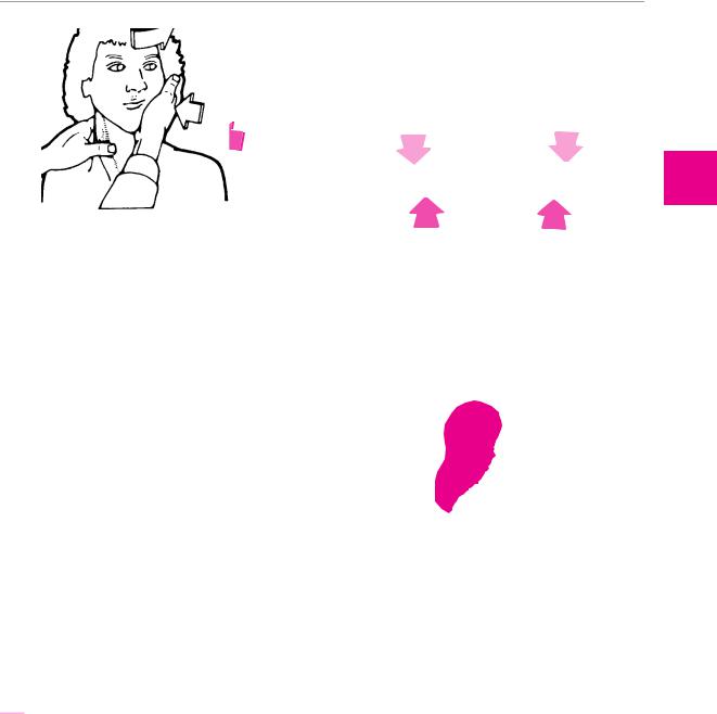
The twelfth cranial nerve is a purely motor nerve to the muscles of the tongue. Lesions of this nerve produce atrophy and weakness of the tongue. A unilateral lesion usually produces a longitudinal furrow; when protruded, the tongue deviates to the weaker side because of the predominant force of the intact contralateral genioglossus m., which “pushes” the tongue across the midline (Fig. 3.16).
Phonation, Articulation, and Speech
Assessment of the patient’s voice and speech is a compulsory part of the neurological examination. The examiner should pay attention to possible hoarseness, to the volume of speech (e. g., hypophonia in Parkinson disease, p. 128), and to possible disturbances of articulation (dysarthria), of the tempo of speech, and of its linguistic form and content (aphasia, p. 41).
Fig. 3.16 Atrophy and weakness of the right half of the tongue in a lesion of the right hypoglossal n.
27
3
The Neurological Examination
Examination of the Upper Limbs
General aspects. The examiner should ask the patient which hand he or she mostly uses, right or left. Only persons who use a pair of scissors, a knife, or a sewing needle with their left hand, or write with the left hand, are true left handers. Any abnormalities of muscle bulk should be noted, in particular isolated atrophy of muscle groups. Fasciculations must be deliberately sought: in our experience, these involuntary contractions of groups of muscle fibers, which induce no movement, can be seen under the skin only by careful observation of the unclothed patient from an adequate distance and
for a sufficient length of time. The trophic state of the skin, the papillary pattern of the fingertips, and the configuration of the nails should also be assessed. Important positive findings include anomalies of finger posture, tremor, or other involuntary movements. The mobility of the larger joints should be tested individually and the pulses in the limbs should be felt. Vascular bruits should be listened for in the supraclavicular fossa when indicated.
|
ARgo |
ARgo leicht |
argo |
Mumenthaler / Mattle, Fundamentals of Neurology © 2006 Thiemealöb auch Argo
All rights reserved. Usage subject to terms and conditions of license.
28 3 The Neurological Examination
Motor Function and Coordination |
smoothly and confidently (Fig. 3.20a). Fluctuating de- |
viation of the finger from the ideal arc is a manifestation |
|
|
of ataxia, indicating either a proprioceptive disturbance |
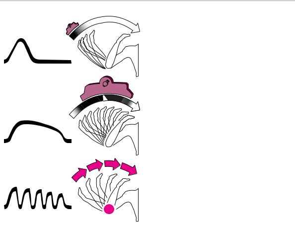
A number of standard tests are used to assess motor |
or a lesion in the ipsilateral cerebellar hemisphere. On |
function and coordination. Diadochokinesis is the abil- |
the other hand, if the deviation first appears when the |
ity to carry out rapid alternating movements, e. g., pro- |
finger is near its target and worsens as it approaches, |
nation and supination of the forearm (Fig. 3.17). Such |
this is called intention tremor (Fig. 3.20c) and is caused |
movements will be abnormally slow (bradydiado- |
by lesions of the dentate nucleus of the cerebellum or of |
chokinesia) or irregular (dysdiadochokinesia) on one side |
its efferent projections. A positive rebound phenome- |
or both in the presence of paresis, extrapyramidal |
non consists of inadequate braking of the normal, small |
processes, and cerebellar diseases. In the postural test, |
rebound movement that occurs when the patient |
the patient extends both arms horizontally in front, in |
isometrically contracts a muscle against the examiner’s |
supination, with eyes closed (Fig. 3.18). An involuntary |
resistance and the resistance is suddenly removed |
sinking, or pronation of one arm (“pronator drift”), or in- |
(Fig. 3.21). If the patient is sitting, the examiner can test |
voluntary flexion at the elbow or wrist, indicates motor |
for rebound of the biceps brachii m. (while taking care |
hemiparesis of central origin; conjugate deviation of |
lest the patient hit himself or herself in the face). If the |
both arms to one side implies an ipsilateral lesion of the |
patient is lying on the examining table, the patient can |
labyrinth or cerebellum. In the arm-rolling test, the |
be asked to stretch out one arm and raise it a short dis- |
patient rapidly rotates the forearms around each other |
tance into the air, then press strongly downward against |
in front of the trunk (Fig. 3.19). Mild hemiparesis is evi- |
the examiner’s resistance. If the examiner suddenly lets |
dent as markedly diminished movement of the affected |
go, a healthy subject will brake the ensuing downward |
limb. In the finger−nose test, the patient keeps his or |
movement of the arm in time, but a patient with hemi- |
her eyes closed and brings the index finger slowly to the |
paresis or cerebellar disfunction will hit the table with |
tip of the nose, in a wide arc. This can normally be done |
it. |
Fig. 3.17 Testing of diadochokinesis by rapid pronation and supi- |
Fig. 3.18 Positional testing of the upper limbs. |
nation of the forearms. |
|
Fig. 3.19 Arm-rolling test. A normal subject rotates both arms to a roughly equal extent, while a patient with central hemiparesis (even if mild!) moves the nonparetic limb much more than the paretic one.
Mumenthaler / Mattle, Fundamentals of Neurology © 2006 Thieme All rights reserved. Usage subject to terms and conditions of license.
Examination of the Upper Limbs 29
Fig. 3.20 Finger−nose test. a Normal, smooth, confident movement. b Ataxic movement c Intention tremor: the closer the finger comes to its target (nose), the more it deviates from the ideal line of approach.
Fig. 3.21 Rebound phenomenon due to a cerebellar lesion. a Method of testing: the examiner’s other hand protects the patient’s face. b When the examiner suddenly releases the patient’s actively flexed arm, the ensuing involuntary flexion should be promptly braked. c Braking is inadequate (the rebound phenomenon is positive) in the presence of an ipsilateral cerebellar lesion
b
a
c
a
b |
c |
3
The Neurological Examination
Muscle Tone and Strength
Muscle tone in the upper limbs can be tested with wide-amplitude passive movement of the radiocarpal joint or of the elbow. The movement should be rapid, but not rhythmic (so that the patient cannot predict its course). Diminished muscle tone (hypotonia) is a characteristic sign of intrinsic muscle lesions, peripheral nerve lesions, ipsilateral cerebellar dysfunction, and hy-
ARgo
perkinetic extrapyramidal diseases. Spasticity is a type of elevated muscle tone produced by lesions of the pyramidal pathway (Fig. 3.22a). The resistance of a spastic upper limb to passive movement is usually strong at first, but may then suddenly give way (“clasp-knife phenomenon”); alternatively, it may increase on continued passive movement. Rigidity is a viscous or waxy resistance to passive movement that can be felt to an equal extent throughout the entire movement; it is
ARgo leicht argo
Mumenthaler / Mattle, Fundamentals of Neurology © 2006 Thiemealöb auch Argo
All rights reserved. Usage subject to terms and conditions of license.
30 3 The Neurological Examination
Spasticity |
Rigidity |
Cogwheel phenomenon |
Fig. 3.22 Abnormalities of muscle tone and the cogwheel phe- |
nomenon. |
Fig. 3.23 Testing for the cogwheel phenomenon in the radiocarpal joint. The examiner fixes the patient’s forearm with one hand, grasps the patient’s fingers with the other, and moves them slowly (but not rhythmically) back and forth.
Table 3.4 Grading of muscle strength.
The 0−5 scale of the British Medical Research Council (MRC).
M0 = no muscle contraction
M1 = visible contraction not resulting in movement
M2 = movement of the body part only when the effect of gravity is eliminated
M3 = movement against gravity
M4 = movement against moderate resistance
M5 = full strength
(Grades M3 and M4 can be optionally subdivided by adding plus or minus signs)
most commonly found in Parkinson disease (Fig. 3.22b). The accompanying parkinsonian cogwheel phenomenon is best appreciated at the radiocarpal joint. The examiner should fix the patient’s forearm with one hand, grasp the patient’s fingertips with the other, and alternately flex and extend the radiocarpal joint, slowly and with a wide excursion, but not in perfect rhythm (Fig. 3.23). The examiner will then feel multiple, brief impulses of resistance at irregular intervals, giving the overall impression of a saccadic movement (Fig. 3.22c). Elevated muscle tone may also result from active opposition to passive movement when the patient apparently cannot relax the muscular contraction. This phenomenon, known by the German term Gegenhalten (“opposition”), is seen in frontal lobe lesions.
Muscle strength is tested in groups of muscles that carry out a single movement, or, if necessary, in individual muscles. The patient is asked to contract the corresponding muscle(s) actively against the examiner’s resistance. The examiner then judges the strength of contraction at the end-point of the related movement. Thus, the examiner tests biceps strength by trying to extend the patient’s flexed elbow against resistance and triceps strength by trying to flex the extended elbow against resistance. The evaluation of potential lesions of individual nerve roots or peripheral nerves requires specific testing of the particular muscles or muscle
groups innervated by these nerves (see p. 208). For the purpose of documentation, muscle strength can be graded semiquantitatively with the MRC scale shown in Table 3.4. Incomplete paralysis is called paresis and complete paralysis is called plegia. Further terms describe the distribution of weakness in the body: hemiparesis or hemiplegia affects one side of the body, paraparesis or paraplegia affects both lower limbs, and quadriparesis or quadriplegia affects all four limbs (less common synonyms for the last two are tetraparesis and tetraplegia). Paralysis of both arms, or brachial diplegia, is a rare occurrence.
Reflexes
Types of reflexes. Reflexes are processes that are induced by a specific stimulus, always take the same course, and cannot be voluntarily influenced by either the patient or the examiner. For the intrinsic muscle reflexes, alternatively called proprioceptive muscle reflexes, the site (muscle) of the eliciting stimulus is the same as that of the reflex contraction; for extrinsic (exteroceptive) reflexes, the stimulus and the response are at different sites and the afferent and efferent arms of the reflex loop, therefore, belong to different peripheral nerves or segmental nerve roots. Extrinsic reflexes become less intense (habituate) on repeated stimulation.
Mumenthaler / Mattle, Fundamentals of Neurology © 2006 Thieme All rights reserved. Usage subject to terms and conditions of license.

Examination of the Upper Limbs 31
Pathological reflexes are usually not seen in normal in- |
important reflexes, and the segmental nerve roots and |
dividuals, or only up to a certain age; they are found in |
peripheral nerves that mediate them, are listed in Tables |
various disease processes affecting the CNS. Some |
3.5−3.7. |
pathological reflexes are of the extrinsic type. The more |
|
Table 3.5 The most important normal intrinsic muscle reflexes
Reflex |
Stimulus |
Response |
Muscle(s) |
Peripheral |
Segment |
|
|
|
|
nerve |
|
|
|
|
|
|
|
Masseter reflex
Trapezius reflex
Scapulohumeral reflex
Biceps reflex
Brachioradialis reflex (“radial periosteal reflex”)
Pectoralis reflex
Triceps reflex
Thumb reflex
Wrist reflex
Finger flexor reflex
Trömner reflex
Adductor reflex
Quadriceps femoris reflex (“patellar tendon reflex,” “knee-jerk reflex”)
Tibialis posterior reflex
Peroneus muscle reflex (foot extensor reflex)
Semimembranosus and semitendinosus reflex
Biceps femoris reflex
Triceps surae reflex (“Achilles reflex,” “ankle-jerk reflex”)
Toe flexor reflex (Rossolimo sign)
tap on the chin or a tongue depressor laid on the lower teeth, with slightly opened mouth
tap on the lateral attachment of the trapezius to the coracoid process
tap on the medial edge of the lower half of the scapula
tap on the biceps tendon with bent elbow
tap on the distal end of the radius with lightly bent elbow and pronated forearm
tap on the scapulohumeral joint from anteriorly
tap on the triceps tendon with bent elbow
tap on the flexor pollicis longus tendon in the distal third of the forearm
tap on the dorsum of the wrist, proximal to the radiocarpal joint
tap on the examiner’s thumb, which is laid in the palm of the patient’s hand; or, tap on the flexor tendons on the volar surface of the wrist
patient’s hand held at the middle finger; tap on the volar side of the distal phalanx of the middle finger
tap on the medial condyle of the femur
tap on the quadriceps tendon below the patella with the knee lightly flexed
tap on the tibialis posterior tendon behind the medial malleolus
foot lightly flexed and supinated; examiner’s finger placed over the distal end of the metatarsal bones; tap on the finger, especially over the 1st and 2nd metatarsal bones
tap on the tendon of the medial knee flexors (patient prone, knee lightly flexed and relaxed)
tap on the tendon of the lateral knee flexors (patient prone, knee lightly flexed and relaxed)
tap on the Achilles tendon (knee lightly flexed, foot in right-angle posture)
tap on the pads of the toes
brief mouth closure |
masseter m. |
trigeminal n. |
V |
movement |
|
|
|
shoulder elevation |
trapezius m. |
accessory n. |
XI C3−C4 |
adduction and ex- |
infraspinatus and |
suprascapular |
C4−C6 |
ternal rotation of |
teres minor mm. |
and axillary nn. |
|
the dependent arm |
|
|
|
elbow flexion |
biceps brachii m. |
musculocu- |
C5−C6 |
|
|
taneous n. |
|
elbow flexion |
brachioradialis m. |
radial and |
C5−C6 |
|
(biceps brachii |
musculocu- |
|
|
and brachialis |
taneous nn. |
|
|
mm.) |
|
|
forward movement |
pectoralis major |
medial and |
C5−T4 |
of the shoulder |
and minor mm. |
lateral pectoral |
|
|
|
nn. |
|
elbow extension |
triceps brachii m. |
radial n. |
C7−C8 |
flexion of the thumb |
flexor pollicis lon- |
median n. |
C6−C8 |
at the inter- |
gus m. |
|
|
phalangeal joint |
|
|
|
extension of the |
wrist extensors |
radial n. |
C6−C8 |
wrist and fingers (in- |
and long exten- |
|
|
constant) |
sors of the fingers |
|
|
flexion of the fin- |
flexor digitorum |
median (ulnar) |
C7−C8 |
gers (and of the |
superficialis m. |
n. |
|
wrist) |
(flexores carpi |
|
|
|
mm.) |
|
|
flexion of the distal |
flexor digitorum |
median (ulnar) |
C7−C8 (T1) |
phalanges of the fin- |
profundus m. |
n. |
|
gers (including the |
|
|
|
thumb) |
|
|
|
leg adduction |
adductors |
obturator n. |
L2−L4 |
knee extension |
quadriceps |
femoral n. |
(L2) L3−L4 |
|
femoris m. |
|
|
supination of the |
tibialis posterior |
tibial n. |
L5 |
foot (inconstant) |
m. |
|
|
dorsiflexion and pro- |
long extensors of |
peroneal n. |
L5−S1 |
nation of the foot |
the foot and toes, |
|
|
|
peronei |
|
|
palpable muscle |
semimembrano- |
sciatic n. |
S1 |
contraction |
sus and semiten- |
|
|
|
dinosus mm. |
|
|
muscle contraction |
biceps femoris m. |
sciatic n. |
S1−S2 |
plantar flexion of |
triceps surae m. |
tibial n. |
S1−S2 |
the foot |
(and other plantar |
|
|
|
flexors) |
|
|
flexion of the toes |
flexor digitorum |
tibial n. |
S1−S2 |
|
and flexor hallucis |
|
|
|
longus mm. |
|
|
|
ARgo |
ARgo leicht |
argo |
Mumenthaler / Mattle, Fundamentals of Neurology © 2006 Thiemealöb auch Argo
All rights reserved. Usage subject to terms and conditions of license.
3
The Neurological Examination

32 |
3 The Neurological Examination |
|
|
|
|
||
|
|
|
|
|
|
||
|
|
Table 3.6 The most important normal extrinsic muscle reflexes |
|
|
|
||
|
|
|
|
|
|
|
|
|
|
Reflex |
Stimulus |
Response |
Muscle(s) |
Peripheral nerve |
Segment |
|
|
|
|
|
|
|
|
|
|
|
|
|
|
|
|
|
|
Pupillary |
incident light, convergence |
constriction |
constrictor |
optic and oculo- |
dien- |
|
|
reflex |
|
|
pupillae m. |
motor nn. |
cephalon, |
|
|
|
|
|
|
|
midbrain, |
|
|
|
|
|
|
|
pons |
Corneal reflex gently touching the cornea from the side, e. g., with a wisp of cotton or piece of tissue paper; the eye looks medially
Bell phenome- attempted active eye closure non (palpebro- while the examiner holds the oculogyric re- upper lids open
flex)
eye closure (and simul- |
orbicularis |
trigeminal and |
midpons |
taneous upward move- |
oculi m. |
facial nn. |
|
ment of the globes: Bell |
|
|
|
phenomenon) |
|
|
|
the globes normally turn |
superior rectus |
trigeminal and |
pons |
upward |
and inferior ob- |
oculomotor nn. |
|
|
lique mm. |
|
|
Auriculopalpe- |
sudden noise from a source that |
bral reflex |
the patient cannot see |
Palatal reflex and |
touching the soft palate or the |
pharyngeal (gag) |
posterior pharyngeal wall with a |
reflex |
tongue depressor |
Mayer reflex of |
forced passive flexion of the |
the proximal |
proximal interphalangeal joints |
interphalangeal |
of the 3rd and 4th fingers |
joint |
|
|
|
Abdominal skin |
rapidly and lightly stroking the |
reflex |
abdominal skin from lateral to |
|
medial |
Cremaster reflex |
stroking the skin on the upper |
(in males) |
medial surface of the thigh (or |
|
pinching the proximal adductor |
|
muscles) |
blink |
orbicularis oculi |
vestibulocochlear |
caudal |
|
m. |
and facial nn. |
pons |
elevation of the palatal veil |
palatal and |
glossopharyngeal |
medulla |
and symmetrical contrac- |
pharyngeal |
and vagus nn. |
|
tion of the posterior |
musculature |
|
|
pharyngeal wall |
|
|
|
adduction and opposition |
adductor polli- |
ulnar and median |
C6−T1 |
of the first metacarpal |
cis m., op- |
nn. |
|
bone |
ponens pollicis |
|
|
|
m. |
|
|
movement of the abdomi- |
abdominal |
intercostal nn., |
T6−T12 |
nal skin, including the |
musculature |
hypogastric n., |
|
umbilicus, toward the side |
|
and ilioinguinal n. |
|
of the stimulus |
|
|
|
retraction of the testes |
cremaster m. |
genital branch of |
L1−L2 |
|
|
the genitofemoral |
|
|
|
n. |
|
Gluteal reflex stroking the skin over the gluteus maximus m.
Bulbocavernosus gently pinching the glans penis; reflex pinprick on the skin of the dor-
sum of the penis
Anal reflex pinprick on the skin of the perianal region or perineum, patient in lateral decubitus position with flexed hip and knee
contraction of the gluteus |
gluteus medius |
superior and infe- |
L4−S1 |
maximus m. (inconstant) |
m., gluteus |
rior gluteal nn. |
|
|
maximus m. |
|
|
contraction of the bulbo- |
bulbo-caverno- |
pudendal n. |
S3−S4 |
cavernosus m. (visible at |
sus m. |
|
|
the root of the penis in the |
|
|
|
perineum, or palpable by |
|
|
|
rectal examination) |
|
|
|
visible contraction of the |
external anal |
pudendal n. |
S3−S5 |
anus |
sphincter |
|
|
Table 3.7 The most important pathological reflexes
Reflex |
Stimulus |
Response |
Significance |
|
|
|
|
Orbicularis oculi reflex |
tap on the glabela or on a fin- |
narrowing of the palpebral |
exaggerated in supranuclear lesions |
(glabellar reflex, |
ger applied to the lateral edge |
fissure by contraction of the |
of the corticopontine pathway and in |
nasopalpebral reflex) |
of the orbit while the orbicu- |
orbicularis oculi m. (possibly |
extrapyramidal diseases |
|
laris oculi m. is contracted |
bilaterally) |
|
Corneomandibular reflex |
like the corneal reflex; mouth |
the jaw deviates to the side |
release of an older functional syn- |
(winking jaw) |
slightly open |
opposite the stimulus |
ergy between the orbicularis oculi m. |
|
|
|
and the lateral pterygoid m.; due to |
|
|
|
an ipsilateral lesion of the corticobul- |
|
|
|
bar pathway, lacunar state, or bulbar |
|
|
|
palsy |
Marcus Gunn phenomenon |
opening the mouth and moving |
a previously ptotic eyelid is very |
(winking jaw) |
the jaw |
strongly elevated |
Bulldog reflex |
placing a tongue depressor |
the patient bites down so hard |
|
between the patient’s teeth |
that the head can be lifted by |
|
|
the tongue depressor |
proof that ptosis is not due to peripheral paresis or myasthenia
release phenomenon (disinhibition) due to diffuse cortical injury, e. g., postanoxic
Continued
Mumenthaler / Mattle, Fundamentals of Neurology © 2006 Thieme All rights reserved. Usage subject to terms and conditions of license.

Examination of the Upper Limbs 33
Table 3.7 The most important pathological reflexes (continued)
Reflex |
Stimulus |
Response |
Significance |
|
|
|
|
Orbicularis oris reflex |
gentle tap on a finger or |
(snout reflex, nasomental |
tongue depressor placed on the |
reflex) |
lateral corner of the mouth or |
|
on the lips (can sometimes also |
|
be elicited from the glabella) |
Suck reflex |
slowly, gently stroking the lips |
Wartenberg reflex |
forceful passive flexion of the |
(“thumb sign”) |
2nd through 5th fingers |
|
|
Palmomental reflex |
intensely stroking the ball of |
(exaggerated or |
the thumb or the palm of the |
asymmetrical) |
hand with a fingernail or |
|
wooden stick |
Grasp reflex |
stroking the palm |
Grasping and groping |
object brought near the palm |
(magnet phenomenon) |
of the (conscious) patient |
contraction of the orbicularis |
absent or only faintly present in nor- |
oris m. with pursing of the lips |
mal individuals; exaggerated as a re- |
|
sult of lesions affecting the supranu- |
|
clear corticopontine pathways (sta- |
|
tus lacunaris, multi-infarct dementia, |
|
extrapyramidal diseases such as |
|
Parkinson disease) |
sucking and, possibly, swallowing movements; occasionally, biting; mouth opening and turning of the head toward the stimulus (termed a magnet reaction if already present when an object is brought near the mouth)
flexion of the thumb
contraction of the ipsilateral chin muscles
severe, diffuse brain injury, decorticate state; e. g., in apallic syndrome after anoxia or severe traumatic brain injury (normal in infants; pathological release phenomenon in later life)
indicates a pyramidal tract lesion
in diffuse cerebral injury (multi-in- farct syndrome, brain atrophy, postanoxic); if unilateral, indicates contralateral brain lesion
finger flexion, possibly grasping normal in infants; later a sign of dif- |
||
of the stimulating object |
|
fuse brain injury (mainly in the fron- |
the hand follows the presented |
tal lobes); seen contralateral to a |
|
|
frontal lobe lesion or ipsilateral to a |
|
object like a magnet, making |
|
|
lesion in the basal ganglia |
||
grasping movements |
|
|
Gegenhalten |
attempted passive stretching of |
the patient actively and in- |
in diffuse frontal lobe disease and |
|
a muscle (e. g., pushing down |
tensely contracts the muscle in |
lesions of the basal ganglia |
|
on the lower jaw, or forcibly ex- |
question, preventing passive |
|
|
tending the flexed fingers) |
stretch (in the absence of |
|
|
|
generalized negativism) |
|
Mass reflexes of the lower e. g., forceful passive flexion of
limbs in paraplegia |
the toes and forefoot (Marie− |
|
|
Foix handgrip) |
|
|
stroking the lateral edge of the |
|
Babinski reflex |
||
(Fig. 3.31) |
foot from the heel to the 5th |
|
|
toe (or else transversely across |
|
|
the plantar arch) |
|
Oppenheim Reflex |
forcefully stroking the anterior |
|
(Fig. 3.31) |
margin of the tibia from proxi- |
|
|
mal to distal (painful) |
|
Gordon Reflex |
forcefully stroking or squeezing |
|
(Fig. 3.31) |
the calf muscles |
|
reveals the intactness of the spinal reflex arc, and therefore of the peripheral nervous system (useful as a “trick” facilitating the nursing care of patients with spastic rigidity)
indicates a lesion of the corticospinal (pyramidal) pathway on the corresponding side
3
The Neurological Examination
Intrinsic muscle reflexes of the upper limb. The intrinsic muscle reflexes are elicited by a rapid, and adequately forceful, blow on the tendon of a muscle or on the bone to which the tendon is attached. The resulting, transient stretching of the muscle excites receptors in the muscle spindles, in which afferent impulses are generated. These travel to the spinal cord and excite the α-motor neurons innervating the stimulated muscle (usually by way of interneurons at the same segmental level). The upper limb reflexes that are usually tested are the triceps, biceps, and radial periosteal reflexes (Table 3.5). The last-named reflex is elicited by a tap on the styloid process of the radius; this is followed by contraction, not only of the brachioradialis m., but also of
ARgo
the biceps and brachialis mm. The elicitation of these reflexes is illustrated in Fig. 3.24.
The two important finger flexor reflexes are essentially variations of the same reflex. The Trömner reflex is elicited by a rapid tap on the pads of the patient’s lightly flexed fingers. The response consists of flexion of the distal interphalangeal joints of the fingers and thumb (only in the hand that was stimulated, not in the other hand). To elicit Hoffmann sign, the examiner gently grasps the distal phalanx of one of the patient’s fingers (usually the middle finger) between his or her own thumb and index finger, then lets it snap back as the thumb slides off the patient’s fingernail. The response is the same as in the Trömner reflex (Fig. 3.25).
ARgo leicht argo
Mumenthaler / Mattle, Fundamentals of Neurology © 2006 Thiemealöb auch Argo
All rights reserved. Usage subject to terms and conditions of license.

34 3 The Neurological Examination
Biceps reflex |
|
Triceps reflex |
Radial periosteal reflex |
|
|
|
|
Fig. 3.24 Elicitation of the intrinsic muscle reflexes of the upper limb.
Fig. 3.25 Elicitation of the Trömner reflex.
The more common abnormalities of the intrinsic muscle reflexes and their significance are presented in Table 3.8.
Facilitating maneuvers. Initially faint or not clearly elicitable intrinsic muscle reflexes can be enhanced with various maneuvers based on the principle that preloading of the intrafusal muscle spindle fibers makes them more sensitive to stretch. Forceful contraction of practically any muscle group in the body results in a generalized sensitization of all muscle spindle fibers. Thus, all of the intrinsic muscle reflexes can be made stronger by having the patient forcefully lift his or her head from the headrest (in the supine position), clench the teeth, make fists, strongly plantar-flex the feet, or interlock the hands and pull hard (this is called the Jendrassik handgrip). These maneuvers are illustrated in Fig. 3.26.
Pyramidal tract signs in the upper limb. Lesions along the pyramidal pathway produce characteristic changes in the pattern of the reflexes that are normally present, as well as other, pathological reflexes that are normally absent. Evidence for a lesion of the pyramidal pathway is generally less obvious in the upper limb than in the
Table 3.8 Significance of the more common abnormalities of the intrinsic muscle reflexes
Abnormality |
Significance |
Remarks |
|
|
|
Apparent absence of all reflexes |
very weak reflexes, or inadequate examining |
facilitation maneuvers, e. g., Jendrassik handgrip |
|
technique |
|
True generalized areflexia |
polyneuropathy, polyradiculopathy |
sensory deficit, perhaps paresis |
|
anterior horn cell disease |
muscle atrophy without sensory deficit |
|
myopathy |
(same) |
|
Adie syndrome |
inspect pupils |
|
congenital areflexia |
often familial |
Absence of an individual reflex |
nerve root lesion |
e. g., triceps reflex (C7), Achilles reflex (S1) |
or reflexes |
peripheral nerve lesion |
e. g., biceps reflex (musculocutaneous n.), |
|
|
Achilles reflex (femoral n.) |
Very weak reflexes |
usually without pathological significance |
often seen in older patients |
Very brisk reflexes |
if generalized, often without pathological sig- |
particularly in younger patients |
|
nificance |
|
Pathologically exaggerated |
“pyramidal tract signs,” spasticity |
compare sides (hemiparesis?) and compare upper |
reflexes |
|
with lower limbs (paraparesis?) |
Positive Hoffmann sign and |
normal if symmetrical and without other, |
|
Trömner reflex |
accompanying “pyramidal tract signs” |
|
|
|
|
Mumenthaler / Mattle, Fundamentals of Neurology © 2006 Thieme All rights reserved. Usage subject to terms and conditions of license.