

clavien_atlas_of_upper_gastrointestinal_and_hepato-pancreato-biliary_surgery2007-10-01_3540200045_springer
.pdf132 |
SECTION 2 |
Esophagus, Stomach and Duodenum |
|
|
|
STEP 2 |
Endoscopy and preparation of the diverticulum |
|
|
|
|
The endoscope is inserted into the esophageal lumen at the level of the diverticulum. By using insufflation and transillumination, the diverticular pouch is defined to allow safe dissection. The pouch is thoroughly dissected until the neck of the diverticulum is completely clear of all adherent tissue, with care to avoid injury to the
pleural sacs.
STEP 3 |
Resection |
|
An endoscopic stapling device is introduced through the operative trocar in the left |
|
|
|
upper quadrant and is advanced to the level of the diverticular neck. The stapler jaws |
|
are closed under simultaneous endoscopic control; the endoscope is advanced into the |
|
stomach to prevent too much mucosa being excised, and it is then withdrawn. Once |
|
this is confirmed, the stapler is fired and mucosal closure is verified. Additional stapler |
|
applications may be necessary to completely incise and close the diverticular neck. |
|
The pouch is removed and the staple line is inspected using the endoscope. |
Epiphrenic Diverticula |
133 |
|
|
STEP 4 |
Myotomy |
|
A myotomy is performed on the opposite esophageal wall, with longitudinal and circular |
|
|
|
muscular fibers divided and the submucosal plane carefully dissected using |
|
the acoustic scalpel. The myotomy is extended proximally above the upper limit of the |
|
diverticulum and distally for almost 1.5cm onto the cardia using either acoustic scalpel |
|
or endo-scissors. |
STEP 5 |
Dor fundoplication |
|
|
The hiatus is closed posteriorly with two or three interrupted sutures. The anterior wrap |
|
|
||
|
is performed by suturing the gastric fundus to the muscular edges of the myotomy |
|
|
(Dor type procedure) and the gastric fundus is also sutured to the superior part of the |
|
|
crura. The fundus is sutured to the right crus and to both the right and left sides of the |
|
|
esophagus. |
|
|
Video-Assisted Thoracoscopic Approach |
|
|
Access |
|
|
Positioning in a right lateral position as for left posterolateral thoracotomy. |
|
|
|
The left lung is deflated using a double lumen endotracheal tube. Ports are inserted |
|
as follows: |
|
|
■ |
10mm at the 6th intercostal space posterior axillary line for the camera |
|
■ |
10mm at the 5th intercostal space at the anterior border of the axillary line for |
|
|
retraction of the lung |
|
■ |
10mm at the 3rd intercostal space for working cannula |
|
■ |
12mm at the mid-axillary line for working cannula |
|
|
The procedure is as for the open procedure. |

134 |
SECTION 2 |
Esophagus, Stomach and Duodenum |
|
|
|
|
|
|
Standard Postoperative Investigations |
|
|
|
■ |
A water-soluble non-ionic contrast swallow is performed on day 5 postoperatively: |
|
|
|
– To exclude suture line leak |
|
|
|
– To verify the position of the wrap |
|
|
|
– To ensure that no significant obstruction has developed |
|
|
|
– To provide an impression of gastric emptying |
|
|
■ |
Carbonated beverages and very large meals must be avoided in the early post- |
|
|
|
operative period |
|
Postoperative Complications
■Early (days to weeks):
–Hemorrhage
–Suture-line leak (if symptomatic then proceed to thoracotomy and repair, if asymptomatic then manage conservatively)
–Pleural effusion
–Empyema
–Subphrenic abscess
■Late (months to years):
–Recurrence of diverticulum. If symptomatic this is managed by open surgery.
–Gastro-esophageal reflux with stricture (treated by dilatation and anti-reflux medication)
–Para-esophageal hernia (may require surgical therapy and repair of the hernia)
Tricks of the Senior Surgeon
■Verification of impermeability of the suture line is confirmed using insufflation of air via the gastroscope in conjunction with insertion of saline via the laparoscope.
■Non-ionic contrast media is preferred to Gastrografin for the postoperative swallow as it provides superior images and is less reactive if aspirated or inadvertently leaks into thorax or abdomen.
■Small diverticula may be treated by plication and myotomy.
■Gastric dysparesis. This usually settles with pro-kinetic agents, but may require pyloroplasty.
■Intraoperative upper gastrointestinal endoscopy facilitates transillumination of the diverticulum, safe staple placement as well as suture line evaluation with insufflation.

|
Techniques of Local Esophagoplasty |
|
|
in Short Esophageal Strictures |
|
|
Asad Kutup, Emre F. Yekebas, Jakob R. Izbicki |
|
|
Introduction |
|
|
Local esophagoplasty is indicated for patients with short esophageal strictures when |
|
|
endoscopic therapies including bougienage and balloon dilatation are not effective. |
|
|
Indications and Contraindications |
|
|
|
Short peptic stricture |
Indications |
■ |
|
|
■ |
Short scarred stenosis |
|
|
Suspicion of malignancy |
Contraindications |
■ |
|
Preoperative Investigations/Preparation for the Procedure
■Esophagogastroscopy with multiple biopsies.
■Special preparation: no peculiarities compared to the previously mentioned operations.
■Anesthesia: endotracheal anesthesia; in case of intrathoracic stenosis, a tracheal tube with the possibility of separate ventilation should be used.
Procedure
Positioning and Access
These depend on the location of the stenosis.
■Collar stenosis: left cervical approach, supination.
■Intrathoracic stenosis: right anterolateral thoracotomy using 4–6th intercostal spaces (ICS).
■Stenosis of the abdominal part of the esophagus: “inverted T-incision” or median laparotomy.
136 |
SECTION 2 |
Esophagus, Stomach and Duodenum |
|
|
|
STEP 1 |
Resection and technique of anastomosis |
|
|
|
|
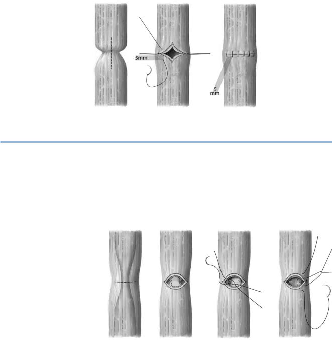
After resection of the stenotic segment, mobilization of the proximal and distal end of the esophagus restores the basis for reconstruction of the continuity with single layer end-to-end anastomosis in single suture technique. The distance of each suture should be 5mm. Each stitch should be placed 5mm distant to the anastomosis.
In stenosis shorter than 1cm, stricturoplasty according to Heineke Mikulicz should be performed. Therefore a longitudinal esophagectomy is performed using a single layer transverse closure with mattress sutures.
STEP 2 |
In case of limited stenosis |
|
In stenosis limited to mucosa and submucosa, a transverse transection of the esophageal |
|
|
|
musculature over the stenosis suffices (A-1). The anterior part of the |
|
stricture is resected (A-2). Reconstruction of the continuity of the mucosa and submu- |
|
cosa in the posterior wall area (A-3) is done followed by transverse closure of the |
|
anterior wall with single layer mattress sutures (A-4). |
A-1 |
|
A-2 |
|
A-3 |
|
A-4 |
Techniques of Local Esophagoplasty in Short Esophageal Strictures |
137 |
|
|
|
|
STEP 3 |
In case of prestenotic dilatation |
|
|
|
|
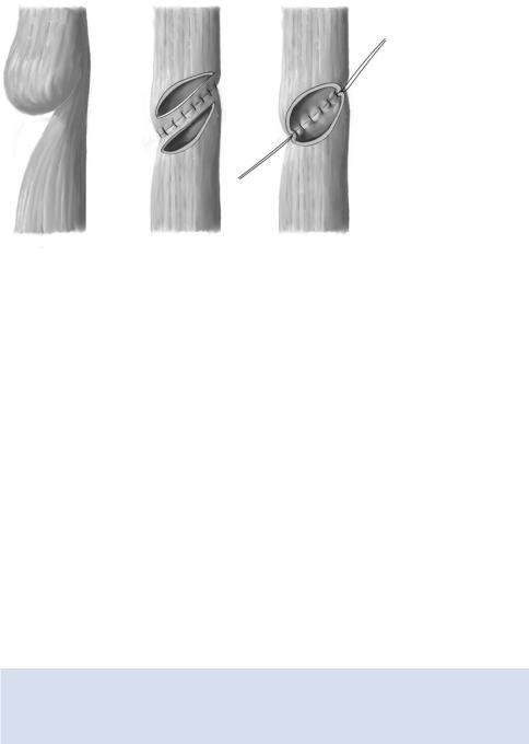
In case of a prestenotic dilatation of the esophagus, side-to side esophagostomy of the preand poststenotic esophageal segment should be performed.
In all local esophagoplasties an intraluminally placed tube (large gastric tube) and abundant external drainage are obligatory.
Standard Postoperative Investigations
■Daily check for insufficiency of the esophagoplasty
■If problems occur after removal of the drain, a contrast study with water-soluble contrast is recommended
■In case of early detected insufficiency: esophagoscopy with the feasibility of stenting
Postoperative Complications
■Leakage
■Mediastinitis
■Pleura empyema
■Peritonitis
■Re-stenosis
Tricks of the Senior Surgeon
■In case of caustic ingestions, organs adjacent to the esophagus, i.e., trachea, may be involved. In these instances, preparation of the esophagus has to be performed carefully.

Operation for Achalasia
Luigi Bonavina, Alberto Peracchia
Introduction
In 1913 Heller reported the first esophageal myotomy for achalasia through a left thoracotomy. Over the years, the transabdominal approach has been extensively adopted, especially in Europe. More recently, laparoscopy has emerged as the initial intervention of choice in several institutions throughout the world.
The operation consists of complete division of the two layers of esophageal muscle (longitudinal and circular fibers) and of the oblique fibers at the esophagogastric junction. A further step of the transabdominal procedure is the construction of an antireflux valve, most commonly an anterior fundoplication according to Dor.
Indications and Contraindications
Indications |
■ |
In patients with documented esophageal achalasia, regardless of the disease’s stage |
|
|
Not deemed fit for general anesthesia and in those with: |
Contraindications |
■ |
|
|
■ |
Significant co-morbidity |
|
■ |
Short life expectancy in whom pneumatic dilation or botulinum toxin injection |
|
|
represent a more reasonable therapeutic option |
|
■ |
Extensive intra-abdominal adhesions from previous upper abdominal surgery can |
|
|
make the laparoscopic approach hazardous |
Preoperative Investigation/Preparation for the Procedure
History and clinical evaluation: |
Duration of dysphagia, nutritional status |
Chest X-ray: |
Atelectasis, fibrosis (s/p aspiration pneumonia) |
Barium swallow study: |
Degree of esophageal dilatation and lengthening |
Esophageal manometry: |
Non-relaxing lower esophageal sphincter, |
|
lack of peristalsis |
Endoscopy: |
Rule out esophageal mucosal lesions, Candida |
|
colonization, associated gastroduodenal disease |
CT scan and/or endoscopic ultrasound (EUS) in selected
patients: Rule out pseudoachalasia (malignancy-induced) Insert a double-lumen nasogastric tube 12h before surgery to wash and clean the esophageal lumen from food debris
Short-term antibiotic and antithrombotic prophylaxis
140 |
SECTION 2 |
Esophagus, Stomach and Duodenum |
|
|
|
|
Laparoscopic Procedure |
|
|
For details on access, see chapter “Laparoscopic Gastrectomy.” |
|
|
|
|
STEP 1 |
Exposure |
|
|
Incision of the phrenoesophageal membrane. Dissection is limited to the anterior |
|
|
||
|
surface of the esophagus and of the diaphragmatic crura to prevent postoperative reflux |
|
|
by preserving the anatomical relationships of the cardia. The cardia is mobilized only in |
|
|
patients with sigmoid esophagus; in such circumstances, it is preferable to reduce the |
|
|
redundancy in the abdomen and to close the crura posteriorly. |
|
|
|
|
STEP 2 |
Heller myotomy |
|
|
The Heller myotomy is started on the distal esophagus using an L-shaped hook until |
|
|
||
|
identification of the submucosal plane. |
|
|
The myotomy is extended on the proximal esophagus for about 6cm using insulated |
|
|
scissors, a Harmonic Scalpel, or Ligasure device. |
|
|
The myotomy is extended on the gastric side including the oblique fibers for about |
|
|
2cm using the L-shaped hook. |
|
Operation for Achalasia |
141 |
|
|
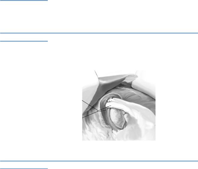
STEP 3
STEP 4
STEP 5
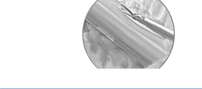
Intraoperative endoscopy
Intraoperative endoscopy aids in evaluating the length of the myotomy, dividing residual muscle fibers, and checking the patency of the esophagogastric junction. This is most helpful in patients previously treated by pneumatic dilation or botulinum toxin injection.
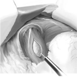
Construction of the Dor fundoplication
The anterior fundic wall is sutured with three interrupted stitches (Prolene for extracorporeal knots, Ethibond for intracorporeal knots) to the adjacent left muscle edge of the myotomy. The most cranial stitch incorporates the diaphragmatic crus.
Security of the fundic wall
A more lateral portion of the anterior fundic wall is secured with three interrupted stitches to the right muscle edge of the myotomy and to the left diaphragmatic crus.

142 SECTION 2 Esophagus, Stomach and Duodenum
Standard Postoperative Investigations
■ Gastrografin swallow on postoperative day 1 to check for esophagogastric transit and absence of leaks
Postoperative Complications
■Persistent dysphagia
■Delayed esophageal emptying
■Recurrent dysphagia
■Esophagectomy may be required in patients with decompensated sigmoid megaesophagus
■Gastroesophageal reflux
■Evidence of stricture may require mechanical dilatation followed by vagotomy and total duodenal diversion
■Leak after undetected perforation
■Consider endoscopic stenting and percutaneous CT-guided drainage
Tricks of the Senior Surgeon
■Once the submucosal plane has been identified, use a pledget swab to create a tunnel before cutting the muscle upward.
■Bleeding from the muscle edges of the myotomy is self-limiting; it is wise to avoid excessive electrocoagulation and to compress with a warm gauze for a few minutes.
■Be careful when the endoscope is advanced into the esophagus and past the cardia after the myotomy is performed; the stomach grasper must be immediately released to prevent iatrogenic perforation.