

Atlas of breast surgery
.pdf34 |
3.4 |
Needle-Localized Biopsy |
3
a |
c |
b
Fig. 3.22a, b Mammogram of the right breast. c Radiograph of the breast tissue specimen with microcalcifications (magnification 2 fold)

Diagnostic Procedures |
Chapter 3 |
35 |
Recommended
1.
2.
Not recommended
3.
Recommended
Fig. 3.23. Guidance for correct approach during needle-localized biopsy. The posterior glandular approach (1 on right) is recommended, whereas periareolar (2 on right) is not recommended. Inframammary approach (3 on right) is only recommended if the tumor is deeply located
36 |
3.4 |
Needle-Localized Biopsy |
3
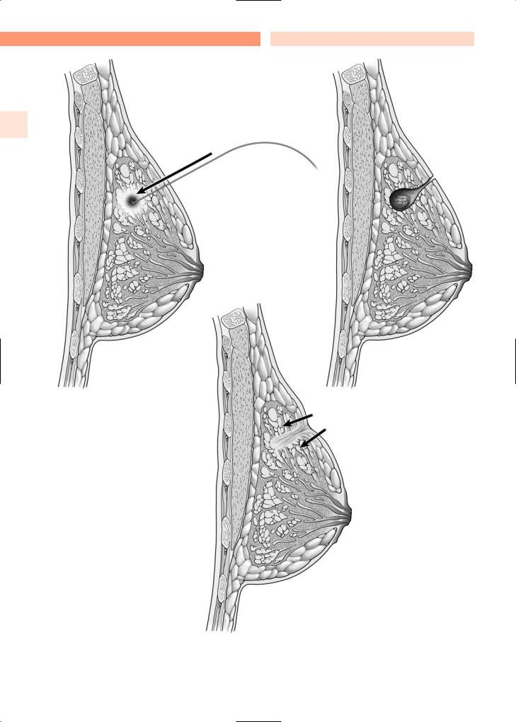
Fig. 3.24. Needle-localized biopsy

Diagnostic Procedures
3.5 Microductectomy
This technique is used to excise a single breast duct, usually to diagnose the cause of nipple discharge from a single duct. If bloody nipple discharge arises from a single duct, it is often attributable to a papilloma.
Surgical procedures around the nipple–areola complex are uncomfortable for the patient, and many surgeons prefer to use a general anesthetic. A catheter attached to a thin butterfly syringe is used to instill blue-dye into the lactiferous duct and sinus (Fig. 3.25a, b). The catheter is removed, and a periareolar incision made. Tissues are dissected down to the duct containing the blue-dye. The duct is excised and
Chapter 3 |
37 |
submitted for pathological evaluation. Afterwards, meticulous attention should be paid to achieving hemostasis with electrocautery. The wound is then irrigated, and the skin edges re-approximated with a running subcuticular absorbable stitch.
Alternatively, a periareolar incision can be made and the areolar lifted up with a skin hook (Fig. 3.26). The area of breast tissue that includes the involved duct (or ducts) is broadly excised, extending the area of excision posteriorly. Once the tissue is excised and hemostasis is obtained,the posterior extent of the excision within the breast can be re-approximated with interrupted absorbable stitches,and the nipple–areo- la complex re-attached to the adjacent skin with a running 3–0 Monocryl subcuticular stitch.
a
b
Fig. 3.25a, b. Microductectomy. a Blue-dye is injected into the lactiferous duct and sinus using a thin butterfly syringe. b Tissue stained by the blue-dye is removed (dotted line)

38 |
3.5 |
Microductectomy |
3
Fig. 3.26. Ductectomy through a periareolar incision

Diagnostic Procedures |
Chapter 3 |
39 |
3.6 Subareolar Dissection
A subareolar dissection is required to extirpate the major ducts immediately below their opening at the nipple (Fig. 3.27). This procedure is often used to diagnose or treat persistent nipple discharge (usually unilateral) arising from many ducts. In many cases, such discharge is attributable to duct ectasia. However, there are other causes of profuse nipple discharge involving many ducts, particularly if the discharge is bilateral, including a prolactin-secreting tumor. Therefore, patients must undergo thorough assessment prior to surgery.
This procedure is usually performed under a general anesthetic. After applying a sterilizing solution over the entire field, a periareolar incision is made, and the major duct system immediately below the nipple–areola complex is excised. Meticulous attention is paid to hemostasis using electrocautery. The
wound is copiously irrigated, and the skin edges are re-approximated with a running absorbable subcuticular stitch.
Fig. 3.27. Subareolar dissection, required to extirpate the major ducts immediately below the nipple

40 |
3.7 |
Mammary Ductoscopy |
3.7 Mammary Ductoscopy
Figures 3.28–3.34 are courtesy of Professor Kefah Mokbel, London, UK.
Mammary ductoscopy is relatively new technolo- 3 gy and its potential applications are still under investigation. However, it has been used to diagnose the source of discharge from a single breast duct (Fig. 3.28). The orifice of the breast duct is dilated, and a small endoscope is passed through it into the ductal system. The small endoscope contains a camera, and this allows for direct visualization of the intraductal system on a television screen. Thus, intraductal pathology can be visualized, allowing for adequate removal of intraductal lesions while preserving
surrounding normal breast tissue.
The ductoscopes that are now frequently used are 1.0 mm in external diameter with a 0.45 mm working channel (Fig. 3.29), allowing air insufflation and saline irrigation during visualization. The working channel provides for ductal dilatation, sampling of intraductal lesions, and also permits irrigation of debris, thereby providing a clear image (Fig. 3.30). Ductoscopes have been utilized in the management of nipple discharge, to help determine the underlying cause of the discharge. Additionally, lavage of the ductal system through the ductoscope (ductal lavage) is under investigation as a means of harvesting epithelial cells, examining these cells for evidence of atypia or malignancy (Figs. 3.31, 3.32, 3.33 and 3.34), and thereby assessing women at high risk for developing breast cancer.
Fig. 3.29. The endoscope, which has an external diameter of 1 mm, a 0.45-mm working channel and a resolution of 10,000 pixels
Fig. 3.30. The procedure
Fig. 3.28. Mammary ductoscopy and pathologic nipple dis- Fig. 3.31. Normal mammary duct visualized on ductoscopy charge (PND)

Diagnostic Procedures |
Chapter 3 |
41 |
Fig. 3.32. Papilloma visualized on ductoscopy
Fig. 3.33. Ductal carcinoma in situ (DCIS) visualized on ductoscopy
Fig. 3.34. Ductoscopic cytology (DCIS)

Chapter 4
Surgery
for Benign Breast Diseases
4.1 Cyst Aspiration
The technique used to aspirate a palpable breast cyst is similar to that used for fine needle aspiration cytology (Fig. 3.2). The skin surface is cleansed with rubbing alcohol. We generally attach a 21-gauge needle attached to a 20-ml syringe. The cyst is secured with the thumb and index fingers or the index and middle fingers. The syringe is held with the other hand, and the cyst aspirated until it is no longer palpable. The contents of a cyst generally contain brown, yellow, or greenish fluid. If such fluid is ob-
tained on aspiration, then there is no need to send it for cytological evaluation. Cytology is necessary only if bloody fluid is obtained on aspiration.
If a cyst is noted on ultrasound but is not palpable, then ultrasound-guided needle aspiration might be indicated. Again, the skin surface is cleansed with rubbing alcohol. The ultrasound probe is held with one hand, identifying the cyst. The syringe is held with the other hand, and the cyst aspirated. Figure 4.1 also shows a cyst aspiration under ultrasound guidance. The cyst is seen in the upper panel (Fig. 4.1a) ultrasound view, and a needle though the cyst is seen in the lower panel (Fig. 4.1b) ultrasound view.

44 |
4.1 |
Cyst Aspiration |
4
a
b
Fig. 4.1a, b. Cyst aspiration under ultrasound guidance