Atlas of breast surgery
.pdf
24 |
3.2 |
Core Needle Biopsy |
3
Fig. 3.7. When taking high-speed core-cut biopsy samples of nonpalpable and palpable breast masses, visualization with ultrasound may be required
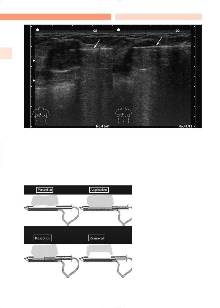
Fig. 3.8. The vacuum-assisted breast biopsy system allows punction, aspiration, resection, and removal of breast lesions that are visualized mammographically throughout the procedure (Ethicon Endo-Surgery)

Diagnostic Procedures |
Chapter 3 |
25 |
Fig. 3.9. A vacuum-assisted percutaneous biopsy device and biopsy needle (brand name Mammotome®, manufactured by Johnson and Johnson Ethicon Endo-Surgery)

26 |
3.2 |
Core Needle Biopsy |
3
Fig. 3.10. Using the vacuum-assisted breast biopsy system, the probe is positioned at the lesion. It vacuums, cuts, and removes tissue samples, which are passed through the probe’s hollow chamber into a collection tray. This allows for multiple samples to be collected while only one incision into the breast is made. At the end hematoma remains visable
Fig. 3.11. The core needle device of the vacuum-assisted breast biopsy system, showing the hollow probe and specimen collection tray

Diagnostic Procedures |
Chapter 3 |
27 |
Fig. 3.12. The vacuum-assisted biopsy system shown in place, below the aperture in the stereotactic table
Fig. 3.13. The surgeon or radiologist can rotate the thumbwheel of the vacuum-assisted biopsy probe, moving it to the correct position for the next biopsy sample, to allow multiple samples to be taken with just one breast incision
Fig. 3.14. Multiple core biopsy samples obtained following one vacuum-assisted biopsy procedure

28 |
3.2 |
Core Needle Biopsy |
3
Fig. 3.15. Ductal carcinoma in situ (DCIS) in a specimen obtained by vacuum-assisted biopsy
Fig. 3.16. Excision biopsy specimen after prior vacuum-assisted biopsy

Diagnostic Procedures |
Chapter 3 |
29 |
Fig. 3.17. Excision biopsy specimen after prior vacuum-assisted biopsy

30 |
3.3 |
Overview of Biopsy Techniques |
3.3 Overview of Biopsy Techniques
As indicated on the previous pages, there are several methods available to biopsy breast tissue (Figs. 3.18a–d, 3.19a–d, 3.20). The optimal method depends
3 on the amount of tissue required and the objective of the biopsy (whether to completely excise a breast lesion or simply obtain a sample) (Fig. 3.19a–d). The greatest amount of tissue is obtained with open biopsy techniques (excisional biopsy or needle-localized
biopsy; Fig. 3.19a), and decreasing amounts are obtained by biopsy with the advanced breast biopsy instrument (ABBI) system (Fig. 3.19b), core biopsy (Fig. 3.19c), and vacuum-assisted biopsy (Figs. 3.8, 3.9, 3.10, 3.19d). The more tissue required, the greater the size of the incision required for the procedure. Thus, a very small incision is required for vacuumassisted biopsy, and increasingly larger incisions are required for the core biopsy, ABBI system, and open biopsy methods.
a |
b |
c |
d |
Fig. 3.18a–d. Comparison of different biopsy techniques, their invasiveness, and the amount of tissue sampled. a Excisional or needle-localized biopsy; b the advanced breast biopsy instrument (ABBI) system; c core biopsy; d the stereotactic vacuum-as- sisted biopsy device

Diagnostic Procedures |
Chapter 3 |
31 |
Excisional or |
ABBI system |
needle-localized |
|
biopsy |
|
a |
b |
Relation biopsylesion
Core biopsy
Breast (stereotactic) biopsy
c |
d |
Relation biopsylesion
Fig. 3.19a–d. Comparison of different biopsy techniques, their invasiveness, and the amount of tissue sampled. a Excisional or needle-localized biopsy; b the advanced breast biopsy instrument (ABBI) system; c core biopsy; d the stereotactic vacuum-as- sisted biopsy device

32 |
3.3 |
Overview of Biopsy Techniques |
3
Fig. 3.20. Examples of core-cut (left) and vacuum-assisted (right) specimens

Diagnostic Procedures
3.4 Needle-Localized Biopsy
A needle-localized biopsy (Fig. 3.21) is performed to assess abnormalities of the breast that are not palpable but are seen on mammogram. To obtain tissue for histological evaluation, the surgeon requires the assistance of the radiologist to localize the mammographic abnormality with a hooked wire.
The patient is first transported to the radiology suite, where the mammographic abnormality is localized with a hooked wire (Fig. 3.22). Proper placement of the wire within the abnormality is essential. Mammograms with two views (anterior–posterior and medio–lateral) should be obtained showing the wire and its relationship to the abnormality. These mammograms guide the surgeon during dissection.
The patient is transported to the operating room with the wire in place. Care should be taken to ensure that the wire is secured on the surface of the breast during transport. The outer part of the wire should be carefully taped on the surface of the breast.
The needle-localized biopsy can be performed using either local or general anesthetic, depending on preferences of the patient and surgeon. The tape is carefully removed from the wire and surface of the breast, and a wide area of the breast is cleaned with a sterilizing solution. A curvilinear incision is made
Chapter 3 |
33 |
immediately adjacent to the wire, along the direction of one of the natural skin crease lines (lines of Langer). Hooks are used to lift up the edges of the skin. With the needle-localized mammograms serving as a guide, the wire and tissue around it is removed en bloc by sharp dissection. Electrocautery should be avoided during dissection, as this can create artifacts that obscure pathological assessment of the specimen. However, once the wire and breast tissue around it are removed, meticulous attention is paid to achieving hemostasis with electrocautery.
It should be emphasized that the incision should be placed near the wire. Periareolar incisions and inframammary incisions are not recommended, unless the wire is immediately adjacent to these areas (Fig. 3.23, 3.24).
Once the breast tissue (with the wire in it) is removed, the specimen is transported to the radiology suite. A specimen film is obtained to confirm excision of the mammographic abnormality. After this is done, the breast wound is irrigated, and skin edges are re-approximated using a running absorbable subcuticular stitch.
Figure 3.22a–c shows: (1) a mammographic abnormality with a wire localizing it (medio-lateral view);
(2) a mammographic abnormality with a wire localizing it (craniocaudal view); and (3) a specimen film of the completely excised mammographic abnormality with the wire through it.
1
2
3
Fig. 3.21. Needle-localized biopsy
