

Color Atlas of Neurology
.pdfPupillary Dysfunction
Amaurosis (right) |
Clivus syndrome |
|
Argyll-Robertson pupils |
|
Ciliary |
|
|
Parinaud syndrome |
|
ganglionitis |
|
|
|
|
|
|
|
Cavernous sinus lesion |
|
|
|
|
Hemispheric lesion |
|
|
|
|
(infarct, hemorrhage) |
|
Parasym- |
|
|
Brain stem lesion |
|
pathetic |
|
|
|
|
dener- |
|
|
Carotid dissection |
|
vation |
|
|
|
|
|
|
|
|
|
|
|
Infiltrating malignant tumor |
Nerves |
|
|
|
Spinal lesion (syringomyelia, |
||
|
|
|
trauma, tumor) |
|
|
|
Indirect light response |
Cranial |
|
|
Direct light response |
Convergence |
||
|
|
|||
|
Spontaneous |
response |
|
|
|
|
|
||
|
Normal |
|
|
|
|
Amaurosis |
|
|
|
Lesion of brachial |
(right) |
|
|
|
Complete right |
|
|
|
|
plexus, thoracic apex, |
|
|
|
|
mediastinum; subclavian |
third nerve palsy |
|
|
|
venous thrombosis |
|
|
|
|
Sympathetic |
Pupillotonia |
|
|
|
|
|
|
|
|
denervation |
|
|
|
|
|
Light-near |
|
|
|
|
dissociation |
|
|
|
|
Atropine eye- |
|
|
|
|
drops, right eye |
|
|
|
|
Clivus syndrome, |
|
|
|
|
intoxications |
|
|
|
|
Parinaud syndrome |
|
|
|
|
Acute pontine |
|
|
|
|
lesion, intoxications |
|
|
93 |
|
Right |
Left |
Pupillary dysfunction |
|
|
|
|||
Rohkamm, Color Atlas of Neurology © 2004 Thieme
All rights reserved. Usage subject to terms and conditions of license.

Cranial Nerves
94
Trigeminal Nerve
Peripheral Connections of the Trigeminal
Ganglion
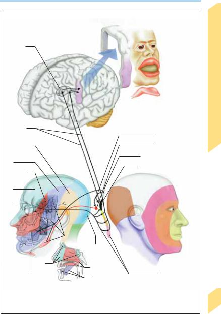
Ophthalmic nerve (V/1). V/1 gives off a recurrent branch to the tentorium cerebelli and falx cerebri (tentorial branch) and the lacrimal, frontal, and nasociliary nerves, which enter the orbit through the superior orbital fissure. The lacrimal nerve supplies the lacrimal gland, conjunctiva, and lateral aspect of the upper eyelid. The frontal nerve divides into the supratrochlear nerve, which supplies the inner canthus, and the supraorbital nerve, which supplies the conjunctiva, upper eyelid, skin of the forehead, and frontal sinus. Finally, the nasociliary nerve gives off branches to the skin of the medial canthus, bridge and tip of the nose, the mucous membranes of the nasal sinus (anterior ethmoid nerve) and sphenoid sinus, and the ethmoid cells (posterior ethmoid nerve).
Maxillary nerve (V/2). Before entering the foramen rotundum, V/2 gives off a middle meningeal branch that innervates the dura mater of the medial cranial fossa and the middle meningeal artery. Other branches innervate the skin of the zygomatic region and temple (zygomatic nerve), and of the cheek (infraorbital nerve). The infraorbital nerve enters the orbit through the inferior orbital fissure, then exits from it again through the infraorbital canal; it innervates the cheek and the maxillary teeth (superior alveolar nerve).
Mandibular nerve (V/3). V/3 gives off a meningeal branch (nervus spinosus) just distal from its exit from the foramen ovale that reenters the cranial cavity through the foramen spinosum to supply the dura mater, part of the sphenoid sinus, and the mastoid air cells. In its further course, V/3 gives off the auriculotemporal nerve (supplies the temporomandibular joint, skin of the temple in front of the ear, external auditory canal, eardrum, parotid gland, and anterior surface of the auricle), the lingual nerve (tonsils, mucous membranes of the floor of the mouth, gums of the lower front teeth, and mucosa of the anterior two-thirds of the tongue), the inferior alveolar nerve (teeth of the lower jaw and lateral gums), the mental nerve (lower lip, skin of the chin, and gums of front teeth), and the buccal nerve (buccal mucosa).
The motor root of CN V contains motor fibers from the trigeminal motor nucleus in the pons
and joins the mandibular nerve to innervate the muscles of mastication (temporalis, masseter, and medial and lateral pterygoid muscles), hyoid muscles (anterior belly of the digastric muscle, mylohyoid muscle), muscles of the soft palate (tensor veli palatini muscle), and tensor tympani muscle.
Central Connections of the Trigeminal
Ganglion
Sensory fibers mediating epicritic sensation terminate in the principal sensory nucleus of the trigeminal nerve, which is located in the pons. Fibers terminating in this nucleus also form the afferent arm of the corneal reflex, whose efferent arm is the facial nerve. Fibers mediating protopathic sensation terminate in the spinal nucleus of the trigeminal nerve, a column of cells that extends down the medulla to the upper cervical spinal cord. The spinal nucleus is somatotopically organized: its uppermost portion is responsible for perioral sensation, while lower portions serve progressively more peripheral areas of the face in an “onion-skin” arrangement. The caudal portion of the spinal nucleus of the trigeminal nerve also receives fibers from cranial nerves VII, IX, and X carrying nociceptive impulses from the ear, posterior third of the tongue, pharynx, and larynx.
Mesencephalic nucleus of trigeminal nerve. This midbrain nucleus, too, contains pseudounipolar neurons, whose long dendrites pass through the trigeminal ganglion without forming a synapse and carry afferent impulses from masticatory muscle spindles and pressure receptors (for regulation of the force of chewing).
Trigeminocortical tracts. Output fibers of the spinal nucleus of the trigeminal nerve decussate in the brain stem and ascend, by way of the trigeminal lemniscus (adjacent to the spinothalamic tract) and the medial lemniscus, to the ventral posteromedial (VPM) and posterior nuclei of the thalamus, where the third neuron of the sensory pathway is located. These thalamic nuclei project via the internal capsule to the postcentral gyrus. The supranuclear innervation of the motor nucleus of the trigeminal nerve is from the caudal portions of the precentral gyrus (bilaterally), by way of the corticonuclear tract.
Rohkamm, Color Atlas of Neurology © 2004 Thieme
All rights reserved. Usage subject to terms and conditions of license.
Trigeminal Nerve
Thalamus
Trigeminal lemniscus
Lesser occipital n. (from C2)
V/3
Trigeminal ganglion
V/1
V/2
V/1
Mylohyoid m., digastric m.
Cortical projections
(postcentral gyrus)
Corticonuclear tract
Motor nucleus of V
Mesencephalic nucleus of V
Principal sensory nucleus of V
C2
C3
Muscles of mastication
Greater occipital n. (from C3)
V/2
Spinal nucleus of V
V/3
Peripheral innervation pattern |
Central innervation pattern |
Cranial Nerves
95
Rohkamm, Color Atlas of Neurology © 2004 Thieme
All rights reserved. Usage subject to terms and conditions of license.

Facial Nerve
Nerve Pathways
|
Central motor pathway. The corticonuclear tract |
|
|
originates in the precentral cortex (area 8), |
|
|
passes in front of the pyramidal tract in the genu |
|
|
of the internal capsule, then travels in the me- |
|
|
dial portion of the ipsilateral cerebral peduncle |
|
|
to reach the facial nucleus in the lower pons. The |
|
|
supranuclear fibers serving the upper facial |
|
|
muscles (frontalis and corrugator supercilii |
|
|
muscles, upper part of orbicularis oculi muscle, |
|
|
superior auricular muscle) decussate in- |
|
|
completely in the pons, so that these muscles |
|
|
have bilateral supranuclear innervation; fibers |
|
|
serving the remaining muscles decussate |
|
Nerves |
completely, so that they have contralateral in- |
|
nervation only. The precentral cortex is re- |
||
sponsible for the voluntary component of facial |
||
expression, while nonpyramidal motor connec- |
||
Cranial |
tions subserve the automatic and emotive com- |
|
ponents of facial expression. These anatomical |
||
|
||
|
facts explain the dissociated functional deficits |
|
|
that set supranuclear facial palsies apart from |
|
|
nuclear or subnuclear palsies, and enable their |
|
|
further differentiation into cortical and subcor- |
|
|
tical types (see below). |
|
|
Peripheral motor pathway. The facial nucleus |
|
|
and its efferent fibers are somatotopically or- |
|
|
ganized. The emerging fibers first run dorsome- |
|
|
dially, then turn anterolaterally to pass around |
|
|
the abducens nucleus (inner genu of facial |
|
|
nerve), and exit the brain stem as the facial |
|
|
nerve in the cerebellopontine angle, near CN VI |
|
|
and VIII. The facial nerve enters the internal |
|
|
acoustic meatus together with the nervus inter- |
|
|
medius and CN VIII, then leaves the meatus to |
|
|
enter the facial canal; it passes between the |
|
|
cochlea and labyrinth, then turns back again |
|
|
(outer genu of facial nerve). After leaving the |
|
|
skull at the stylomastoid foramen, it continues |
|
|
inside the parotid gland and gives off motor |
|
|
branches to all muscles of facial expression as |
|
|
well as the platysma, ear muscles, stapedius, di- |
|
|
gastric (posterior belly), and stylohyoid muscles. |
|
|
Sensory and parasympathetic fibers (nervus in- |
|
|
termedius). Sensory fibers from the geniculate |
|
|
ganglion travel to the superior salivatory nu- |
|
|
cleus, nucleus of the tractus solitarius (p. 78), |
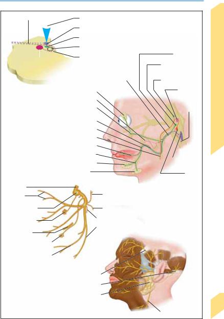
96and spinal nucleus of the trigeminal nerve (p. 94). Taste fibers from the anterior two-thirds
of the tongue (lingual nerve) and the soft palate
(greater petrosal nerve) join the chorda tympani. Preganglionic parasympathetic fibers travel in the greater petrosal nerve to the pterygopalatine ganglion, from which postganglonic fibers pass to the lacrimal, nasal, and palatine glands; other preganglionic fibers travel in the chorda tympani to the submandibular ganglion, from which postganglionic fibers pass to the sublingual and submandibular glands. Connections via the contralateral medial lemniscus to the thalamus and postcentral gyrus, and to the hypothalamus, subserve reflex salivation in response to the smell and taste of food. The facial nerve carries sensory fibers from the external auditory canal, eardrum, external ear, and mastoid region (posterior auricular nerve), as well as proprioceptive fibers from the muscles it innervates.
Functional Systems
The voluntary component of facial expression is mediated by the precentral cortex, in which the face is somatotopically represented. Only the upper facial muscles have bilateral supranuclear innervation; thus, a central supranuclear facial palsy does not affect eye closure or the ability to knit one’s brow. Yet facial palsy that spares the upper face is not necessarily of supranuclear origin: because the facial nucleus and nerve are also somatotopically organized, incomplete lesions of these structures may also produce a similar appearance. An important and sometimes helpful distinguishing feature is that a supranuclear palsy may affect facial expression in the lower face in a dissociated fashion. Supranuclear facial palsy due to a cortical lesion impairs voluntary facial expression, but tends to spare emotional expression (laughing, crying); that due to a subcortical lesion (e. g., in Parkinson disease or hereditary dystonia) does just the opposite.
The following reflexes are of clinical significance (A = afferent arm, E = efferent arm): orbicularis oculi reflex (blink reflex; A: V/1; E: VII); corneal reflex (A: V/1; E: VII); sucking reflex (A: V/2, V/3, XI; E: V, VII, IX, X, XII), palmomental reflex (A: thenar skin/muscles; E: VII), acoustic blink reflex (A: VIII; E: VII), visual blink reflex (A: II; E: VII), orbicularis oris reflex (snout reflex; A: V/2; E: VII).
Rohkamm, Color Atlas of Neurology © 2004 Thieme
All rights reserved. Usage subject to terms and conditions of license.
Facial Nerve
Peripheral pathway |
|
|
|
|
|
|
|
|
|
Corticonuclear tract |
||||||||||||
|
|
|
|
|
|
|
|
|
|
|
|
Nucleus of the solitary tract |
||||||||||
|
|
|
|
|
|
|
|
|
|
|
|
|||||||||||
|
|
|
|
|
|
|
|
|
|
|
|
Superior salivatory nucleus |
||||||||||
|
|
|
|
|
|
|
|
|
|
|
|
|
Nucleus abducens |
|||||||||
|
|
|
|
|
|
|
|
|
|
|
|
Inner genu of facial nerve |
||||||||||
Motor nucleus of VII |
||||||||||||||||||||||
Nuclear region |
Lacrimal gland |
|
|
|
Nervus intermedius |
|
|
|||||||||||||||
|
|
|||||||||||||||||||||
|
|
|
|
|
|
|
|
|
|
|
|
|
|
|||||||||
|
|
|
|
|
|
|
|
|
|
|
|
|||||||||||
|
|
|
Otic ganglion |
|
|
|
|
|
|
|
|
|||||||||||
|
|
|
|
|
|
|
|
|
||||||||||||||
|
|
|
Pterygopalatine |
|
|
|
|
|
|
|
||||||||||||
|
|
|
|
|
|
|
|
|||||||||||||||
|
|
|
ganglion |
|||||||||||||||||||
|
|
|
Chorda tympani |
|
|
|
|
|
|
|||||||||||||
|
|
|
|
|
|
|
||||||||||||||||
|
|
|
Lingual nerve |
|
|
|
|
|
|
|||||||||||||
|
|
|
|
|
|
|
||||||||||||||||
|
|
|
Motor fibers |
|
|
|
|
|
|
|||||||||||||
|
|
|
|
|
|
|
||||||||||||||||
|
|
|
Taste fibers |
|
|
|
|
|
|
|||||||||||||
|
|
|
|
|
|
|
||||||||||||||||
|
|
|
Submandibular ganglion |
|||||||||||||||||||
|
|
|
Sublingual gland |
|
|
|
|
|
||||||||||||||
|
|
|
|
|
|
|||||||||||||||||
|
|
|
Submandibular gland |
|||||||||||||||||||
Peripheral |
|
|
|
|
|
|
|
|
|
|
|
|
|
|
|
|
|
|
|
|
||
pathway |
|
|
|
|
|
|
|
|
|
|
|
|
|
|
|
|
|
|
|
|
||
Temporal |
|
|
|
|
|
|
|
|
|
|
|
|
|
|
|
|
|
Posterior |
||||
branches |
|
|
|
|
|
|
|
|
|
|
|
|
|
|
|
|
|
auricular n. |
||||
External genu of |
|
|
|
|
|
|
|
|
|
|
|
|
|
|
|
|
|
Digastric branch |
||||
facial nerve |
|
|
|
|
|
|
|
|
|
|
|
|
|
|
|
|
|
Stylohyoid branch |
||||
Pterygopalatine |
|
|
|
|
|
|
|
|
|
|
|
|
|
|
|
|
|
|||||
|
|
|
|
|
|
|
|
|
|
|
|
|
|
|
|
|
||||||
|
|
|
|
|
|
|
|
|
|
|
|
|
|
|
|
|
|
|
|
|||
ganglion |
|
|
|
|
|
|
|
|
|
|
|
|
|
|
|
|
|
Cervical branch |
||||
Lingual nerve |
|
|
|
|
|
|
|
|
|
|
|
|
|
|
|
|
|
|||||
|
|
|
|
|
|
|
|
|
|
|
|
|
|
|
|
|
|
|
|
|||
Subman- |
|
|
|
|
|
|
|
|
|
|
|
|
|
|
|
|
|
|
|
|
|
|
|
|
|
|
|
|
|
|
|
|
|
|
|
|
|
|
|
|
|
|
|
|
|
dibular ganglion |
|
|
|
|
|
|
|
|
|
|
|
|
|
|
|
|
|
|
|
|
||
Marginal mandibular |
|
|
|
|
|
|
|
|
|
|
|
|
|
|
|
|
|
|
|
|
||
|
|
|
|
|
|
|
|
|
|
|
|
|
|
|
|
|
|
|
|
|||
branch |
Branches of facial nerve |
|||||||||||||||||||||
|
|
|
||||||||||||||||||||
|
|
|
Temporal branches |
|
|
|
|
|
|
|
|
|
||||||||||
|
|
|
|
|
|
|
|
|||||||||||||||
|
|
|
Posterior auricular n. |
|
|
|
|
|||||||||||||||
|
|
|
|
|
||||||||||||||||||
|
|
|
Parotid plexus, |
|
|
|
|
|
|
|
|
|
|
|
|
|
||||||
|
|
|
parotid gland |
|||||||||||||||||||
|
|
|
|
|
|
|
|
|
|
|
|
|
|
Motor branches |
||||||||
Motor fibers
Superior salivatory nucleus
Motor nucleus of V
Abducens
nucleus
Spinal nucleus
Nucleus of the
solitary tract
Motor nucleus of VII
Peripheral tracts
 Cervical branch
Cranial Nerves
97
Rohkamm, Color Atlas of Neurology © 2004 Thieme
All rights reserved. Usage subject to terms and conditions of license.

Cranial Nerves
98
Facial Nerve Lesions
Examination. Motor function is assessed at rest (asymmetry of face/skin folds, atrophy, spontaneous movements, blink rate) and during voluntary movement (forehead, eyelids and brows, cheeks, mouth region, platysma). Trigeminal nerve dysfunction (V/1) causes unilateral or bilateral absence of the blink reflex; facial palsy may impair or abolish the blink response, but lagophthalmos persists, because the extraocular muscles are unimpaired. Similar logic applies to
Facial Nerve Lesions
other facial nerve reflexes (p. 96). If the patient complains of loss of taste, it is tested accordingly (p. 78). Lacrimation can be tested with the Schirmer test, which, however, is positive only if tear flow is minimal or absent. The salivation test is used to measure the flow of saliva from the submandibular and sublingual glands. The stapedius reflex is tested by measuring the contraction of the stapedius muscle in response to an acoustic stimulus.
Site of Lesion |
Clinical Features |
|
|
Cortex or internal capsule |
Contralateral central facial palsy (+ pyramidal tract lesion, p. 46). Emotional |
|
component of facial expression is unimpaired |
Brainstem, facial nucleus |
Pontine syndrome (p. 70, 72, 359), myokymia |
Cerebellopontine angle |
Ipsilateral peripheral facial palsy (+V/1–2, VI, VIII; p. 74). Hemifacial spasm |
|
ipsilateral. |
Base of skull, internal acoustic |
Peripheral facial palsy (+ other cranial nerve palsies; p. 74) |
meatus |
|
Geniculate ganglion |
Peripheral facial palsy, dysgeusia, hyposalivation, diminished lacrimation, ear |
|
ache, hyperacusis (due to absence of stapedius reflex) |
Facial canal distal to genicu- |
Peripheral facial palsy, dysgeusia, hyposalivation (but normal lacrimation), |
late ganglion |
hyperacusis |
Proximal to stylomastoid fora- |
Peripheral facial palsy, dysgeusia, hyposalivation, intact stapedius reflex |
men |
|
Stylomastoid foramen |
Purely motor peripheral facial palsy |
Parotid gland, facial region |
More or less complete, purely motor facial palsy; palsy due to lesions of in- |
|
dividual branches of the facial nerve |
|
|
For signs and symptoms of facial nerve lesions, see Table 7 on p. 362.
Rohkamm, Color Atlas of Neurology © 2004 Thieme
All rights reserved. Usage subject to terms and conditions of license.
Facial Nerve Lesions
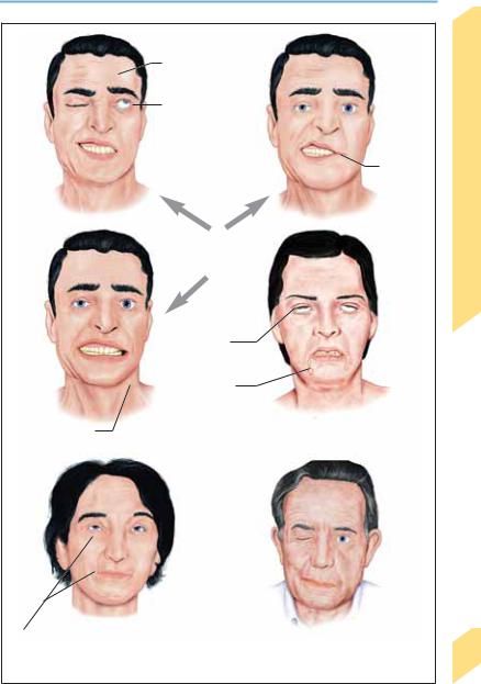
Unilateral paresis of frontalis m.
Lagophthalmos
Paresis at corner of mouth
Left peripheral facial palsy
Bilateral absence of lid closure
Drooling
Paresis of platysma |
Bilateral peripheral facial palsy |
Cranial Nerves
Involuntary associated movements
99
Synkinesia |
Right hemifacial spasm |
Rohkamm, Color Atlas of Neurology © 2004 Thieme
All rights reserved. Usage subject to terms and conditions of license.

Cranial Nerves
100
Hearing
Perception of Sound
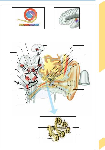
Sound waves enter the ear through the external acoustic meatus and travel through the ear canal to the tympanic membrane (eardrum), setting it into vibration. Vibrations in the 20–16 000 Hz range (most sensitive range, 2000–5000 Hz) are transmitted to the auditory ossicles (malleus, incus, stapes). The base of the stapes vibrates against the oval window, creating waves in the perilymph in the vestibular canal (scala vestibuli) of the cochlea; these waves are then transmitted through the connecting passage at the cochlear apex (helicotrema) to the perilymph of the tympanic canal (scala tympani). (Oscillations of the round window compensate for volume changes caused by oscillations of the oval window. Sound waves can also reach the cochlea by direct conduction through the skull bone.) Migrating waves are set in motion along the basilar membrane of the cochlear duct; they travel from the stapes to the helicotrema at decreasing speed, partly because the basilar membrane is less tense as it nears the cochlear apex. These waves have their amplitude maxima at different sites along the basilar membrane, depending on frequency (tonotopicity): there results a frequency-specific excitation of the receptor cells for hearing—the hair cells of the organ of Corti, which is adjacent to the basilar membrane as it winds through the cochlea.
Cochlear Nerve
The tonotopicity of the basilar membrane causes each hair cell to be tuned to a specific sound frequency (spectral analysis). Each hair cell is connected to an afferent fiber of the cochlear nerve inside the organ of Corti. The cochlear nerve is formed by the central processes of the bipolar neurons of the cochlear ganglion (the first neurons of the auditory pathway); it exits from the petrous bone at the internal acoustic meatus, travels a short distance in the subarachnoid space, and enters the brain stem in the cerebellopontine angle. Central auditory processing involves interpretation of the pattern and temporal sequence of the action potentials carried in the cochlear nerve.
Auditory Pathway
As it ascends from the cochlea to the auditory cortex, the auditory pathway gives off collateral projections to the cerebellum, the oculomotor and facial nuclei, cervical motor neurons, and the reticular activating system, which form the afferent arm of the acoustically mediated reflexes.
Axons of the cochlear nerve originating in the cochlear apex and base terminate in the anterior and posterior cochlear nuclei, respectively. These nuclei contain the second neurons of the auditory pathway. Fibers from the posterior cochlear nucleus decussate in the floor of the fourth ventricle, then ascend to enter the lateral lemniscus and synapse in the inferior colliculus (third neuron). The inferior colliculus projects to the medial geniculate body (fourth neuron), which, in turn, projects via the acoustic radiation to the auditory cortex. The acoustic radiation passes below the thalamus and runs in the posterior limb of the internal capsule. Fibers from the anterior cochlear nucleus also decussate, mainly in the trapezoid body, and synapse onto the next (third) neuron in the olivary nucleus or the nucleus of the lateral lemniscus. This branch of the auditory pathway then continues through the lateral lemniscus to the inferior colliculus and onward through the acoustic radiation to the auditory cortex.
The primary auditory cortex (area 41: Heschl’s gyrus, transverse temporal gyri) is located in the temporal operculum (i.e., the portion of the temporal lobe overlying the insula and separated from it by the sylvian cistern). Areas 42 and 22 make up the secondary auditory cortex, in which auditory signals are further processed, recognized, and compared with auditory memories. The auditory cortex of each side of the brain receives information from both ears (contralateral more than ipsilateral); unilateral lesions of the central auditory pathway or auditory cortex do not cause clinically relevant hearing loss.
Rohkamm, Color Atlas of Neurology © 2004 Thieme
All rights reserved. Usage subject to terms and conditions of license.
|
|
Hearing |
|
|
Cochlear |
|
|
|
|
duct |
|
|
|
|
|
|
Frequency |
|
|
|
|
bands |
|
|
20 000Hz |
20 Hz |
|
|
|
Migrating wave, spectral analysis, |
Auditory cortex |
|
||
tonotopicity |
|
|
|
|
Superior colliculus |
Areas 41, 42 |
Cochlea |
|
|
|
|
|||
Inferior colliculus |
Acoustic radiation |
Oval window |
|
|
Medial |
Nerves |
|||
|
Stapes |
|||
geniculate |
|
|||
body |
|
Vestibular system |
||
|
|
|||
Nucleus |
|
Cranial |
||
|
|
|||
of lateral |
Lateral |
Malleus, |
||
lemniscus |
lemniscus |
incus |
||
Olivary nuclei |
|
|
||
|
|
|
||
Anterior |
|
|
|
|
cochlear |
|
|
|
|
nucleus |
|
|
|
|
Cochlear |
|
|
|
|
nerve |
|
|
|
|
Posterior |
|
External |
|
|
cochlear nucleus |
|
|
||
|
auditory canal |
|
||
|
Tensor |
|
||
Trapezoid body |
|
|
||
tympani m. |
Tympanic |
|
||
|
|
|||
Medullary striae |
|
membrane |
|
|
|
|
|
||
Auditory tube |
Conduction of Sound; auditory pathway |
|
||
(eustachian tube) |
|
|||
|
Cochlear n. |
Cochlear |
|
|
|
|
ganglion |
|
|
|
Cochlear duct |
Scala vestibuli |
|
|
|
|
Scala tympani |
|
|
|
Organ of Corti, |
|
|
|
basilar membrane, |
|
|
||
|
hair cell |
|
101 |
|
|
Cochlea |
|
||
|
|
|
||
Rohkamm, Color Atlas of Neurology © 2004 Thieme
All rights reserved. Usage subject to terms and conditions of license.

Cranial Nerves
102
Disturbances of Deglutition
Impairment of swallowing (deglutition) is called dysphagia; pain on swallowing is called odynophagia. Dysphagia or vomiting due to neurological disease often causes aspiration (entrance of solid or liquid food into the airway below the vocal cords). Globus hystericus is a foreign-body sensation in the swallowing pathway independent of the act of swallowing. Despite its name, it is not always psychogenic; organic causes include Zenker diverticulum and gastroesophageal reflux.
Deglutition
Mechanism. The food is ground by the teeth and moistened with saliva to form chyme, which is molded by the tongue into an easily swallowed bolus (oral preparatory phase). The tongue pushes the bolus into the oropharynx (oral phase) to initiate the reflex act of swallowing (pharyngeal phase). The lips and jaw close, the soft palate rises to seal off the nasopharynx, and the bolus bends the epiglottis backward. The bolus is pushed further back by the tongue, respiration briefly ceases, and the raised larynx occludes the airway. The upper esophageal sphincter slackens (cricopharyngeus, inferior pharyngeal constrictor, smooth muscle of upper portion of esophagus). Pressure from the tongue and pharyngeal peristalsis push the bolus past the epiglottis and into the esophagus (esophageal phase). The larynx is lowered, respiration is reinstated, and esophageal peristalsis propels the bolus into the stomach.
Nerve pathways. Fibers of CN V/2, VII, IX, and X to the nucleus ambiguus and the nucleus of the tractus solitarius (p. 78) make up the afferent arm of the swallowing reflex. The motor swallowing center (one on each side) lies adjacent to these nuclei and is associated with the upper medullary reticular formation; it coordinates the actions of the numerous muscles involved in swallowing. Efferent signals reach these muscles through CN V/3, VII, IX, X, and XII. Crossed and uncrossed supranuclear innervation is derived from the cerebral cortex (precentral and postcentral gyri, frontoparietal operculum, premotor cortex, and anterior insular region). Spinal motor neurons also participate (C1–C4).
Neurological Disturbances of Deglutition
(See Table 8 on p. 362)
The disturbance usually manifests itself at the beginning of the act of swallowing (e. g., a feeling of food stuck in the throat, the escape of liquid or solid food through the nose, choking, coughing). Associated inflammation of the swallowing pathway may cause odynophagia. Chronic dysphagia causes inadequate nutrition and weight loss. Neurogenic dysphagia usually impairs the swallowing of liquids more than solids; soft, chilled foods (like pudding or yogurt) are often easier to swallow. Sensory disturbances in the larynx and trachea, a diminished cough reflex, and muscle weakness may cause aspiration, sometimes unremarked by the patient (silent aspiration). The diagnostic evaluation of dysphagia may require special tests such as radiocinematography, video endoscopy, manometry, and pH measurement.
Rohkamm, Color Atlas of Neurology © 2004 Thieme
All rights reserved. Usage subject to terms and conditions of license.