

192 ORTHOPEDICS PROSTHESIS FIXATION FOR
example of bioactive material will be hydroxyapatite, Ca10 (PO4)6 (OH)2, which is similar in composition to the inorganic part of natural bone. The terms biodegradable, bioresorbable, and bioabsorbable are often used interchangeably. For most orthopedic devices, bioactive surfaces are ideal for better cell–materials attachment.
In vivo cell–materials interaction is a fairly complex process. In simple terms, when orthopedic devices are placed inside, our body will try to isolate the device by forming a fibrous tissue around it, which is particularly true for devices made with bioinert materials. If the material is bioactive, bone cells will first attach to the implant surface and then grow or proliferate. A material shows good biocompatibility when cells will attach and grow quickly on the device surface. Growth factors, such as bone morphogenic proteins (BMP), are sometimes used to stimulate cell attachment and growth behavior in vivo. After proliferation, cells will differentiate or produce new bones, which will repair the site and anchor the device. All three stages, attachment, proliferation, and differntiation of bone cells are important for repair and reconstruction of musculoskeletal disorders.
PATIENT SPECIFIC ORTHOPEDIC DEVICE: A FUTURE TREND
Patient specific device is the future trend for repair and reconstruction of musculoskeletal disorders. Due to the advancement of CAD and RP based small volume manufacturing technologies, orthopedic devices for people with special needs will be designed to meet the specific requirements. Such activities are currently pursued in academic research and hope to translate into standard industrial practice in the next one or two decades. Three-dimensions (3D) visualization of disorders using computer images and physical models, and follow-up discussion between patients and physicians will help to better educate the patient population about their problems and possible options. These options can then be transformed to physical models for trials before placing them in vivo. The goal is to reduce revision surgeries and increase device lifetime while maintaining the quality of life for the patient population. Various innovative scientific and engineering advancements toward novel materials and design options are helping us to make significant developments to achieve this goal.
BIBLIOGRAPHY
Cited References
1.Musculoskeletal Disorders and the Workplace: Low Back and Upper Extremities. New York: National Academic Press; 2001.
2.Materials science. Encyclopædia Britannica. 2005. Encyclopædia Britannica Premium Service.
3.Charnley J, Kamangar A, Longfield MD. The optimum size of prosthetic heads in relation to the wear of plastic sockets in total replacement of the hip. Med & Biol Eng 1969;1:31–39.
4.Hench LL. Bioceramics: From concept to clinic. J Amer Ceram Soc 1991;74(7):1487–510.
5.Burg KJL, Porter S, Kellam JF. Biomaterials development for bone tissue engineering. Biomaterials 2000;21:2347–2359.
6.Berndt CC, Haddad GN, Farmer AJD, Gross KA. Thermal spraying for bioceramic applications. Mater Forum 1990 14:161–173.
7.Mayor MB, Merritt K, Brown SA. Metal allergy and the surgical patient. Am J Surg 1980;139:447–479.
8.Chao EYS, Coventry MB. Fracture of the femoral component after total hip replacement. J Rone Joint Surg 1981;63A:1078– 1094.
9.Roodman GD. Mechanisms of bone metastasis. N Eng J Med 2004;350(16):1655–1664.
10.Ravaglioli A, Krajewski A. Bioceramics: Materials, Properties, Application. London: Chapman and Hall, 1992. pp. 156–197.
11.Fontanna MG, Greene ND. Corrision Engineering. New York: McGraw-Hill; 1978.
12.Evans EM, Freeman MAR, Miller AJ, Vemon-Roberts B. Metal sensitivity as a cause of bone necrosis and loosening of prostheses in total joint replacements. J Bone Joint Surg 1974;56B:626–642.
13.Halpin DS. An unusual reaction in muscle in association with Vitallium plate: A report of possible metal hypersensitivity. J Bone Joint Sur Br 1975;57(4):451–453.
14.Garcia DA. Biocompatibility of dental implant materials measured in an animal model. J Dental Res. 1981;60(1):44–49.
15.Roberts WE. Osseous adaptation to continuous loading of rigid endosseous implants. Am J Orthod 1984;86(2):95–111.
16.Pilliar RM. Powder metal-made orthopaedic implants with porous surfaces for fixation by tissue ingrowth. Clin Orthop 1983;176:42–51.
17.Ducheyne P, Martens M. Orderly oriented wire meshes as porous coatings on orthopaedic implants II: The pore size, interfacial bonding and microstructure after pressure sintering of titanium OOWM. Clin Mats 1986;1:91–98
18.Ducheyne P, Qiu Q. Bioactive ceramics: the effect of surface reactivity on bone formation and bone cell function. Biomaterials 1999;20:2287–2303.
19.Darsell J, Bose S, Hosick H, Bandyopadhyay A. From CT Scans to Ceramic Bone Grafts. J Am Ceramic Soc 2003; 86(7):1076–1080.
20.Bose S, et al. Pore Size and Pore Volume Effects on Calcium Phosphate Based Ceramics. Mat Sci Eng 2003; C 23:479– 486.
ORTHOPEDICS PROSTHESIS FIXATION FOR
PATRICK J. PRENDERGAST
Trinity Centre for
Bioengineering
Dublin, Ireland
INTRODUCTION
The fixation of an orthopedic implant should secure it rigidly to the underlying bone. The ideal fixation will sustain high forces, pain free, for the remaining lifetime of the patient. Difficulties in achieving this objective arise because (1)
1.The loads are often several times body weight in the lower extremity.The loads are fluctuating, or cyclic, and furthermore extremely high loads can occur occasionally (2).

ORTHOPEDICS PROSTHESIS FIXATION FOR |
193 |
2.The presence of the implant alters the stress transfer to the underlying bone leading to bone remodelling or fibrous tissue formation at the bone/implant interfaces. This can threaten the long-term mechanical integrity of the prosthetic replacement.
3.The range of materials that can be placed in contact with bone is limited by biocompatibility issues.
The fixation of an orthopedic implant may be catagorized as either cemented fixation or biological fixation.
Cemented fixation involves securing the implant into the bone with a ‘‘bone cement.’’ By far the most common bone cement is based on the (polymer polymethylmethacrylate (PMMA)). PMMA bone cement is polymerized in situ during the surgery. It contains radiopacificiers in the form of particles of barium sulphate (BaSO4) or zirconia (ZrO2), which make it visible in radiographs (3). It also contains an inhibitor (hydroquinone) to prevent spontaneous polymerization and an initiator (benzoyl peroxide) to allow polymerization at room temperature. Antibiotics to prevent infection (e.g., gentimacin) may also be added. Table 1 lists typical components of bone cement and their roles. Polymerization begins when a powder of the PMMA polymer is mixed with the MMA monomer liquid. The mixing can either be done by hand in a mixing bowl just before to its use in the surgery or a mechanical mixing system may be used; these have the advantage of reducing the porosity of the bone cement and increasing its fatigue life. The cement is applied in a doughy state to the bone before placement of the implant.
In biological fixation, the implant is secured to the bone by a process known as ‘‘osseointegration.’’ Osseointegration occurs by bone ingrowth onto the surface of the implant. The surface of the implant must have a structure so that, when the bone grows in, sufficient tensile and shear strength is created. Bone ingrowth requires a mechanically stable environment and an osteoconductive surface. An osteoconductive surface can be achieved by various treatments, e.g., plasma spraying with hydroxyapatite. Ingrowth occurs over approximately 12 weeks, and during this period, implant stability is required: Initial stability can be achieved by press-fitting the implant into the bone, or by using screws.
Hybrid fixation refers to the use of both cemented and biological techniques for the fixation of a prosthesis. For example, a hip replacement femoral component may be
fixated using cement, whereas the acetabular cup may be fixated into the pelvic bone by osseointegration.
Failure of prosthesis fixation is observed as loosening and pain for the patient. If loosening occurs without infection it is called aseptic loosening. Loosening is a multifactorial process and does not have just one cause. Loosening of cemented fixation often occurs by fatigue failure of the bone cement, but loosening can have several root causes: fatigue from pores in the cement and stress concentrations at the implant/cement interface, debonding at the prosthesis/cement interface or cement/bone interface, or bone resorption causing stresses to rise in the cement. Loosening of biological fixation occurs if the relative micromotion between the bone and the implant is too high to allow osseointegration, i.e., if the initial stability of the implant is insufficient. Huiskes (4) proposed the concept of failure scenarios as a method for better understanding the multifactorial nature of aseptic loosening. The failure scenarios are
1.Damage accumulation failure scenario: the gradual cracking of bone cement, perhaps triggered by interface debonding, pores in the cement, or increased stresses due to peripheral bone loss.
2.Particulate reaction failure scenario: wear particles emanating from the articulating surfaces or from metal/metal interfaces in modular prostheses (fretting wear) can migrate into the interfaces causing bone death (osteolysis).
3.Failed ingrowth failure scenario: High micromotion of the implant relative to the bone can prevent bone ingrowth, as can large gaps (> 3 mm). If the area of ingrowth is insufficient, then the strength of the fixation will not be high enough to sustain loading when weight-bearing commences.
4.Stress shielding failure scenario: Parts of the bone can be ‘‘shielded’’ from the stresses they would normally experience because of the rigidity of the implant. This can lead to resorption of the bone and degeneration of the fixation.
5.Stress bypass failure scenario: In biological fixation, ingrowth can be patchy leading to stress transfer over localized areas. When this happens, some bone tissue is ‘‘bypassed,’’ and in these regions, bone atrophy can occur because the stress is low.
Table 1. Components of Bone Cement and Their Roles
Components |
Role |
Amount |
|
|
|
Liquid |
|
20 mL |
Methyl methacrylate (monomer) |
Wetting PMMA particles |
97.4 v/o |
N,N,-dimethyl-p-toluidine |
Polymerization accelerator |
2.6 v/o |
Hydroquinone |
Polymerization inhibitor |
75 þ 15 ppm |
Solid powder |
|
40 g |
Polymethyl methacrylate |
Matrix material |
15.0 w/o |
Methyl methacrylate-styrene-copolymer |
Matrix material |
75.0 w/o |
Barium sulphate (BaSO4), USP |
Radiopacifying agent |
10.0 w/o |
Dibenzoyl peroxide |
Polymerization initiator |
0.75 w/o |
From Park (3).
Note: v/o: % by volume; w/o: % by weight.
194ORTHOPEDICS PROSTHESIS FIXATION FOR
6.Destructive wear failure scenario: In some joint replacement prostheses, e.g., hip and knee, wear can occur to such a degree that the component eventually disintegrates.
CEMENTED FIXATION
It is common to classify cementing techniques according to ‘‘generation’’: The first generation involved hand-mixing and finger packing of the cement, and the second generation improved the procedure by using a cement gun and retrograde filling of the canal, with a bone-plug to contain the cement within the medullary canal. This allows pressurization of the cement and therefore better interdigitation of the cement into the bone. Third generation (called modern cementing) uses, in addition, mechanical mixing techniques for the cement to remove pores and pulsative lavage to clean the bone surface of debris. The most common mechanical mixing technique is ‘‘vacuum mixing,’’ where the powder and monomer are placed together in a mixing tube and the air is removed under pressure; often the tube can then be placed into an injection gun from which it can be extruded into the bone cavity. Another mechanical mixing technique is centrifugation (i.e., spinning the polymerizing bone cement in a machine), which is found to remove pores and increase the fracture strength
(3). Precoating the implant with a PMMA layer or addition of a roughened surface strengthens the implant/bone cement interface.
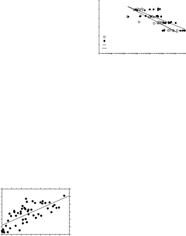
Fixation strength using bone cement relies on an interdigitation of the bone cement with the bone; i.e., it is a mechanical interlock between the bone and the solidified cement that maintains the strength and not a chemical bond. Good interdigitation requires that the bone bed be rough. Creating a rough surface is done by appropriate broaching during preparation of the bony bed; it also requires lavage to clean the bed of loose debris and marrow tissue. Mann et al. (5) found the strength of the bone cement/bone interface to be positively correlated with the degree of interdigitation (Fig. 1). To achieve superior
(MPa) |
3 |
θ = 0˚ |
|
|
|
|
|
|
2.5 |
|
|
|
|
|
|
||
|
|
|
|
|
|
|
|
|
app |
2 |
|
|
|
|
|
|
|
s |
|
|
|
|
|
|
|
|
strength, |
1.5 |
|
|
|
|
|
|
|
|
|
|
|
|
|
|
|
|
Apparent |
1 |
|
|
|
|
|
|
|
.5 |
|
|
|
|
|
|
|
|
|
|
|
|
|
|
|
|
|
|
0 |
|
|
|
|
|
|
|
|
0 |
100 |
200 |
300 |
400 |
500 |
600 |
700 |
|
|
|
|
qint (mg/cc-mm) |
|
|
||
Figure 1. Interdigitation of the bone cement into the bone increases the strength of the bone cement interface. qint is the product of the average value of the thickness of the interdigitated region and the density of the interface region measured using a CT scan. See Mann et al. (5) for details.
|
30 |
|
|
(MPa) |
25 |
|
|
20 |
|
||
|
|
||
Stress |
15 |
|
|
10 |
Hand-mixed |
||
|
|||
|
|
Vacuum-mixed
5Hand-mixed Vacuum-mixed
0
0 |
10 |
102 |
103 |
104 |
105 |
106 |
Cycles to Failure (Nf)
Figure 2. A comparison of the fatigue strength of hand-mixed and vacuum-mixed bone cement. After Murphy and Prendergast (10).
interdigitation, it was thought useful to develop ‘‘low viscosity’’ cements, and although higher penetration was achieved, the clinical outcomes using low viscosity cements in hips were not superior (6).
PMMA bone cement undergoes an exothermic polymerization reaction. This means that heat is produced on polymerization and this can cause necrosis of the surrounding bone tissue. Another consequence of heating is that the cement expands and contracts on cooling. As solidification occurs before to full cooling, residual stresses are generated in the cement (7). This is one reason to minimize the thickness of the cement layer. Also, metallic stems, because they conduct heat, can minimize the peak temperature transmitted to the bone, cooling the metallic implant before implantation has also been suggested. Bioactive cements have also been proposed; see the review by Harper (8). These cements have filler particles added to create a bioactive surface on the cement; fillers can be hydroxyapatite powder or fibers, bone particles, or human growth hormone. Alternatives to PMMA are bisphenol-a- glycidyl methacrylate (BIS-GMA) or poly(ethylmethacrylate) (PEMA)/n-butylmethacrylate (nBMA) cement. However, these cements are not yet widely used.
The mechanical strength depends on the brand of cement used and on the mixing technique (9). To prevent the damage accumulation failure scenario (see above), sufficient fatigue strength is required. This has been measured as a function of mixing technique (Fig. 2) (10). Being a polymer operating close to its melting temperature, bone cement is also subject to creep, i.e., viscoplasticity, and the creep strain as a function of stress has been measured under dynamic loading (11). However, it is clear that the in vitro testing conditions may not account for many of the extremely complex in vivo conditions, so these results should be interpreted with caution (12).
OSSEOINTEGRATION (CEMENTLESS FIXATION)
There is no simple definition of osseointegration, although Albrektsson (13) advocates the following: Osseointegration means a relatively soft-tissue-free contact between implant
Figure 3. Bone ingrowth into a multilayer of a proximal part of a femoral hip prosthesis. After Eldridge and Learmonth (15).
and bone, leading to a clinically stable implant. Early in the study of the osseointegration concept, Skalak (14) found osseointegration was promoted by a micro-rough surface more so than a smooth one. Since then, many animal experiments investigating the effect of plasma spraying the surface and various methods of creating a porous surface have been reported. For orthopedic fixation, porous surfaces with beads in one or more layers have been used, as have wire meshes attached to surfaces, and plasma spraying the surface with hydroxyapatite.
Figure 3 shows bone ingrowth into a multilayer of a proximal part of a femoral hip prosthesis (15). It can be observed that ingrowth is patchy; this is what is commonly found, even with successful implants retrieved at autopsy (16); it is evident, therefore, that ingrowth is not required everywhere on the prosthesis for a successful fixation. Ingrowth is controlled by a combination of the mechanical environment and the size of the pores; the spacing between the pores should not be greater than the degree of micromotion or else the new bone ingrowth path will be continuously sheared as the tissue attempts to grow in. In experiments in dogs, Søballe (17) studied the relationship between implant coating and micromotion and found that hydroxyapatite coating increased the rate of bone ingrowth, and that a relative motion between implant and bone of 150 mm allowed osseointegration, whereas a relative motion of 500 mm inhibited it. The mechanobiolo-
ORTHOPEDICS PROSTHESIS FIXATION FOR |
195 |
gical consequences of these different shearing magnitudes was analyzed by Prendergast et al. (18). The depth of the porosity will also affect the strength, with multilayer beaded surfaces having the potential for greater tensile strength (1).
FIXATION OF PROSTHESIS DESIGNS
Each implant design has specialized fixation features. In the following sections, examples are provided of the fixation approaches used in the main orthopedic implant categories.
Hip Prostheses
Although hip arthroplasty may involve replacement of the femoral side only, total hip arthroplasty (THA) involves replacement of the proximal femur and the acetabular socket. Both cemented and cementless fixation is used for both the femoral component (the ‘‘stem’’) and the acetabular component (the ‘‘cup’’). Selection is a matter of surgeon choice, although there is some agreement that the cementless fixation is preferable in younger patients because cementless implants are easier to revise than cemented where complete removal of the cement mantle may be problematic.
Considering the femoral side first, cemented fixation takes two categories: stem designs in which a bond is encouraged between the stem and the cement (referred here as bonded stems) and designs that discourage a bond (referred here as unbonded stems). Stem bonding can be achieved through roughening of the stem surface to create a mechanical interlock between the metallic stem or cement or through use of a PMMA precoat to create a chemical bond between the precoat/cement interface. Bonded stems usually contain a collar that rests on the bone surface preventing subsidence and often containing ridges, dimples, and undercoats to provide additional interlock with the cement. As long as the bonded stems remain bonded, they have the theoretical benefit of reducing the stress levels in the cement. However, if the bonded stems fail, the roughened surface could generate debris particles and initiate a loosening process. In contrast to the bonded stems, unbonded stems discourage a bond between the stem and the cement through use of a smooth, polished stem surface in combination with a stem design that typically has no collar or macrofeatures to lock into the cement. With the lack of a bond, the polished stems facilitate some stem subsidence within the cement mantle and thereby allow wedging of the implant within the medullary canal. Lennon et al. (19) compared the damage accumulation around polished with matt stems and did not find a difference in the damage accumulated in their cement mantles. Another point of comparison between cemented and cementless fixation is that cementless stems will have a larger cross-sectional area than cemented stems because they must fill the medullary canal; this will make cementless hip prostheses stiffer and predispose them to the stress shielding failure scenario. Recognizing this, it is usual for the osseointegration surface to be on the proximal part of cementless stems to ensure proximal load transfer;
196 ORTHOPEDICS PROSTHESIS FIXATION FOR
furthermore, patches of osseointegrative surface may be limited to the posterior and anterior faces of the stem.
Considering the acetabular side, the cup is either made from ultra-high-molecular-weight polyethylene (UHMWPE), ceramic, or metal. UHMWPE cups may be metal-backed. As the head can be either ceramic (a modular head can be connected to a metal femoral component using a Morse taper) or metal (modular or monobloc), this means that several combinations of bearing materials are possible. Polyethylene cups and metal heads are the most common, but the others, such as metal-on-metal, are advocated as well. The selection of bearing materials is important for the fixation because a high frictional torque predisposes to loosening of the cup or stem and because the wear particles produced can provoke the particulate reaction failure scenario. Polyethylene cups are cemented into the acetabulum using bone cement. Metal-backing of the cup is designed to decrease stresses in the polyethylene ‘‘liner,’’ which should lead to lower wear rates although it is also predicted to increase stress concentrations in the fixation at the periphery of the cup (20). Metal, ceramic, and metal-backed UHMWPE cups may be threaded on the outside so that they can be screwed into the acetabelum, or they may be fixated by osseointegration.
The interrelationship between design factors and fixation of hip implants is complicated and involves maximizing strength of the cement/metal interface, the cement itself, and the bone/cement interface. According to the design philosophy of polished stems, it is better to safeguard the vital bone/cement interface by allowing the cement/metal interface to fail first and facilitating subsidence (21). Not only should the interfaces have the required strength, but the stresses should be minimized to ensure the most durable fixation (22)— the measures to achieve this are listed in Table 2.
Knee Prostheses
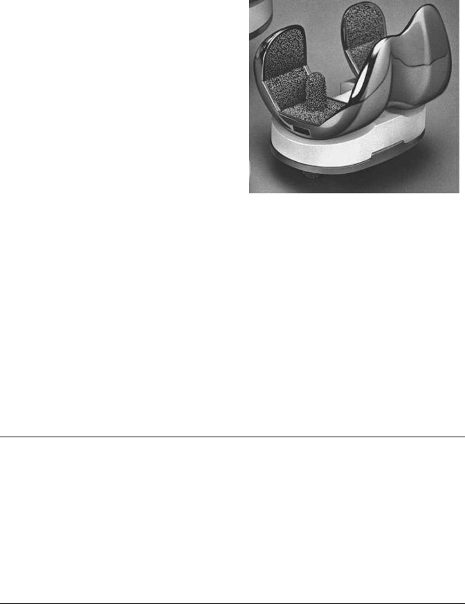
Total knee replacement involves femoral and tibial components, and a component for patellar resurfacing (a patellar ‘‘button’’) is also often used. Both cemented and cementless
Figure 4. A knee replacement prosthesis showing porous-coating for osseointegration and posts for fixation. From Robinson (23).
fixation is used in knee arthroplasty. The femoral component may be fixated with an intramedullary stem that may be cemented, or it may have a porous surface for osseointegration with medial and lateral ‘‘posts’’ to aid initial stability. The tibial component consists of a metal ‘‘tray’’ and a polyethylene insert; the tray may also be fixated with an intramedullary stem cemented into the tibia, perhaps accompanied by medical and lateral posts/pegs for rotational stability. Figure 4 shows a design fixated by osseointegration (23). Walker (24) gives a thorough description of the options available for knee prostheses.
Upper Extremity and IVD Prostheses
Upper extremity prostheses include the shoulder, elbow, and wrist (1). Total shoulder arthroplasty (TSA) consists of a humeral component with an intermedullary stem and a
Table 2. Measures that Maximize Strength and Minimize Stress in Total Hip Replacement Structures
|
Cement/Metal |
|
|
|
Interface |
Cement |
Cement/Bone Interface |
|
|
|
|
Maximize |
Grit-blasted metal |
Optimal preparation |
Careful reaming |
strength |
PMMA-coated metal |
Pressurization |
Pressurization |
|
|
Cement restrictor |
Minimal polymerization heat |
|
|
|
Minimal monomer |
|
|
|
Bone lavage |
|
|
|
Minimal wear debris |
Minimize stress |
Reduce patient weight |
|
|
|
Reduce patient activity |
|
|
|
Anatomical reconstruction of the femoral head |
|
|
|
Minimal friction |
|
|
|
No impingement or subluxation |
|
|
Bonded cement/metal interface
Optimal implant and cement mantle design
Optimal implant material
Optimal (reproducible) placement
Adapted from Huiskes (22).
glenoid component inserted into the glenoid cavity of the scapula. The glenoid component is either all-polyethylene; in which case, it is cemented; or metal-backed; in which case, it may be fixated by osseointegration. Glenoid components may have several pegs, or they may have one central ‘‘keel’’ for fixation (25). Elbow prostheses consist of humeral, ulnar, and radial components, all which may be fixated with or without cement. Wrist prostheses replaces the radial head and the schapoid and lunate bones of the wrist and may be cemented and uncemented (1). Intervertebral disk (IVD) prostheses replace the degenerated disk with a polymer; there are several strategies for fixation: The endplates may be porous coated and plasma sprayed for osseointegration to the cancellous bone with vertical fins to increase stability. IVD prostheses may also be fixed to adjacent vertebral bodies with screws (26).
EVALUATION OF FIXATION AND FUTURE STRATEGIES
One of the key issues in orthopedic implant fixation is whether to use cemented fixation or biological fixation, with surgeons on both sides of the debate (16,27). Cemented fixation has the advantage of immediate postoperative, stability whereas concerns may be raised about the reliability of bone cement’s fatigue strength; furthermore, there is a school of thought that the exothermic polymerization reaction should be avoided if at all possible. Biological fixation by osseointegration has the advantage of avoiding the use of the PMMA cement but runs the risk of the failed ingrowth failure scenario; furthermore, immediate postoperative weight-bearing is not possible. Finally cementless implants are easier to revise if they fail.
Another key issue in orthopedic implant fixation is that of preclinical testing and regulatory approval of new fixation technologies. Considerable challenges exist in achieving consensus around regulatory tests that safeguard patients against ineffective devices while still allowing innovation (4). Preclinical tests can use either (1) finite element models of the direct postoperative situation, e.g., for the hip (28), knee (29), or shoulder (25), or computer simulations of a failure scenario, e.g., damage accumulation (30); (2) physical model ‘‘bench’’ testing with simulators (24,31); or (3) animal testing. Animal testing is not ideal for testing the biomechanical efficacy of orthopedic implant fixation because the implant geometry must be modified to fit the animal skeleton. Furthermore, an important emerging concept is that of patient-specific implants based on computational analysis of a patient’s medical images (32).
One useful clinical method to assess implant fixation is through the use of radiostereometric analysis (RSA). With this approach, the migration of the implant relative to the bone can be determined and is used to determine designs that may be at risk of early loosening. Retrospective and prospective clinical studies are also very useful to determine designs or materials that have promising or poor clinical results. On a larger scale, implant registries performed in many countries in Western Europe can provide information on how designs, materials, and surgical tech-
ORTHOPEDICS PROSTHESIS FIXATION FOR |
197 |
niques rank in terms of risk of failure. All of these clinical tools can aid in understanding the role of implant fixation in success of joint replacements.
A final issue is the degree to which broader technological innovations in surgery and medicine will affect orthopedics. For example, minimally invasive therapy (33) requires special implants and associated instrumentation. Tissue-engineering and regenerative medicine also has the potential to change the nature of orthopedics, not only by reducing the need for joint arthroplasty implants but by integrating tissue engineering concepts with conventional implant technologies, for example, cell seeding into implant surfaces to promote biological fixation.
ACKNOWLEDGMENTS
Research funded by the Programme for Research in ThirdLevel Institutions, administered by the Higher Education Authority. Dr. A. B. Lennon and Ms. S. Brown are thanked for their comments.
BIBLIOGRAPHY
1.Prendergast PJ. Bone prostheses and implants. In: Cowin SC, editor. Bone Mechanics Handbook. Boca Raton: CRC Press; 2001; 35(1)–35(29).
2.Prendergast PJ, van der Helm FCT, Duda G. Analysis of joint and muscle loading. In: Mow VC, Huiskes R, editors. Basic Orthopaedic Biomechanics and Mechanobiology. Philadelphia: Lippincott Williams & Williams; 2005;29–89.
3.Park JB. Orthopaedic prosthesis fixation. In: Bronzino JD, editor. The Biomedical Engineering Handbook. Boca Raton: CRC Press; 1995. pp. 704–723.
4.Huiskes R. Failed innovation in total hip replacement. Diagnosis and proposals for a cure. Acta Orthopaedica Scandinavica 1993;64:699–715.
5.Mann KA, Mocarski R, Damron LA, Allen MJ, Ayers DC. Mixed-mode failure response of the cement-bone interface. J Orthop Res 2001;19:1153–1161.
6.Balderston RA, Rothman RH, Booth RE, Hozack WJ. The Hip. New York: Lea & Febiger; 1992.
7.Lennon AB, Prendergast PJ. Residual stress due to curing can initiate damage in porous bone cement: experimental and theoretical evidence. J Biomechan 2002;35:311–321.
8.Harper EJ. Bioactive bone cements. Proc Inst Mech Eng Part H J Eng Med 1998;212:113–120.
9.Lewis G. The properties of acrylic bone cement: A state-of-the- art review. J Biomed Mater Res 1997;38:155–182.
10.Murphy BP, Prendergast PJ. On the magnitude and variability of fatigue strength in acrylic bone cement. Int J Fatigue 2000;22:855–864.
11.Verdonschot N, Huiskes R. The dynamic creep behaviour of acrylic bone cement. J Biomed Mater Res 1995;29:575– 581.
12.Prendergast PJ, Murphy BP, Taylor D. Discarding specimens for fatigue testing of orthopaedic bone cement: A comment on Cristofolini et al. (2000). Fatigue Fracture Eng Mater Structures 2002;25:315–316.
13.Albrektsson T. Biological factors of importance for bone integration of implanted devices. In: Older J, editor. Implant Bone Interface. New York: Springer; 1990;7–19.
14.Skalak R. Biomechanical considerations in osseointegrated prostheses. J Prosthetic Dentistry 1983;49:843–860.