

Fundamentals of Biomedical Engineering
.pdf222 |
FUNDAMENTALS OF BIOMEDICAL ENGINEERING |
THE ULTRASOUND TRANSDUCER
1. The transducer is a device capable of changing one form of energy into another. In ultrasound, the transducer is both sender and receiver of ultrasound pulses and echoes. The transducer converts electrical impulses into ultrasound waves and vice versa. Generally a piezoelectrical crystal is used to create the ultrasound waves. As the receiver, the transducer has many functions like amplification, compensation, demodulation, compression and rejection. Man-made lead zirconate and lead titanate are also used as transducer. The electricity is applied to the transducer at a specific pulse rate which allows waves to travel and echo back to the receiver. The transducer sends pulse of one microsecond duration with the interval of 999 microseconds before sending next pulse. Hence the ultrasound scan head is in the “listening” mode for the echoes for most of the time. The beam emitted by the transducer has two fields viz. near field and far field. Near field has width from the transducer’s face to the focal point beyond which the beam diverges and is called far field. Due to divergence, resolution region is bad in the far field as compared to the near field.
IMAGE RESOLUTION
1.Resolution is the ability of the system to separate and define small and closely separated structure. The resolution can be: (1) lateral and (2) axial. Lateral resolution is the ability of the system to separate and define small structures in the plane perpendicular to the beam axis. Lateral resolution can be optimised by focussing the beam at the area of interest and then slowly increasing the frequency. If the beam width
is greater than the separation between two objects then these objects can not be resolved. Axial resolution is the measure of the system to separate and define structures along the axis of the beam. It depends upon the pulse duration. Two neighbouring structures can be resolved by beam if the wavelength of the beam is less than their axial distance between them. However the average ultrasound pulse contains two wave lengths. Therefore higher frequency transducer has to be used to improve the axial resolution. The frequency can not be made much higher to have better resolution as then the penetration of wave falls with increased frequency.
WORKING OF ULTRASOUND
1.The ultrasound image is formed from the useful information contained by the echoes of the ultrasound which are reflected back while traversing and interacting with the tissues of the body. These interaction contributes to image formation and images vary as the tissues vary themselves. It is important to have known values of acoustic impedance (z) and speed of ultrasound in the particular tissue. The acoustic impedance is a function of the elasticity and density of a particular tissue. Materials with high acoustic impedance can transmit sound faster than others. The acoustic impedance of some materials are :
(a) |
Blood |
— 1.6 × |
105 gm /cm2 |
|
(b) |
Bone |
— 7.8 × |
105 |
gm /cm2 |
(c) |
Fat |
— 1.4 × 10⁵ |
gm /cm2 |
|
(d) |
Soft tissue |
— 1.6 × |
105 |
gm /cm2 |
(e) |
Air |
— 0.004 |
× 105 gm /cm2 |
|
(f) |
Water |
— 1.5 × 10⁵ |
gm /cm2 |
|

ULTRASOUND IMAGING |
223 |
2.The ultrasound beam is attenuated while traversing through the tissues. The beam may be partly scattered, reflected, refracted or absorbed. The amount of intensity removed from the beam per unit depth is expressed as the attenuation coefficient
I2
dB = 10 log10 I1 which is a logarithmic
expression of the ratio of intensity of the returning echoes to the intensity of the original sound beam. As frequency increases, the attenuation coefficient also increases. The beam is generally attenuated one dB per mhz and per cm of tissue traversed. When the beam reaches perpendicular to the tissue interface within the body, the energy is reflected back towards the transducer cum receiver. The amount of energy reflected is proportional to the difference in acoustic impedance between the structures forming the tissue interface. These echoes are manipulated for the reconstruction of the diagnostic image.
figure. It consists of a computer with a display unit, circuitory and a hand held transducer. The transducer has the shape of a microphone. It is meant to send out the ultrasound beam and to receive the reflected sound waves.The reflected sound waves are fed to a computer which with the help of algorithms, process them to create the images.
3.Another reason of the loss of beam energy DOPPLER ULTRASOUND is absorption by the way of heat. In order to
visualise internal structure, some form of compensation has to be employed. The difference in the intensity and amplitude of returning ehocs are compensated by the methods known as time gain compensation and depth gain compensation. On application of these compensation, equal amplitudes of echoes are displayed for the tissues having same impedances irrespective of the depth traversed or time elapsed between pulse transmission and listening of echo.
ULTRASOUND MACHINE
1.A bed side ultransound machine is about the size of a small cupboard as shown in the
1.Doppler ultrasound is based on the principle that sound reflected by a moving target like blood has a different frequency from the incident sound wave. The difference in frequencies is known as Doppler shift which is proportional to the velocity of the target. Doppler shift is the useful information with the echoes which helps in the detection of flowing blood. It also enables to quantify the velocity of the blood. It is possible to give colour coding to the doppler information and superimpose it on a real time B-mode image facility which can help in identification of blood vessels or blood vessels having abnormal flow. This technique can also be used to diagnose coronary stenosis.

224 |
FUNDAMENTALS OF BIOMEDICAL ENGINEERING |
ADVANTAGES OF ULTRASOUND
1.Ultrasound is relatively inexpensive and non invasive. It does not expose patients to ionizing radiation and hence it is safe. It is preferrd for children and pregnant women. The machine is also comparatively inexpensive.
DISADVANTAGES OF ULTRASOUND
1.Ultrasound imaging system is highly operator dependent. It cannot be used for full body survey. It can not image air containing organs or bones. The resolution of the ultrasound image is inversely related to the depth of penetration. The quality of image decreases in the case of obese patients.
Doppler ultrasound
Sonar |
|
|
Am plitude |
|
|
Am plitude to |
|
|
Two dim ensional cross |
|
|
m ode |
|
|
|
|
|||
(m arine) |
|
|
|
|
time m ode |
|
|
sectional real-tim e |
|
|
|
|
|
|
|
||||
|
|
im aging |
|
|
|
|
|||
|
|
|
|
|
|
|
|
|
|
|
|
|
|
|
|
|
|
|
|
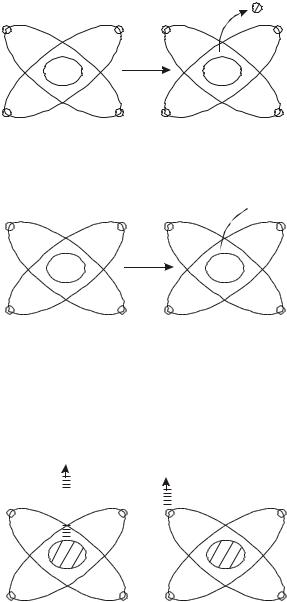
Evolution of Ultrasound System
OBJECTIVE TYPE QUESTIONS
Fill in the gaps |
|
|
|
1. |
The application of ultrasound is based on |
||
|
the --------- |
principle. (a) Sonar (b) ultra |
|
2. |
Acoustic waves are easily --------- |
in water. |
|
|
(a) replaced (b) transmitted |
|
|
3. |
Ultrasound has frequency --------- |
than |
|
20,000 hertz. (a) greater (b) lesser
4.Ultrasound wave travels in the form of ----
----- wave. (a) transverse (b) longitudinal
5.The average speed in tissues of ultrasound
is taken as --------- |
(a) 1400 m/s (b) 1540 |
m/s |
|
6. Incident wave at the interface is partly
transmitted and partly --------- |
. (a) absorbed |
(b) reflected |
|
7.The intensity of echoes depends upon the characteristic of meduim known as --------
-. (a) specific density (b) specific impedance
8.Specific impedances depends upon density
and --------- |
in the meduim. (a) speed |
(b) pressure |
|
9.If meduim 1 and 2 have impedance of z1 and z2 respectively, then percent reflected beam depends upon ---------. (a) z1 + z2 (b) z1 – z2
10. |
Penetration of ultrasound wave in the tissue |
|
|
increases with |
--------- of frequency. |
|
(a) increase (b) decrease |
|
11. |
A-mode is --------- |
mode. (a) amplification |
|
(b) amplitude |
|
12. |
B-mode is --------- |
mode. (a) brightness |
|
(b) biotissue |
|
13. |
M-mode is --------- |
mode. (a) mobile |
|
(b) motion |
|
14. Piezoelectic transducer can convert electric
energy into --------- |
energy and vice versa. |
(a) ultrasound (b) heat
ULTRASOUND IMAGING |
225 |
15. |
When incident sound wave is reflected from |
|
|
moving target, its frequency is changed |
|
|
which is known as --------- |
shift. (a) hobbler |
|
(b) doppler |
|
16. |
Ultrasound is a --------- |
imaging system. |
|
(a) invasive (b) non invasive |
|
17. |
The quality of image --------- |
the case of |
|
obese patients. (a) increases (b) decreases |
|
18. |
Gel is applied between skin and transducer |
|
|
to --------- |
impedance. (a) lower |
|
(b) increase |
|
19. |
The change of impedance at bone interface |
|
|
is --------- |
and ultrasound wave is |
|
completely --------- |
. (a) small, transmitted |
(b) large, reflected
|
|
|
|
|
|
ANSWERS |
|
|
|
|
|
||
1. |
(a) |
2. |
(b) |
3. |
(a) |
4. |
(b) |
5. |
(b) |
6. |
(b) |
7. |
(b) |
8. |
(a) |
9. |
(b) |
10. |
(a) |
11. |
(b) |
12. |
(a) |
13. |
(b) |
14. |
(a) |
15. |
(b) |
16. |
(b) |
17. |
(b) |
18. |
(a) |
19. |
(b) |
|
|
|
|
226 |
FUNDAMENTALS OF BIOMEDICAL ENGINEERING |
RADIOISOTOPES |
& |
|
AND RADIOTHERAPY |
||
|
||
|
|
|
|
|
There is no greatness where there is no simplicity, goodness and truth
INTRODUCTION
1.When a combination of neutrons and protons which does not exist in nature, is produced artificially, then the atom is unstable. Such atom is called a radioisotope. The nucleus of radioisotope tries to become stable by emitting alpha and /or beta particle, which may be accompanied by gamma rays (photons radiation). This process is called radioactive decay. The photons radiation emitted by radioisotopes can be easily imaged by gamma cameras. Suitable radioisotopes as labelled tracers can participate in the metabolism or other body functions and therefore are carried or concentrated in target organ. Image quality depends on the tracer concentration in the target area and on the emission dynamics of the radioisotope
used. Hence radioisotopes are invaluable tool in the field of medical diagnosis and radiotherapy.
TYPES OF RADIOACTIVE DECAY
1.Radioisotops can be decayed by: (1) beta decay (2) gamma decay (3) alpha particle emission and (4) decay by electron capture. Beta decay takes place by emission of beta particles from radioisotope. Beta particles have a charge and mass equal to those of high speed electrons. Beta particle can be positrions (β+) or negatrons (β–) as shown in the figure. The equation of formation of beta particle can be :–
(a) Neutron → proton + β – (negatron)
Example : 14 C6 → 14 N7 + β – (b) Proton → neutron + β+ (positron)
Example : 22 Na11 → 22 Na10 + β+
RADIOISOTOPES AND RADIOTHERAPY |
|
|
227 |
e– |
e– |
e–1 |
β+e–1 |
11P |
|
Decay |
10P |
11N |
|
|
12N |
e– |
e– |
e–1 |
e–1 |
Positron Decay (β+)
 β–
β–
6 P |
Decay |
7 P |
|
||
8 N |
|
7 N |
Imgatron Decay (β–)
2.Gamma emission involves electromagnetic radiation similar to x-rays but it has a shorter wavelength as shown in figure. Gamma radiation takes place with transformation of
Gam m a
e– |
|
e– |
|
||
|
||
|
||
|
|
|
|
|
|
|
|
|
|
|
|
nucleus of a radioisotope with emission of alpha or beta particles. The equation of gamma radiation (x) is:
131 I61 → 131 × C64 + 3β–1 + γ
x-rays
e– |
e– |
Nucleus |
|
|
Nucleus |
e– |
e– |
e– |
e– |
X-Rays and Gamma Radiation

228 |
FUNDAMENTALS OF BIOMEDICAL ENGINEERING |
3.Alpha particle consists of helium nucleus with two neutrons and two protons. Radioisotopes having higher atomic weight generally decay by emitting alpha particle which is known as alpha particle emission. Emission of an alpha particle results in decrease of atomic number by two units and
atomic mass by four units. The equation of
alpha emission is: 226Ra88 → 222 Rn86 + 4He2++
|
|
|
2P |
|
|
|
2N |
e– |
e– |
e– |
e– |
88P |
|
Decay |
86P |
138N |
|
|
136N |
e– |
e– |
e– |
e– |
Alpha Particle Radiation
4.The number of protons in a nucleus can also be reduced by the process of electron capture. This is called decay by electron capture. In this radioactive decay, one of the inner orbital electron is attracted into the nucleus where it combines with the proton in nucleus to form a neutron i.e., e– + p+ = n. Combining results in the loss of one proton and gain of one neutron in the nucleus. Though there is no emission of any particle but x-rays are emitted due to electron moving from inner orbit to nucleus. Iron having atomic number 55 usually decays by this mode.
e– |
G am m a e– |
e– |
Electron Capture |
e– |
|
|
INTERACTION OF NUCLEAR RADIATION WITH MATTER
1.Radioistopes are used as tracers which emit α particle, β-particles or γ-rays. Since these radiations have different physical characteristics, the manner inwhich these interact with other matters also differ. Gamma rays are high energy photons having neither charge nor mass. The penetrating power of gamma rays is much greater than that of alpha or beta particles. However ionizing power of gamma rays is less. Gamma rays can produce: (1) photo-ionization (2)
compton effect and (3) pair production. In photoionization, γ-rays interact with orbital electron which is ejected as negatron with energy equal to gamma rays. The negatron interact with other atoms to ionize them. Gamma rays having more energy can be scattered by electron after absorbing energy for ejection. This results into ejection of a negatron and a new photon of lowered energy which moves in altered direction after scattering. In pair production, gamma rays interact with an electron and a positron of nucleus resulting into emission of negatron and of positron. Alpha particle is a helium nucleus having two protons and two

RADIOISOTOPES AND RADIOTHERAPY
neutrons. Alpha particles interact with matter in two ways. In one case, alpha particles impart energy to orbital electrons of the atom of the matter but without any ejection. Excited electrons emit excess energy as photons. In second case, alpha particles cause ionization of matter by ejection of orbital electrons and leaving behind positive charged atoms. Alpha emitting isotopes are not commonly used as tracer in imaging as these isotopes have higher atomic number. Beta particles (positrons and negatrons) are very small. They have high velocity resulting higher penetrating power. Beta particles dissipate their energy largely by ionisation or excitation of the atoms with which they interact.
RADIOACTIVE DECAY
1.The number of atoms in a radioisotope that disintegrate during a given time interval decreases exponentially with time as shown in the figure. If λ = decay constant,
No = number of atoms present originally, N = number of atoms present after time t
then N = Noe–λt. The time for the half the number of atoms to disintegrate is given by t1/2 =0.693/λ
Number of atom s (N)
Time 
MEASUREMENT OF RADIOACTIVITY
1.The measurement of radioactivity is determination of the rate of emission of
229
alpha, beta and gamma rays from the radioisotope. These radiations are also known as ionizing radiations as they are capable of causing ionization directly or indirectly. The methods commonly used for detection and measuring radioacitivity are based on the ionization of the gases like in Geiger-Mullier counter and proportional counter, or on the excitation of solids or solution as in scintiallation counter or on induction of specific chemical reaction in certain emulsion as in auto-radiography. In the Geiger-Mullier counter, alpha and beta particles enter in the counter tube through mica window having gases under pressure with anode and cathode as shown in figure with potential difference of 800 - 2500 volts. The alpha and beta particles ionize molecules of the gases which move towards appropriate electrode under the voltage gradient. The process produced a continuous flow of ions which produces discharge pulses of 10 volt amplitude with duration of 50 to 100 microseconds. These pulses are counted which is a measurement of radioactivity. In proportional counters, the gradient voltage is kept lower than GeigerMullier counter and it requires a preamplifier to avoid reducing the pulse size. Scintillator counter uses a chemical to convert radiation energy into light. When an ionizing particle is absorbed in the scintillator, some of the energy acquired by the scintillator is emitted as a pulse of visible light or near ultraviolet radiation. The light falls on a photomultiplier tube resulting in pulse of electrons which is counted to measure radioactivity.

230 |
FUNDAMENTALS OF BIOMEDICAL ENGINEERING |
+
–
Tungston anode
Glass jacket
High pressure argon and m ethane gas
Copper cylinder Glass (cathode)
bead
 M ica window
Geiger-Mullier Counter
RADIOTHERAPY
1.Surgery, radiotherapy and /or chemotherapy are used for the treatment of cancer. It must be ensured in radiotherapy that the radiation dose delivered to a patient should be optimally focused to produce a maximum effect in the volume of cancerous tissue and a minimal effect in the neighbouring healthy tissues. Radiotherapy is carefully planned, simulated, executed and verified. Treatment planning is carried by modelling to match the absorption characteristics of the radiation
within the anatomy of the patient. Radiation data is obtained from dosimetry and patient anatomy is acquired from CT scanners. Algorithms are used with these data and patient’s anatomy in a computer for modelling. To obtain an optimum treatment plan, the cancerous cells are irradiated from several directions and for certain duration for two weeks or so. The outcome obtained from this radiotherapy is a good indicator of further requirements of radiotherapy for the patient.
OBJECTIVE TYPE QUESTIONS
Fill in the gaps |
|
|
1. |
Radioisotope has -------- |
nucleus. |
|
(a) unstable (b) stable |
|
2. |
Photon radiation can be easily imaged by |
|
|
--------- camera. (a) Alpha (b) Gamma |
|
3. |
Alpha particle consists of --------- |
nucleus |
|
with two neutrons and |
two protons. |
|
(a) helium (b) hydrogen |
|
4. |
Gamma radiation is --------- |
radiation similar |
|
|
to x-rays. (a) light (b) electomagnetic |
||
5. |
Beta particle has charge and mass equal to |
||
|
that of high speed --------- |
. (a) proton |
|
|
(b) electron |
|
|
6. |
The --------- |
is time during which half |
|
|
number of atoms disintegrates. (a) half time |
||
|
(b) half life |
|
|
RADIOISOTOPES AND RADIOTHERAPY
7. Radioisotopes are used as --------- |
. (a) tracer |
(b) medicine |
|
8.Radiotherapy is used for the treatment of
---------. (a) cancers (b) malfunctioning
9.The radiation dose delivered to a patient
|
231 |
should be --------- |
focused on cancerous |
tissues. (a) optimally (b) maximumally |
|
10.--------- uses chemical to convert radiation energy into light when an ionizing particle is absorbed. (a) Geiger-mullier (b) scintillator
|
|
|
|
|
|
ANSWERS |
|
|
1. |
(a) |
2. |
(b) |
3. |
(a) |
4. (b) 5. (b) |
6. (b) |
7. (a) |
8. |
(a) |
9. |
(a) |
10. |
(b) |
|
|
|