

11
2
Anatomy and
Physiology
Julia Robertson and Andy Mead
•Introduction
•Skeletal system
•Muscular system
•Fascia
•Nervous system
•Other systems
•Comparative human and canine anatomy
Introduction
A balance is required in the body on a cellular level, as it is in every part of nature. The basic requirement of every cell must be met in order for it to survive, in the same way that soil must be correctly balanced in order for a plant to grow well. If one part of a system is not working properly, it will have an effect on another part of the system and the overall performance will be compromised. This chapter will aim to show how the body works in this holistic way and how each system interacts with another. In order to understand how massage and physical therapy affect the body, it is important to understand the systems within the body, how they function, and, most importantly, how they inter-relate.
12 Chapter 2
Skeletal system
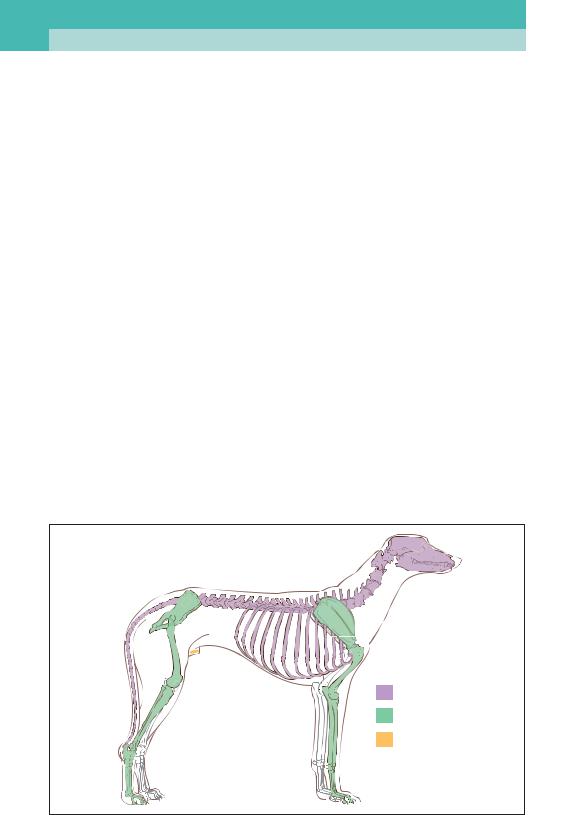
The skeleton can be defined as the stiff hardened tissues that form the supporting framework of an animal’s body. The skeleton is often divided into three distinct portions: the axial, the appendicular, and the visceral skeletons (2).
•The axial skeleton comprises the bones of the skull, the hyoid apparatus (larynx), the
vertebral column, and the ribcage. It principally performs a protective role.
•The appendicular skeleton comprises the bones of the limbs and the bones that connect the limbs to the axial skeleton, e.g. the pelvis joining onto the sacrum.
•The visceral skeleton comprises the bones that develop in soft tissue structures. In dogs, this includes only one bone, the os penis.
It is not the intention to describe the anatomy of the canine skeleton in great detail here, as there are many other texts on the subject. Instead, an overview is presented.
Bones are a vital part of the skeletal system. They provide support and give shape to the body, as well as protecting vital organs including the heart and the brain. Above all, bones enable movement.
Bones comprise semi-rigid connective tissue and a mineral component, hydroxyapatite. The connective tissue is formed of type 1 collagen, the most common protein of connective tissue, which is made up of three polypeptide strands interlinked by hydrogen bonds into a lefthanded helix. This provides an essentially brittle matrix of bone; what little capacity it has to withstand compressive and tensile forces is provided by weak hydrogen bonds between strands.
Axial
Appendicular
Visceral
2 Major divisions of the canine skeleton.
|
|
Anatomy and Physiology |
13 |
|
|
Hydroxyapatite is essentially calcium and phosphorus (Ca10[PO4]6[OH]2) and constitutes approximately 70% of bone. It usually crystallizes in a very organized hexagonal pattern, providing rigidity to the structure of bone, as well as giving bone its smooth, white appearance. Bone also contains many different types of cells supported within the matrix, which allow the structure to grow, repair, and, perhaps more importantly, provide a scaffold for the performance of vital functions, such as red and white blood cell production.
Bone is the body’s mineral store, providing a reservoir of, for example, calcium and phosphorus, which can be mobilized for the purposes of:
•Maintaining calcium homeostasis.
•Detoxifying heavy metals.
•Producing blood cells within the marrow.
•Transferring sound though the bones of the ear.
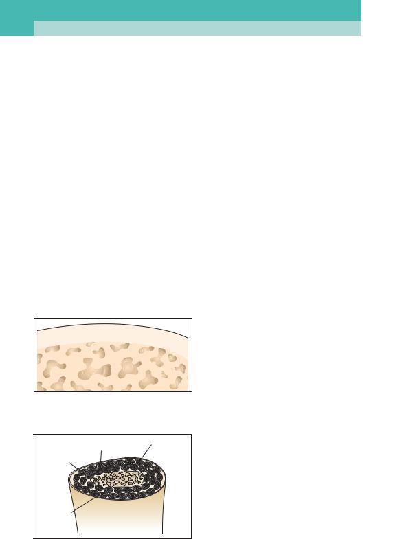
The structure of bone can be categorized into two types, compact and trabecular.
Compact bone
This forms the hard outer layer of bone, which is both smooth and well-ordered. There are minimal gaps here between collagen fibres and the mineral matrix. Compact bone is often referred to as cortical bone (3).
Osteone
Blood vessels |
Osteocyte |
|
Haversian canal |
3 Diagram of cortical bone showing osteones and structural organization.
14 Chapter 2
Trabecular bone
This forms the rather more disorganized, spongy, central portion of bone (4, 5). Trabecular bone reduces the total weight of a bone, as well as providing a framework for blood vessels and nerves. Without the presence of this type of bone, mammalian species would be greatly restricted in terms of size, due to the fact that greater muscle mass would be required to lift and move heavier bones. Without trabecular bone, bones of the axial skeleton would need to be as wide as they were long in order to withstand the forces put on them. Trabecular bone is often harvested surgically to aid with fracture repair.
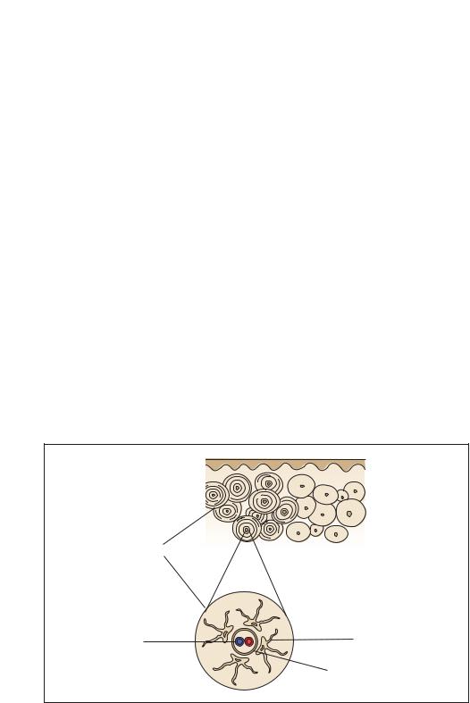
Cellular structure of bone
It is necessary to discuss in some detail the cellular structure of bone in order to understand better the process of bone development, and thereby understand how, if disturbed, these processes can lead to malformation and disease which may become significant in later life.
The main building blocks of bone at the cellular level are osteoblasts, osteocytes, and osteoclasts. These cells are present within bone throughout life. Bone is constantly being remodelled, as it is subject to ever-present stresses and strains, even at a microscopic level. Each of these constituent cells of bone has a different role in the development and maintenance of its structure, and of calcium homeostasis. All are given the prefix ‘osteo’, which pertains to bone.
4 Trabecular bone showing disorganization of structure.
Periosteum Osteone
Cortical
bone
Spongy
bone
Osteoblasts
These are bone-forming cells that produce osteoid, which is principally made up of collagen type 1. Osteoblasts have roles in the production of hormones and enzymes, most notably the enzyme alkaline phosphatase, which, among other functions, enables the mineralization of bone.
Osteocytes
Osteocytes are essentially osteoblasts which become trapped in the collagen/ hydroxyapatite matrix and occupy a space within the structure called a lacuna. These cells communicate with one another through special channels and help bone to manage forces and stress, as well as playing a part in calcium metabolism. Calcium homeostasis is imperative within the mammalian body, in order to allow muscle contraction, nerve communication, and maintenance of the correct structure and rigidity of bone.
Osteoclasts
5 A section through bone showing the position of trabecular and cortical bones.
These are cells engaged in the remodelling of bone during growth or after injury.
|
|
Anatomy and Physiology |
15 |
|
|
Bone development
The process of bone development is complex, and will not be covered in great detail within this text. The embryo undergoes the process of gastrulation, which involves the early fetal cells differentiating into three distinct layers – ectoderm, mesoderm, and endoderm. It is the mesoderm layer that produces the rudimentary origins of limbs, and therefore bony tissue. The process by which bone develops from this mesoderm is known as ossification; the process is subdivided into endochondral ossification and intramembranous ossification.
Endochondral ossification
Endochondral ossification commences within the embryo and only finishes when the animal reaches maturity. Put simply, an initial scaffold of cartilage is gradually replaced with both cortical and trabecular bone tissue. Such a process accounts for the development of every long bone of the body.
The cartilage template formed within the embryo by the mesodermal cells is replaced by bone at three distinct ossification centres: the diaphysis (or middle) of each long bone, and two epiphyses, one at each end of the long bones. This process occurs from the centre outwards; long bones end up capped by cartilage which has not been ossified. This forms the articular cartilage of joints. Two discoid areas develop between the epiphyses and the diaphysis, known as growth plates or metaphyses (6). Lengthening of bone always occurs at the side of the growth plate that is closer to the epiphysis.
As previously discussed, the inner core of a long bone is spongy or less solid. It is the osteoclasts that remodel and reshape bone which results in the presence of a cavity in the centre of long bones known as the medullary cavity.
The growth plates, which remain after the differentiation of the ossification
Epiphysis
Metaphysis
Epiphyseal
line
Diaphysis
Metaphysis
Epiphysis
Area of growth
(Epiphyseal slot of growth plate)
6 Endochondral ossification showing the position of diaphysis, metaphysis, and epiphysis.
centres, allow long bones to lengthen as the animal grows, long after it has left the womb. This process only stops once these growth plates close, i.e. are also ossified, a process that occurs only when an animal reaches maturity. However, these growth plates vary in the age at which they close. To the untrained eye, they can often be mistaken for fractures on radiographs.

16 Chapter 2
Intramembranous ossification
This differs from endochondral ossification in that there is no cartilaginous scaffold, bone being formed directly from fibrous connective tissue. This occurs in bones such as the frontal bone, and others that form the skull.
Bone construction
The form/shape of any long bone is determined by the cortex, the outer strong layer of the bone. The cortex is arranged in a uniform manner comprising a central channel, the Haversian canal, through which blood vessels, nerves, and lymphatic vessels flow. Around this, a matrix of hydroxyapatite and collagen form concentric tubes known as lamellae. The whole system is known as an osteone (3). Within the lamellae are spaces (lacunae) which are home to the osteocytes. The whole system of lamellae, lacuna, and Haversian canals is referred to as the Haversian system, which, as previously discussed, can be densely packed, as in compact bone, or separated by much larger spaces, as in cancellous bone.
The entire surface of bone, apart from the joint capsule, is covered by a tough connective tissue, the periosteum, which has the primary role of forming attachments for muscles, tendons, and ligaments. The periosteum also has bone-forming properties which may be called upon on in the event of a fracture.
Like any other cells of the body, the cells of bone require nutrients and the removal of waste products. These are supplied and removed by an extensive skeletal blood supply which comprises 5% or more of the cardiac output. Such blood vessels, known as nutrient arteries, penetrate the cortex of bone via nutrient foramina and go on to form the tiny blood vessels of the Haversian systems.
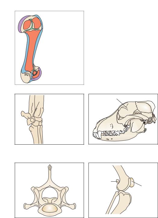
Classification of bones
Long bones
Long bones are longer than they are wide, and consist of two epiphyses covered in articular cartilage and a diaphysis (7). These are the main bones of the limbs and their shape means that they act as levers.
Short bones
These are roughly cube shaped and consist of a core of cancellous bone covered by a layer of compact bone. Short bones are only found within the carpus and tarsus (8).
Flat bones
Flat bones consist of two plates of compact bone with a core of cancellous bone. Examples include the majority of bones in the skull and the ribs (9).
Irregular bones
These are similar to flat bones, except that they are far more complex in shape and arrangement. The vertebrae are examples of irregular bones (10).
Sesamoid bones
These are small bones buried within tendons. They reduce friction and, therefore, tendon damage, by altering the angle at which a tendon passes over a joint, allowing greater ease of movement and acting as a pivot. These specialized bones prevent tendon wear and increase leverage. The most obvious example of a sesamoid bone is the patella (11).
|
|
Anatomy and Physiology |
17 |
|
|
 Epiphyseal cartilage
 Articular cartilage
 Spongy bone
 Cortical bone
7 Diagram of a long bone, showing portions of spongy and cortical bone, and articular and epiphysial cartilage.
|
|
Carpal bones |
1 |
|
1: Accessory carpal |
|
bone |
|
|
|
|
2 |
3 |
2: Ulnar carpal |
|
|
3: Intermediate carpal |
8 Short bones of the carpus.
Frontal bone |
9 The frontal bone of the canine skull, a flat bone.
Fabella
(Sesamoid)
Patella (Sesamoid)
10 A lumbar vertebra, an irregular bone.
11 Sesamoid bones such as the patella and fabella in the canine stifle joint act as pivots and allow flexion and extension.
18 Chapter 2
Biomechanics of bone
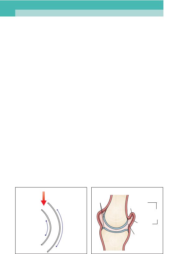
When weight or stress is applied to bone, it acts like a beam of wood or steel – the force is concentrated in the outer surface, which explains why the medulla need not be solid like the cortex. Similarly, this accounts for the fact that bones are often tubular in shape. The cortex has the ability to recover from considerable deformation; however, should the osteones bend or stretch too far, stress fractures will soon appear, even if there is not enough force to break the bone in two. The majority of fractures are caused by excessive bending, stress being applied to both sides of the bone equally. However, different types of forces will affect opposite sides of the bone: one side will be affected by tensile forces pulling the cells, lamellae, collagen, and mineral components apart, and the other will be affected by compressive forces pushing structures together and crushing them (12). Generally speaking, the former are more easily dealt with by bone.
Joints
Joints are defined as the junction between two or more bones and are classified as simple or compound. Joints provide motion and flexibility to the skeleton and, perhaps most importantly, allow growth.
Different types of joints allow different amounts of movement. Indeed some joints allow no movement at all; for example, the joints of the skull; these are called synarthroses. For the purposes of physical therapy, the movable or synovial joints, by far the most common joints of the body, are the most important (13). The structure of the joint determines the type and direction of movement allowed. This provides a logical system for classification: they can be either plane, ball and socket, hinge, pivot, condylar, or saddle joints.
Movements of joints
These are divided into the following categories:
•Flexion − decreasing the inner angle between two bones, or bending the joint.
•Extension − increasing the angle, or straightening the joint.
•Abduction − pulling away from the centre or axis; for example, moving the pelvic limb laterally.
•Adduction − pulling towards the centre or axis; for example, moving the pelvic limb medially.
•Rotation − movement around an axis.
•Circumduction − circular movement of a limb.
Force (weight) |
Articular |
|
|
exerted |
|
cartilage |
Synovial |
|
|
|
|
|
|
|
membrane |
|
|
|
Joint |
|
|
|
capsule |
Compression |
Tensile |
|
Fibrous |
|
layer |
||
forces |
forces |
|
|
|
|
||
|
|
|
Joint |
|
|
|
cavity |
12 Forces involved in the bending of bone. 13 Synovial joint.
|
|
Anatomy and Physiology |
19 |
|
|
•Protraction – in this context, moving a limb forward.
•Retraction – pulling a limb backwards.
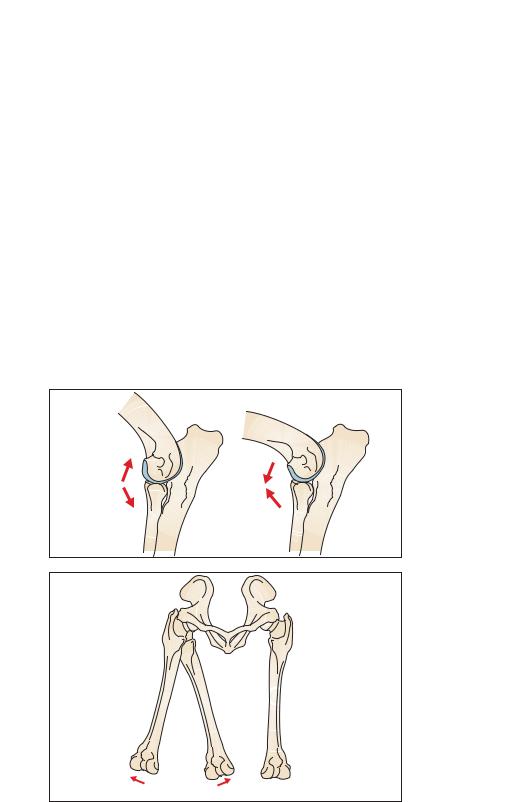
In the case of the elbow, a hinge joint permits flexion and extension (14). In the hip, a ball and socket joint allows flexion, extension, abduction, adduction, rotation, and circumduction (15).
Joint structure
For the purposes of this text, we shall concentrate on the structure of synovial joints. The importance of structure will
become clear when discussing diseases associated with synovial joints in subsequent chapters.
In a synovial joint, the two articulating bones are divided by a fluid-filled structure, the joint cavity. The fluid (synovial fluid) is found within the joint capsule and is surrounded by a thin layer of synovial or joint fluid-producing cells, known as the synovial membrane. This is a weak structure and is often, but not always, given strength by various additional ligaments and tendons, which are designed to prevent movement in
14 An elbow joint undergoing flexion and extension, medial view.
Extension |
Flexion |
15 Dog pelvis and femora – cranial view. Rotation, adduction, and abduction of the hip joint.
 Rotation
Abduction Adduction
20 Chapter 2
certain directions and limit the amount of movement that can be achieved by a joint (16). At either end of the opposing bones and within the joint capsule is articular cartilage, a specialized form of cartilage that reduces the friction and compressive forces of locomotion. The purpose of articular cartilage is to cushion the bones and prevent wear and tear. It is smooth, elastic, and thick in the areas which require it most, i.e. the points at which the compression and friction forces are greatest.
In dogs, the articular cartilage is only 1mm thick in places; therefore, it is fragile and subject to damage from an early age. Articular cartilage has no nerve or blood supply and only receives nutrient support by diffusion from the surrounding structures, particularly the synovial fluid. Surrounding the articular cartilage within the joint capsule is the synovial fluid, which is derived from the synovial cells of the synovial membrane. The synovial membrane is a connective tissue sheet that produces the lubricant of the joint, called the glycosaminoglycans. The synovial membrane has a rich vascular supply and innervation. The fluid it produces is very viscous, its viscosity often being measured to establish the cause of various disease processes. The synovial fluid acts as a lubricant, reducing the friction between joints, and as a nutritional source for the surrounding articular cartilage.
Surrounding and encapsulating all of the above is the joint capsule, a fibrous layer. This attaches to the bone on either side of the joint and provides congruity. Proprioceptive nerves innervate these structures. The ligaments provide strength to the joints themselves.
Skeletal terminology
In order to describe the position of bony lesions accurately, it is important to use the correct anatomical terminology. The various parts of a bone’s surface are called:
•Crest − a ridge of bone, e.g. the occipital crest of the skull.
•Condyle − a rounded projection of bone at a joint, e.g. the femoral condyle.
•Epicondyle − a separate, smaller prominence close to the condyle.
•Foramen − a natural opening through a bone, through which muscles, nerves, blood vessels, etc. pass, e.g. the obturator foramen of the pelvis.
•Fossa − a hollow or depression, e.g. the olecranon fossa of the elbow.
•Head − a rounded articular surface, e.g. the head of the femur.
•Tuberosity − a protuberance of a bone, e.g. the tibial tuberosity.
•Trochanter − a type of tuberosity specific to the femur and a large rough process, e.g. the greater, lesser, and third trochanters of the femur.
•Trochlea − a depression in the bone where major tendons lie forming a type of hinge, e.g. the trochlear groove of the knee joint.
•Tubercle − a small rounded process, e.g. the greater tubercle of the femur.
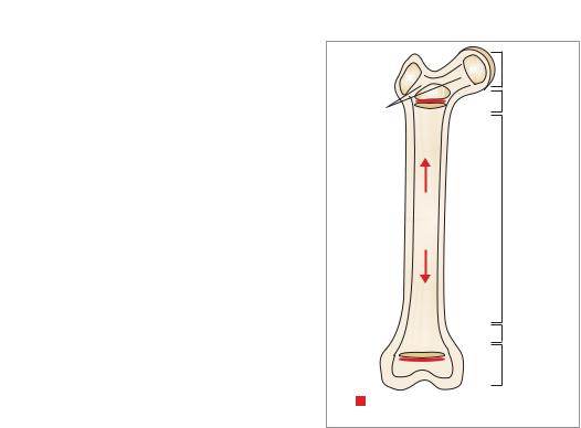
For the practitioner, it is important to be able to identify the palpable bony prominences of the canine, for example, the greater trochanter of the femur (17), the vertical spinous processes the ilium of the pelvis, and so on. These points of reference are far easier to palpate in the fit athletic patient and within certain breeds, for example the Greyhound. These landmarks are best described diagrammatically (18).
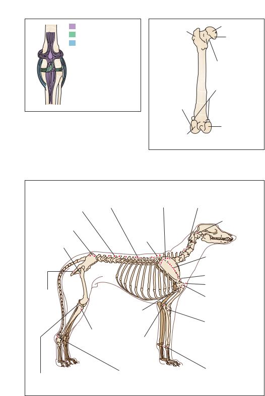
|
|
Anatomy and Physiology |
21 |
|
|
Patella tendon
Cruciate ligaments
Collateral ligaments
16 Cranial view of a simplified stifle joint, showing ligaments and tendons.
Femoral head
Greater
trochanter 

 Fovea of femoral head
Neck of femur
Sesamoid bones (fabellae)
Lateral epicondyle
Lateral |
Medial |
|
condyle |
||
condyle |
||
|
17 Palpable points of the canine femur, caudal view.
|
Anticlinal |
Dorsal border |
Spinous process |
vertebra |
of the scapula |
of the lumbar |
|
Spinous |
vertebra |
|
|
|
process of |
|
Iliac crest/cranial |
|
|
|
the thoracic |
|
dorsal iliac spine |
|
vertebra |
Greater |
|
|
trochanter |
|
|
Ischiatic
tuberosity 
 Patella
Deltoid tuberosity
Lateral condyle of the tibia
Lateral epicondyle of the femur (medial aspect, medial epicondyle of the femur)
Olecranon process
Calcaneous and calcaneal tuberosity
Transverse process of the cervical vertebra
Atlas
Spine of the scapula
Acromion process Manubrium
Greater tubercle of the humerus
Lateral epicondyle of the humerus (medial aspect, medial epicondyle of the humerus)
Carpus
18 Palpable canine bony landmarks.