


62 |
2 Skull Base Segment |
|
|
2.2 Anatomic Pictures
ICAp
Fig. 2.1 Horizontal segment. Lateral vision of the right cavernous sinus and horizontal portion of the internal carotid artery
ET eustachian tube, ICAh horizontal portion of the internal carotid artery, ICAi intracranial portion of the internal carotid artery, ICAp parapharyngeal portion of the internal carotid artery, TC tentorium cerebelli, VN vidian nerve, V1 first branch of the trigeminal nerve, V2 second branch of the trigeminal nerve, V3 third branch of the trigeminal nerve, IIIcn oculomotor nerve, IVcn trochlear nerve

2.2 Anatomic Pictures |
63 |
|
|
Fig. 2.2 Lateral vision of the right jugulomastoid region
ASC anterior semicircular canal, CC carotid canal, ET eustachian tube, ICAp parapharyngeal portion of the internal carotid artery, IJV internal jugular vein, JB jugular bulb, LSC lateral semicircular canal, ME middle ear, MMA middle meningeal artery, PSC posterior semicircular canal, SS sigmoid
sinus, ZR zygomatic root, VIIcn VIIcn facial nerve, IXcn glossopharyngeal
nerve, V vestibule, yellow arrow stapes, black arrow incus
LCS |
TTM |
|
|
|
|
||
PSC |
RW |
ICAh |
|
|
VIIcn |
|
|
|
JB |
ET |
ICAh |
|
|
|
|
|
IXcn |
|
LSC |
|
|
|
|
|
XIcn Xcn |
ASC |
VIIcn |
|
|
||
LSC
PSC
RW |
TTM |
|
VIIcn |
||
MMA |
||
|
||
JB |
|
|
|
ET |
|
IJV |
ICAp |
|
|
Fig. 2.3 Right exposure of the middle ear and jugular foramen. Note the intimate relationship between the eustachian tube and the carotid canal
ASC anterior semicircular canal, ET eustachian tube, ICAp parapharyngeal portion of the internal carotid artery, ICAh horizontal portion of the internal carotid artery, IJV internal jugular vein, JB jugular bulb, LSC lateral semicircular canal, MMA middle meningeal artery, PSC posterior semicircular canal, RW round window, TTM tensor tympani muscle, VIIcn facial nerve, IXcn glossopharyngeal nerve, Xcn vagus nerve, XIcn accessory nerve, red arrow cochlea
The posterior bend of the ICAh within the petrous bone is located just below and anterior to the cochlea. It is separated from the tympanic cavity by a thin bone that may be dehiscent.

64 |
2 Skull Base Segment |
|
|
VIIcn
Fig. 2.4 Lateral view of the tympanojugular region
CJS carotidojugular spine, ET eustachian tube, ICAp parapharyngeal portion of the internal carotid artery, IJV internal jugular vein, JN Jacobson’s nerve, ME middle ear, MMA middle meningeal artery, TTM tensor tympani muscle, TVPM tensor veli palatini muscle, V3 third branch of the trigeminal nerve, VIIcn facial nerve, IXcn glossopharyngeal nerve, yellow arrow lesser petrosal nerve, red arrow inferior part of the inferior tympanic nerve (Jacobson’s nerve), black asterisk round window, black circle malleus, arrowhead incudostapedial joint
At the level of the jugular foramen area, the ICA moves away from the jugular bulb below the middle ear, and it is separated from it by the CJS. Jacobson’s nerve is joined by the carotidotympanic nerves from the pericarotid sympathetic plexus to form the lesser petrosal nerve (Janfaza and Nadol 2001b).

2.2 Anatomic Pictures |
65 |
|
|
Fig. 2.5 Axial view at the level of the horizontal portion of the internal carotid artery
BA basilar artery, Cl clivus, Co cochlea, ET eustachian tube, IAC internal acoustic canal, ICAh horizontal portion of the internal carotid artery, LPM lateral pterygoid muscle, ME middle ear, MPM medial pterygoid muscle, SS sphenoid sinus, TM temporal muscle, TVPM tensor veli palatini muscle, red arrow bony segment of the eustachian tube (musculo-tubal canal)
Fig. 2.6 Axial view at the level of the horizontal portion of the internal carotid artery
Co cochlea, ICAh horizontal portion of the internal carotid
artery, MMA middle MMA meningeal artery, TTM tensor
tympani muscle, V vestibule, VN vidian nerve, V3 third branch of the trigeminal nerve, VII–VIIIcns acousticfacial bundle, red arrow deep petrosal nerve, black arrow greater petrosal nerve
A venous plexus surrounds the ICAh within the carotid canal. A nerve plexus, coming from the superior cervical ganglion, accompanies the artery.
66 |
2 Skull Base Segment |
|
|
FO
FS FL
SPF
PAp
CI
CC
CR
PF
JF
OC
CF
SP
SMF
Fig. 2.7 Skull base from below
CC
CR
°
° ° ° °
PF
JF
CC carotid canal, CF condylar fossa, Cl clivus, CR carotid ridge, FL foramen lacerum, FO foramen ovale, FS foramen spinosum, JF jugular fossa, PAp petrous apex, PF pyramidal fossa, SMF stylomastoid foramen, SP styloid process, SPF sphenopetrosal fissure, dark green arrow external opening of the cochlear aqueduct, violet arrow mastoid canaliculus, red arrow opening of the musculo-tubal canal, blue arrow foramen lacerum region, green arrow tympanic canaliculus, black circles intrajugular ridge, black arrow hypoglossal canal, blue-sky line groove for the auricular branch of vagus nerve (Arnold’s nerve)
The FL is a hole in the dry skull; it is closed by fibrocartilaginous tissue in living patients. It is passed by meningeal branches of the ascending pharyngeal artery and emissary veins. The FO transmits the V3 and accessory meningeal artery and sometimes the lesser superficial petrosal nerve. The FS transmits the middle meningeal artery and veins and the recurrent branch of V3 (for the dura of the middle cranial fossa). The mastoid canaliculus transmits Arnold’s nerve (of the vagus nerve). This nerve passes through the mastoid and exits at the level of the tympanomastoid suture. The tympanic canaliculus transmits Jacobson’s nerve and the inferior tympanic artery (from the ascending pharyngeal artery).

2.2 Anatomic Pictures |
67 |
|
|
LWS
°
FO
|
|
|
|
FL |
FS |
|
|
|
|
|
|
PS |
OC |
|
FO FS |
|
CC |
|
|
|
|||
OS |
|
|
SPF |
||
|
|
|
|
|
|
|
* |
ACP |
ICAh |
* |
|
|
|
|
|||
TS |
|
|
|
||
PF |
|
|
TI |
|
TI |
|
DS |
|
|
|
* |
|
Cl |
|
|
|
|
Fig. 2.8 Right middle cranial fossa and clivus from above
ACP anterior clinoid process, CC carotid canal, Cl clivus, DS dorsum sellae, FL foramen lacerum, FO foramen ovale, FS foramen spinosum, ICAh horizontal portion of the internal carotid artery, LWS lesser wing of the sphenoid, OC optic canal, OS optic strut, PF pituitary fossa, PS planum sphenoidale, SPF sphenopetrosal fissure, TI trigeminal impression, TS tuberculum sellae, yellow drawing petrolingual ligament, black circle lingual process of the sphenoid, red asterisk groove for the greater petrosal nerve, black asterisk middle clinoid process
The petrolingual ligament can be considered as a continuation of the periostium of the carotid canal (Osawa et al. 2008). In most cases, the medial aspect of the horizontal portion of the internal carotid artery is not covered by bone, but simply by dura. The hiatus of the facial canal transmits the greater superficial petrosal nerve and the superficial petrosal branch of the middle meningeal artery (MMA) to the facial nerve.
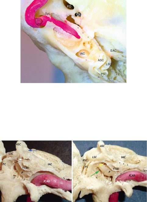
68 |
2 Skull Base Segment |
|
|
SPF
ICAh
*
Fig. 2.9 Relationship between the horizontal portion of the internal carotid artery and the cochlea (vision from above)
ASC anterior semicircular canal, EAC external auditory canal, FO foramen ovale, FS foramen spinosum, IAC internal auditory canal, ICAh horizontal segment of the internal carotid artery, LSC lateral semicircular canal, PSC posterior semicircular canal, SPF sphenopetrosal fissure, SpL lingula of the sphenoid, blue arrowhead vestibular aqueduct, black asterisk cochlea, black circles groove for the greater petrosal nerve
The cochlea is posterosuperiorly and laterally located in respect to the posterior bend of the horizontal portion of the internal carotid artery.
Fig. 2.10 Relationship between the horizontal portion of the internal carotid artery and the inner ear (anterior view)
ASC anterior semicircular canal, FC facial canal (mastoid segment), IAC internal acoustic canal, ICAh horizontal portion of the internal carotid artery, LSC lateral semicircular canal, P promontory, PSC posterior semicircular canal, TTMC tensor tympani muscle canal, blue arrow canal for the subarcuate artery, white arrow tympanic segment of the facial canal, yellow arrow oval window, green arrow round window, black asterisks groove for the greater petrosal nerve
The subarcuate artery usually originates medial to the internal acoustic meatus, and penetrates the subarcuate canal (petromastoid canal). It can send branches to the petrous apex and to the bone of the semicircular canals and of the vestibule.

2.2 Anatomic Pictures |
69 |
|
|
ON
Fig. 2.11 Right middle cranial fossa from above. The gasserian ganglion has been reflected upward to show the position of the petrous portion of the internal carotid artery
ACA anterior cerebral artery, AchA anterior choroidal artery, BA basilar artery, GG gasserian ganglion, GPN greater petrosal nerve, ICAc cavernous portion of the internal carotid artery, ICAh horizontal portion of the internal carotid artery, ILT inferolateral trunk, MCA middle cerebral artery, MMA middle meningeal artery, OA ophthalmic artery, ON optic nerve, PcomA posterior communicating artery, SPS superior petrosal sinus, TI trigeminal impression, TR trigeminal root, V1 first branch of the trigeminal nerve, V3 third branch of the trigeminal nerve, IIIcn oculomotor nerve, IV trochlear nerve, VIcn abducens nerve
Usually, the superior petrosal sinus is superior to the roots of the trigeminal nerve. Sometimes, the sinus divides and lies superior and inferior to the roots (Janfaza and Nadol 2001a).
It connects the cavernous sinus to the sigmoid sinus. It drains blood from the veins of the brainstem (including the petrosal vein of Dandy), inferior cerebral vein and superior cerebellar vein.

70 |
2 Skull Base Segment |
|
|
Fig. 2.12 Close vision of the middle cranial fossa. The gasserian ganglion has been removed
DPN deep petrosal nerve, ET eustachian tube, GPN greater petrosal nerve, ICAh horizontal portion of the internal carotid artery, LWS lesser wing of the sphenoid, MMA middle meningeal artery, VN vidian nerve, V1 first branch of the trigeminal nerve, V2 second branch of the trigeminal nerve,
V3 third branch of the trigeminal nerve, IIIcn oculomotor nerve, IVcn trochlear nerve, VIcn abducens nerve, yellow arrow a tentorial branch of the inferolateral trunk
Fig. 2.13 Right middle cranial fossa floor (view through a temporal craniotomy) and reconstruction on petrous bone
GG geniculate ganglion, GPN greater petrosal nerve, FO foramen ovale, FS foramen spinosum, ICAh horizontal portion of the internal carotid artery, JN Jacobson’s nerve, LPN lesser petrosal nerve, MMA middle meningeal artery, P promontory, TLD dura of the temporal lobe, TTM tensor tympani muscle, V3 third branch of the trigeminal nerve, yellow arrow petrous apex
The LPN crosses the floor of the middle cranial fossa (MCF) anterior to the GPN (Kakizawa et al. 2007) and exits the cranial cavity most of the time through an innominate canal. The outer surface of this canaliculus is usually located posteriorly to the foramen ovale and spinosum (Kakizawa et al. 2007). This nerve conveys the parasympathetic fibres derived from the superior and inferior salivary nucleus (Porto et al. 1978). It is formed by fibres from cranial nerves VII (nervus intermedius), IX (Jacobson’s nerve), and X (Arnolds’s nerve). The most important part is due to cranial nerve IX (Jacobson’s nerve). This emerges on the MCF posterior to the TTM. While the GPN courses above the ICAh, the LPN courses above the tensor tympani muscle. This muscle occupies a bony canal below the floor of the middle fossa, anterior to the ICAh and above the eustachian tube (ET).

2.2 Anatomic Pictures |
|
|
71 |
|
|
|
|
|
|
TC |
IV cn |
GG |
|
CS |
V1 |
V2 |
|
GG |
V3 |
|
|
|
|
|
V3 |
|
V2 |
|
|
GPN |
SS |
|
MMA |
ICAh |
VN |
|
|
|
|
PA |
IJV |
ICAp |
ET |
|
|||
|
|
Fig. 2.14 Right anterior petrous apex
CS cavernous sinus, ET eustachian tube, GG gasserian ganglion, GPN greater petrosal nerve, ICAh horizontal portion of the internal carotid artery, ICAp parapharyngeal portion of the internal carotid artery, IJV internal jugular vein, MMA middle meningeal artery, PA petrous apex, SS sphenoid sinus, TC tentorium cerebelli, VN vidian nerve, V1 first branch of the trigeminal nerve, V2 second branch of the trigeminal nerve, V3 third branch of the trigeminal nerve, IVcn trochlear nerve, blue arrow lesser petrosal nerve, yellow arrow greater petrosal nerve
The lesser petrosal nerve (LPN) leaves the cranial cavity either through the foramen ovale or via its own canal. Note that in the left picture the medial portion of the ICAh is not covered by bone. This condition is not rare. The LPN is also accompanied by a branch of the middle meningeal artery: the superior tympanic artery. This branch supplies the tensor tympani muscle and part of the mucosa of the tympanic cavity (Lang 1995). LPN and superior tympanic artery enter the tympanic cavity via the superior tympanic canaliculus.

72 |
2 Skull Base Segment |
|
|
H
Fig. 2.15 Endoscopic views of sphenoid sinus with particular regard to the so-called front door to Meckel’s cave
Cl clivus, CR clival recess, CS cavernous sinus, H hamulus, ICAc cavernous portion of the internal carotid artery, ICAc/pr protuberance of the cavernous portion of the internal carotid artery, ICAh horizontal portion of the internal carotid artery, LPP lateral pterygoid plate, LRSS lateral recess of the sphenoid sinus, MC Meckel’s cave, MPP medial pterygoid plate, OC occipital condyle, PAp petrous apex, VN vidian nerve, yellow line course of the vidian nerve, red lines profile of the carotid artery, white asterisk greater petrosal nerve, black asterisk vidian canal

2.2 Anatomic Pictures |
73 |
|
|
Fig. 2.16 Endoscopic vision of the “front door” to Meckel’s cave
CR clival recess, ET eustachian tube, ICAc cavernous portion of the internal carotid artery, ICAh horizontal portion of the internal carotid artery, PAp petrous apex, PLL petrolingual ligament, VN vidian nerve, V2 second branch of the trigeminal nerve, red arrow artery for the foramen rotundum, yellow arrow greater petrosal nerve
The petrolingual ligament connects the petrous apex and the lingula of the sphenoid. It can be considered the border between the horizontal and cavernous portions of the internal carotid artery.

74 |
2 Skull Base Segment |
|
|
***
V3
Fig. 2.17 Endoscopic vision of the suprapetrous window. The dura of the middle cranial fossa has been displaced upward, and the greater petrosal nerve coming out from the geniculate ganglion is evident. The black arrow in the small picture indicates the perspective of the vision in the bigger image
ET eustachian tube, GPN greater petrosal nerve, MCFd dura of the middle cranial fossa, MMA middle meningeal artery, SPS superior petrosal surface, TI trigeminal impression, V3 third branch of the trigeminal nerve, yellow arrow accessory middle meningeal artery, white asterisks greater petrosal nerve groove
The skull base given by the sphenoid bone has been drilled away, and the third branch of the trigeminal nerve and the MMA have been freed from their canals. An accessory MMA is seen in close relationship to V3. When present, it passes through the foramen ovale.

2.2 Anatomic Pictures |
75 |
|
|
Fig. 2.18 Endoscopic view of the relationship between the eustachian tube and the horizontal segment of the internal carotid artery
CS cavernous sinus, ET eustachian tube, ICAh horizontal portion of the internal carotid artery, ICAp parapharyngeal portion of the internal carotid artery, IJV internal jugular vein, MMA middle meningeal artery, OC occipital condyle, SCG supracondylar groove, TVPM tensor veli palatini muscle, V3 third branch of the trigeminal nerve, XIIcn hypoglossal nerve, green arrow glossopharyngeal nerve, blue arrow accessory nerve, blue-sky arrow isthmus of the ET (musculo-tubal canal), black arrow inferior petrosal sinus, red arrow vagus nerve
CS
ICAh
V3
ET
MMA
ICAp
ICAh
MMA
XIIcn SCG
TVPM
ICAp OC
IJV
The second genu of the internal carotid artery (ICA) occupies the upper portion of the foramen lacerum. The lower aspect of the foramen lacerum (FL) is covered by fibrocartilaginous tissue, which is firmly attached to the ICA, eustachian tube, clivus, and petrous portion of the temporal bone (Falcon et al. 2011). To reach the skull base attachment of the ET, the insertion of the tensor veli palatine muscle (at the level of the anterior wall of the ET and the spine of the sphenoid) has to be removed. Into the carotid canal, the ICAh is surrounded by a loose venous plexus and by sympathetic plexus.
From the ventral perspective the medial margin of the jugular tubercle is postero-medially located to the lower portion of the foramen lacerum (Fernandez-Miranda 2012).
76 |
2 Skull Base Segment |
|
|
CC
FS
MTC
SCG
SpS JF
OC
SP
ICAh
MMA
XIIcn SCG
TVPM
ICAp OC
IJV
Fig. 2.19 Relationship between the bony eustachian tube and the horizontal segment of the internal carotid artery
CC carotid canal, FS foramen spinosum, ICAh horizontal portion of the internal carotid artery, ICAp parapharyngeal portion of the internal carotid artery, IJV internal jugular vein, JF jugular foramen, MMA middle meningeal artery, MTC musculo-tubal canal, OC occipital condyle, SCG supracondylar groove, SP styloid process, SpS spine of the sphenoid, TVPM tensor veli palatini muscle, XIIcn hypoglossal nerve, green arrow glossopharyngeal nerve, blue arrow accessory nerve, red arrow vagus nerve, blue-sky arrow isthmus of the ET (musculo-tubal canal), black arrow inferior petrosal sinus, yellow arrow extracranial orifice of the hypoglossal canal

2.2 Anatomic Pictures |
77 |
|
|
SPF
*
GG
CP
TTMC°°°°°
MTC
Fig. 2.20 Relationship between the bony eustachian tube and the horizontal segment of the internal carotid artery
CC carotid canal, CP cochleariform process, FL foramen lacerum, FO foramen ovale, GG geniculate ganglion, JF jugular foramen, M mastoid, MTC musculo-tubal canal, OC occipital condyle, PSF petrosquamous fissure, SP styloid process, SPF sphenopetrosal fissure, SpS spine of the sphenoid, TMF tympanomastoid fissure, TTMC tensor tympani muscle canal, dark green arrow isthmus of the eustachian tube, blue arrow stapes, black asterisk stylomastoid foramen, black circles groove for the greater petrosal nerve, white asterisk foramen spinosum
Note that the eustachian tube indicates the carotid canal only approximately. In other words, it lies on a different plane in respect of the vessel, and from an anterior viewpoint, it covers the vessel for all its length. Surgeons should have in mind that the external orifice of the carotid canal is not on the same coronal plane of the foramen lacerum (anterior genu). It is by far more posteriorly located.

78 |
2 Skull Base Segment |
|
|
V3 |
|
V3 |
|
|
ET |
|
RP |
|
|
|
|
MA |
|
|
|
ICAp |
RP |
MA |
ICAp |
|
|
|
|
LN |
|
|
|
MPM |
|
IJV |
LCapM |
|
SP |
|
|
MCFd
VN ICAc
V3 |
ICAh |
FCB |
ICAp
APA
Fig. 2.21 Endoscopic vision of the infratemporal fossa and upper parapharyngeal space
ET eustachian tube, FCB fibrocartilago basalis, ICAc cavernous portion of the internal carotid artery, ICAh horizontal portion of the internal carotid artery, ICAp parapharyngeal portion of the internal carotid artery, IJV internal jugular vein, LCapM longus capitis muscle, LN lingual nerve, MA maxillary artery, MCFd middle cranial fossa dura, MMA middle meningeal artery, MPM medial pterygoid muscle, RP rhinopharynx, SP soft palate, VN vidian nerve, V3 third branch of the trigeminal nerve, white arrow bony portion of the ET (just anterior to the carotid canal), yellow arrow petrous apex and petroclival region
Although not perfectly, the tube acts like an arrow and helps in the identification of the most cranial part of the parapharyngeal portion of the internal carotid artery. Given the variability of the carotid artery within the parapharyngeal space, the surgeon should be aware of such relationship.

2.2 Anatomic Pictures |
79 |
|
|
Fig. 2.22 Endoscopic view of the petrous and upper parapharyngeal portion of the internal carotid artery
ICAh horizontal portion of the internal carotid artery, ICAp parapharyngeal portion of the internal carotid artery, blue-sky arrows periosteal branches, black asterisk posterior genu of the horizontal portion of the internal carotid artery
The internal carotid artery within the carotid canal offers some branches: vidian, periosteal, and caroticotympanic arteries. The caroticotympanic artery arises from the vertical part of the ICAh and supplies the tympanic cavity. The vidian artery may arise from the horizontal segment of the ICAh, enter the pterygoid canal, and anastomose with the same-named branch from the maxillary artery. Periosteal branches can arise from the ICAh at different levels.