

22 The Female Genital Tract*
Development
The development of the female genital tract is relevant to both anomalies in this region and the histogenesis of various tumors. The primordial germ cells arise in the wall of the yolk sac by the fourth week of gestation; by the fifth or sixth week they migrate into the urogenital ridge. The mesodermal epithelium of the urogenital ridge then proliferates, to eventually produce the epithelium and stroma of the gonad. The dividing germ cells, which are of endodermal origin, are incorporated into the proliferating mesodermal epithelium to form the ovary.1
A second component of female genital development is the müllerian duct. At about the sixth week, invagination and subsequent fusion of the coelomic lining epithelium forms the lateral müllerian (or paramesonephric) ducts. Müllerian ducts progressively grow caudally to enter the pelvis, where they swing medially to fuse with the urogenital sinus at the müllerian tubercle (Fig. 22-1A). Further caudal growth brings these fused ducts into contact with the urogenital sinus, formed when the cloaca is subdivided by the urorectal septum. The urogenital sinus eventually becomes the vestibule of the external genitalia (Fig. 22-1B). Normally the unfused portions mature into the fallopian tubes, the fused caudal portion develop into the uterus and upper vagina, and the urogenital sinus forms the lower vagina and vestibule. Consequently, the entire lining of the uterus and tubes as well as the ovarian surface is ultimately derived from coelomic epithelium (mesothelium). This close embryologic relationship between the mesothelium and müllerian system may be reflected in adult life in the form of benign (endometriosis) and malignant (endometrioid and serous neoplasia) lesions, which may arise in both the surface of the ovaries and the peritoneal surfaces. In addition, it explains the morphologic overlap of tumors arising in the various parts of the female genital tract (e.g., serous, endometrioid, clear cell).
The epithelium of the vagina, cervix, and urinary tract is formed by induction of basal cells from the underlying stroma, which undergo squamous and urothelial differentiation.2 A portion of these cells remains uncommitted, forming the reserve cells of the cervix. The latter are capable of both squamous and columnar cell differentiation.3
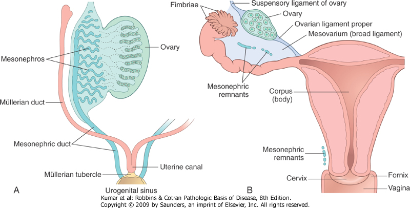
Figure 22-1 Embryology and anatomy of the female genital tract. A, Early in development the mesonephric (blue) and müllerian (red) ducts merge at the urogenital sinus to form the müllerian tubercle. B, By birth the müllerian ducts have fused to form the fallopian tubes, uterus, and endocervix (red), merging with the vaginal squamous mucosa. The mesonephric ducts regress but may be found as a remnant in the ovary, adnexa, and cervix (Gartner duct). (Adapted from Langman J: Medical Embryology. Baltimore, Williams and Wilkins, 1981.)
In males, müllerian inhibitory substance4 from the developing testis causes regression of the müllerian ducts, and the paired wolffian (or mesonephric) ducts form the epididymis and the vas deferens. Normally the mesonephric duct regresses in the female, but remnants may persist into adult life as epithelial inclusions adjacent to the ovaries, tubes, and uterus. In the cervix and vagina these rests may be cystic and are termed Gartner duct cysts. Many of the events in the formation of the internal and external genitalia and their epithelial coverings result from reciprocal epithelial-stromal signaling, leading to mesenchymal remodeling and changes in epithelial cell fate.2,5