

Pocket Atlas of Sectional Anatomy, Volume 2 Thorax, Abdomen and Pelvis
.pdf
|
|
|
sagittal |
33 |
1 |
|
|
22 |
|
2 |
|
|
23 |
|
3 |
|
|
24 |
|
4 |
|
|
25 |
|
5 |
|
|
26 |
|
6 |
|
|
27 |
|
7 |
|
|
|
|
|
|
28 |
|
|
8 |
|
|
|
|
|
|
29 |
|
|
9 |
|
|
|
|
|
|
30 |
|
|
10 |
|
|
|
|
|
|
31 |
|
|
|
|
|
|
|
11 |
|
|
32 |
|
12 |
|
|
|
|
|
|
33 |
|
|
13 |
|
|
|
|
|
|
34 |
|
|
14 |
|
|
|
|
|
|
35 |
|
|
15 |
|
|
|
|
16 |
|
|
36 |
|
17 |
|
|
37 |
|
18 |
|
|
38 |
|
19 |
|
|
39 |
|
20 |
|
|
|
|
|
|
|
|
|
21 |
|
|
|
|
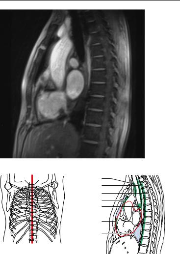
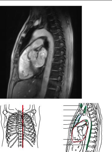
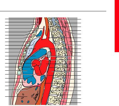
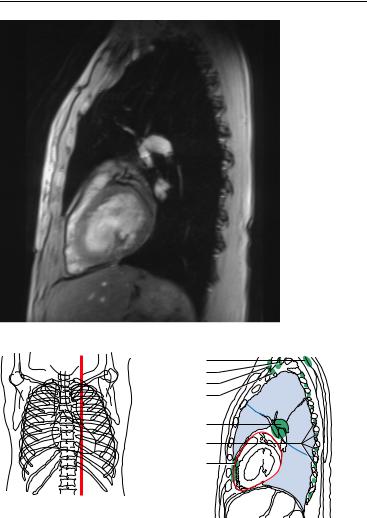
12. |
Pectoralis major muscle |
28. |
Right lung |
|
13. |
Right auricle |
29. |
Azygos vein |
|
14. |
Right coronary artery |
30. |
Trachea |
|
15. |
Right ventricle |
31. |
Right pulmonary artery |
|
16. |
Right atrioventricular (tricuspid) |
32. |
Vertebra |
|
|
valve |
33. |
Left atrium |
|
17. |
Right coronary artery (posterior |
34. |
Intervertebral space |
|
|
interventricular branch) |
35. |
Right atrium |
|
18. |
Right coronary artery (terminal |
36. |
Foramen |
|
|
branch) |
37. |
Inferior vena cava |
|
19. |
Diaphragm |
38. |
Superior articular process |
|
20. |
Rectus abdominis muscle |
39. |
Hepatic veins |
|
21. |
Liver |
40. |
Thyroid lymph nodes |
|
22. |
Splenius cervicis and capitis mus- |
41. |
Deep cervical lymph nodes |
|
|
cle |
42. |
Supraclavicular lymph nodes |
|
23. |
Semispinalis capitis muscle |
43. |
Paratracheal lymph nodes |
|
24. |
Longus colli muscle |
44. |
Anterior mediastinal lymph nodes |
|
25. |
Erector spinae muscle |
45. |
Parasternal lymph nodes |
|
26. |
Trapezius muscle |
46. |
Paravertebral lymph nodes |
|
27. |
Semispinalis thoracis muscle (mul- |
47. |
Subpericardial adipose tissue |
|
|
tifidus muscle) |
|
|
|
Moeller, Pocket Atlas of Sectional Anatomy, Vol. 2 © 2001 Thieme All rights reserved. Usage subject to terms and conditions of license.
34 MRI of the Thorax
41
42
43
44
45
46
47
|
|
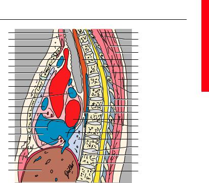
—— = Borders of lung segments |
|
|
|
—— = Pericardium |
|
1. |
Trachea |
6. |
Left brachiocephalic vein |
2. |
Sternocleidomastoid muscle |
7. |
Sternum (manubrium) |
3. |
Sternohyoid, sternothyroid, omo- |
8. |
Pericardium |
|
hyoid muscle |
9. |
Right lung |
4. |
Clavicle |
10. |
Ascending aorta |
5. |
Brachiocephalic trunk |
11. |
Right pulmonary artery |
Moeller, Pocket Atlas of Sectional Anatomy, Vol. 2 © 2001 Thieme All rights reserved. Usage subject to terms and conditions of license.
|
|
|
sagittal |
35 |
1 |
|
|
21 |
|
2 |
|
|
22 |
|
3 |
|
|
23 |
|
4 |
|
|
24 |
|
5 |
|
|
25 |
|
6 |
|
|
26 |
|
7 |
|
|
27 |
|
8 |
|
|
28 |
|
9 |
|
|
29 |
|
10 |
|
|
30 |
|
11 |
|
|
31 |
|
12 |
|
|
32 |
|
13 |
|
|
33 |
|
14 |
|
|
34 |
|
15 |
|
|
35 |
|
16 |
|
|
36 |
|
|
|
9 |
|
|
17 |
|
|
|
|
|
|
37 |
|
|
13 |
|
|
|
|
|
|
|
|
|
18 |
|
|
38 |
|
19 |
|
|
39 |
|
20 |
|
|
40 |
|
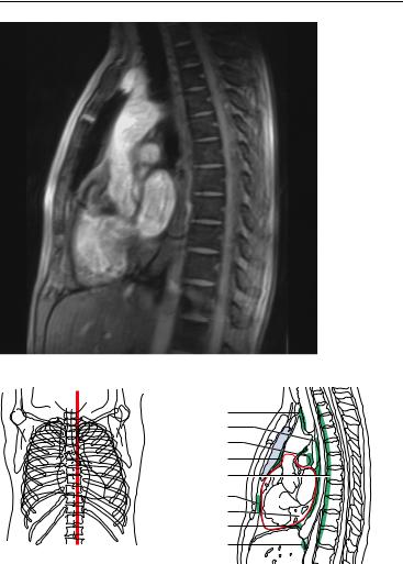
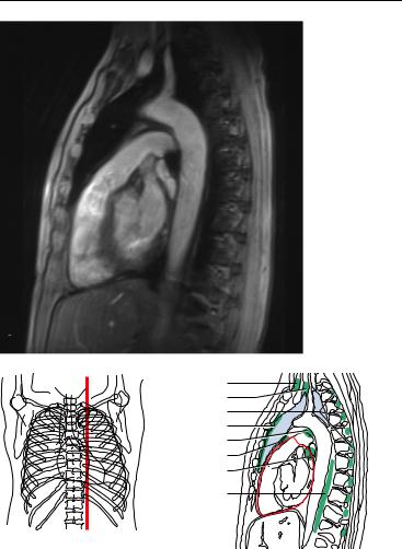
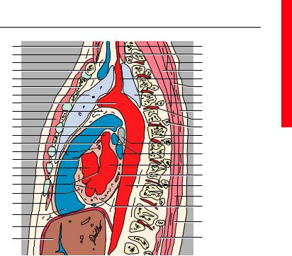
12. |
Right auricle |
30. |
Spinosus process (T6) |
|
13. |
Right coronary artery |
31. |
Ligamenta flava |
|
14. |
Interartricular septum |
32. |
Azygos vein |
|
15. |
Right atrioventricular (tricuspid) |
33. |
Sinus of pericardium |
|
|
valve |
34. |
Left atrium |
|
16. |
Right ventricle |
35. |
Thoracic vertebra (T9) |
|
17. |
Xyphoid process of sternum |
36. |
Right atrium |
|
18. |
Diaphragm |
37. |
Intervertebral space |
|
19. |
Rectus abdominis muscle |
38. |
Anterior longitudinal ligament |
|
20. |
Liver |
39. |
Inferior surface of vertebral body |
|
21. |
Semispinalis capitis muscle |
|
(T12) |
|
22. |
Splenius cervicis muscle and sple- |
40. |
Superior surface of vertebral body |
|
|
nius capitis muscle |
|
(L1) |
|
23. |
Serratus posterior superior muscle |
41. |
Paratracheal lymph nodes |
|
24. |
Rhomboid major muscle |
42. |
Juxtaesophageal lymph nodes |
|
25. |
Esophagus |
43. |
Paravertebral lymph node |
|
26. |
Spinal cord |
44. |
Anterior mediastinal lymph nodes |
|
27. |
Semispinalis thoracis muscle and |
45. |
Tracheobronchial lymph nodes |
|
|
multifidus muscle |
46. |
Prepericardial lymph nodes |
|
28. |
Erector spinae muscle |
47. |
Subpericardial adipose tissue |
|
29. |
Trapezius muscle |
|
|
|
Moeller, Pocket Atlas of Sectional Anatomy, Vol. 2 © 2001 Thieme All rights reserved. Usage subject to terms and conditions of license.
36 MRI of the Thorax
|
|
36 |
|
|
|
37 |
|
|
|
38 |
3 |
|
|
39 |
|
|
|
40 |
|
|
|
41 |
|
|
|
42 |
|
|
|
43 |
|
|
|
44 |
|
|
|
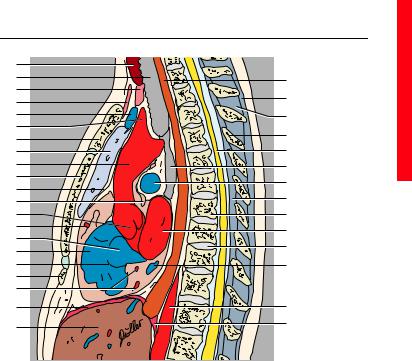
—— = Borders of lung segments |
|
|
|
—— = Pericardium |
|
|
|
(Segments of the lungs, see page 2) |
|
1. |
Thyroid gland |
6. |
Brachiocephalic trunk |
2. |
Trachea |
7. |
Sternum (manubrium) |
3. |
Sternothyroid muscle |
8. |
Left lung |
4. |
Sternohyoid muscle and omohyoid |
9. |
Ascending aorta |
|
muscle |
10. |
Sternum (body) |
5. |
Left brachiocephalic vein |
11. |
Pericardium |
Moeller, Pocket Atlas of Sectional Anatomy, Vol. 2 © 2001 Thieme All rights reserved. Usage subject to terms and conditions of license.
|
sagittal |
37 |
1 |
|
|
2 |
22 |
|
3 |
|
|
23 |
|
|
4 |
|
|
|
|
|
5 |
24 |
|
6 |
25 |
|
7 |
|
|
26 |
|
|
8 |
|
|
9 |
27 |
|
10 |
28 |
|
11 |
|
|
29 |
|
|
12 |
|
|
13 |
30 |
|
14 |
31 |
|
15 |
|
|
32 |
|
|
16 |
|
|
|
|
|
17 |
33 |
|
18 |
|
|
19 |
|
|
20 |
34 |
|
21 |
35 |
|
|
|
12. |
Transverse sinus of pericardium |
27. |
Bifurcation of trachea |
13. |
Aortic bulb and aortic valve |
28. |
Right pulmonary artery |
14. |
Right coronary artery |
29. |
Ligamenta flava |
15. |
Right atrium |
30. |
Thoracic vertebra |
16. |
Right ventricle |
31. |
Left atrium |
17. |
Right atrioventricular (tricuspid) |
32. |
Intervertebral space |
|
valve |
33. |
Coronary sinus |
18. |
Xyphoid process of sternum |
34. |
Descending aorta |
19. |
Right coronary artery (posterior |
35. |
Diaphragm (lumbar part) |
|
interventricular branch and right |
36. |
Paratracheal lymph nodes |
|
marginal branch) |
37. |
Juxtaesophageal lymph nodes |
20. |
Diaphragm |
38. |
Tracheobronchial lymph nodes |
21. |
Liver |
39. |
Anterior mediastinal lymph nodes |
22. |
Esophagus |
40. |
Paravertebral lymph nodes |
23. |
Supraspinous ligament |
41. |
Parasternal lymph nodes |
24. |
Interspinal ligaments |
42. |
Subpericardial adipose tissue |
25. |
Spinous process |
43. |
Superior phrenic lymph nodes |
26. |
Spinal cord |
44. |
Inferior phrenic lymph nodes |
Moeller, Pocket Atlas of Sectional Anatomy, Vol. 2 © 2001 Thieme All rights reserved. Usage subject to terms and conditions of license.
38 MRI of the Thorax
45
46
47
48
49
50
51
52
|
|
—— = Borders of lung segments |
|
|
|
—— = Pericardium |
|
1. |
Thyroid gland |
9. |
Left lung |
2. |
Sternothyroid, sternohyoid, omo- |
10. |
Ascending aorta |
|
hyoid muscles |
11. |
Pectoralis major muscle |
3. |
Sternocleidomastoid muscle |
12. |
Pulmonary trunk |
4. |
Common carotid artery |
13. |
Left coronary artery |
5. |
Clavicle |
14. |
Aortic bulb |
6. |
Sternoclavicular joint |
15. |
Aortic valve |
7. |
Left brachiocephalic vein |
16. |
Right ventricle |
8. |
Sternum (manubrium) |
17. |
Left ventricle |
Moeller, Pocket Atlas of Sectional Anatomy, Vol. 2 © 2001 Thieme All rights reserved. Usage subject to terms and conditions of license.
|
sagittal |
39 |
1 |
23 |
|
2 |
24 |
|
3 |
|
|
25 |
|
|
4 |
|
|
26 |
|
|
5 |
|
|
27 |
|
|
6 |
|
|
28 |
|
|
7 |
|
|
8 |
29 |
|
9 |
30 |
|
10 |
31 |
|
11 |
32 |
|
12 |
33 |
|
13 |
34 |
|
14 |
35 |
|
15 |
36 |
|
16 |
37 |
|
17 |
38 |
|
18 |
|
|
39 |
|
|
19 |
|
|
25 |
|
|
20 |
|
|
40 |
|
|
21 |
|
|
41 |
|
|
22 |
42 |
|
43 |
|
|
|
|
|
|
44 |
|
18. |
Right atrioventricular (tricuspid) |
34. |
Transverse sinus of pericardium |
|
valve |
35. |
Left atrium |
19. |
Anterior papillary muscle |
36. |
Pericardium |
20. |
Rectus abdominis muscle |
37. |
Descending aorta |
21. |
Diaphragm |
38. |
Coronary sinus |
22. |
Liver |
39. |
Erector spinae muscle |
23. |
Semispinalis capitis muscle |
40. |
Intervertebral space |
24. |
Splenius cervicis and capitis mus- |
41. |
Anterior longitudinal ligament |
|
cle |
42. |
Inferior surface of vertebral body |
25. |
Esophagus |
43. |
Superior surface of vertebral body |
26. |
Trapezius muscle |
44. |
Lumbar vertebra (L1) |
27. |
Posterior serratus muscle |
45. |
Thyroid lymph nodes |
28. |
Subclavian artery |
46. |
Deep cervical lymph nodes |
29. |
Rhomboid major muscle |
47. |
Parasternal lymph nodes |
30. |
Semispinalis thoracis muscle and |
48. |
Anterior mediastinal lymph nodes |
|
multifidus muscle |
49. |
Bronchopulmonal lymph nodes |
31. |
Aorticopulmonary window |
50. |
Tracheobronchial lymph nodes |
32. |
Ligamenta flava |
51. |
Subpericardial adipose tissue |
33. |
Left main stem bronchus |
52. |
Prevertebral lymph nodes |
Moeller, Pocket Atlas of Sectional Anatomy, Vol. 2 © 2001 Thieme All rights reserved. Usage subject to terms and conditions of license.
40 MRI of the Thorax
|
|
44 |
|
|
|
45 |
|
|
|
46 |
3 |
|
|
47 |
|
|
|
48 |
|
|
|
49 |
|
|
|
50 |
|
|
|
51 |
|
|
|
—— = Borders of lung segments |
|
|
|
—— = Pericardium |
|
|
|
(Segments of the lung, see page 2) |
|
1. |
Thyroid gland |
9. |
Aorticopulmonary window |
2. |
Sternocleidomastoid muscle |
10. |
Pectoralis major muscle |
3. |
Left internal jugular vein |
11. |
Left pulmonary artery |
4. |
Clavicle |
12. |
Left auricle |
5. |
Left brachiocephalic vein |
13. |
Left coronary artery (anterior in- |
6. |
Sternum (manubrium) |
|
terventricular branch) |
7. |
Left lung |
14. |
Left coronary artery (circumflex |
8. |
Aortic arch |
|
branch) |
Moeller, Pocket Atlas of Sectional Anatomy, Vol. 2 © 2001 Thieme All rights reserved. Usage subject to terms and conditions of license.
|
|
|
sagittal |
41 |
1 |
|
|
24 |
|
2 |
|
|
25 |
|
3 |
|
|
26 |
|
4 |
|
|
27 |
|
5 |
|
|
28 |
|
6 |
|
|
29 |
|
7 |
|
|
7 |
|
8 |
|
|
30 |
|
9 |
|
|
31 |
|
10 |
|
|
32 |
|
11 |
|
|
33 |
|
12 |
|
|
34 |
|
13 |
|
|
35 |
|
14 |
|
|
36 |
|
15 |
|
|
|
|
|
|
37 |
|
|
16 |
|
|
|
|
|
|
38 |
|
|
17 |
|
|
|
|
18 |
|
|
39 |
|
19 |
|
|
40 |
|
20 |
|
|
|
|
|
|
41 |
|
|
21 |
|
|
|
|
|
|
42 |
|
|
22 |
|
|
|
|
|
|
|
|
|
23 |
|
|
43 |
|
15. |
Valve of pulmonary trunk |
32. |
Posterior intercostal artery |
|
16. |
Left ventricle |
33. |
Head of rib |
|
17. |
Conus arteriosus |
34. |
Thoracic vertebra |
|
18. |
Left atrioventricular (mitral) valve |
35. |
Left main stem bronchus |
|
19. |
Interventricular septum |
36. |
Pulmonary veins |
|
20. |
Rectus abdominis muscle |
37. |
Left atrium |
|
21. |
Right ventricle |
38. |
Coronary sinus |
|
22. |
Diaphragm |
39. |
Descending aorta |
|
23. |
Liver |
40. |
Erector spinae muscle |
|
24. |
Vertebral artery |
41. |
Myocardium |
|
25. |
Longus colli muscle |
42. |
Latissimus dorsi muscle |
|
26. |
Splenius cervicis and capitis mus- |
43. |
Intercostal muscle |
|
|
cle |
44. |
Superficial cervical lymph nodes |
|
27. |
Semispinalis cervicis muscle |
45. |
Deep cervical lymph nodes |
|
28. |
Subclavian artery |
46. |
Posterior intercostal lymph nodes |
|
29. |
Serratus posterior superior muscle |
47. |
Parasternal lymph nodes |
|
|
and rhomboid major muscle |
48. |
Aortopulmonal lymph nodes |
|
30. |
Semispinalis thoracis muscle and |
49. |
Anterior mediastinal lymph nodes |
|
|
multifidus muscle |
50. |
Bronchopulmonal lymph nodes |
|
31. |
Trapezius muscle |
51. |
Paravertebral lymph nodes |
|
Moeller, Pocket Atlas of Sectional Anatomy, Vol. 2 © 2001 Thieme All rights reserved. Usage subject to terms and conditions of license.
42 MRI of the Thorax
33 |
|
|
34 |
|
|
35 |
|
1/2 |
36 |
|
|
37 |
3 |
6 |
38 |
|
|
|
|
39 |
10 |
|
—— = Borders of lung segments —— = Pericardium
Left Lung
1+2.Apicoposterior segment of upper lobe
3. Anterior segment of upper lobe
6. Superior segment of lower lobe
10. Posterior basal segment of lower lobe
Moeller, Pocket Atlas of Sectional Anatomy, Vol. 2 © 2001 Thieme All rights reserved. Usage subject to terms and conditions of license.