

Учебник по анатомии (для англ.яз)
.pdfThorax
Branches from both the parasympathetic and sympa thetic systems contribute to the formation of the cardiac plexus. This plexus consists of a superficial part, inferior to the aortic arch and between it and the pulmonary trunk (Fig. 3.76A), and a deep part, between the aortic arch and the tracheal bifurcation (Fig. 3.76B).
From the cardiac plexus, small branches that are mixed nerves containing both sympathetic and parasym pathetic fibers supply the heart. These branches affect nodal tissue and other components of the conduction system, coronary blood vessels, and atrial and ventricular musculature.
|
|
Cardiac nerves from |
|
|
|
A |
|
sympathetic trunk |
|
|
Right vagus nerve |
|
|
|
|
|
|
|
|
Left vagus nerve |
|
|
|
|
|
|
|
|
||||
|
|
|
|
|
|
|
|
|
||
|
|
|
|
|
|
|
|
|
||
Vagal cardiac branches |
|
|
|
|
|
|
|
|
|
Vagal cardiac branches |
|
|
|
|
|
|
|
|
|||
|
|
|
|
|
|
|
|
|||
|
|
|
|
|
|
|
|
|
|
|
|
|
|
|
|
|
|
|
|
|
Superficial cardiac plexus |
|
|
|
|
|
|
|
|
|
|
|
Arch of aorta
Superior vena cava
Pulmonary trunk
Cardiac nerves from sympathetic trunk
B
Right recurrent laryngeal nerve
Left recurrent laryngeal nerve
Right vagus nerve
Left vagus nerve
Vagal cardiac branches 
Vagal cardiac branches
Deep cardiac plexus
3-82 |
Fig. 3.76 Cardiac plexus. A. Superficial. B. Deep. |
|
Drake_ch03_main.indd 82 |
|
|
8/25/2008 4:13:39 PM |
|
|
||
|
|
|
|
Parasympathetic innervation
Stimulation of the parasympathetic system:
■decreases heart rate;
■reduces force of contraction; and
■constricts the coronary arteries.
The preganglionic parasympathetic fibers reach the heart as cardiac branches from the right and left vagus nerves. They enter the cardiac plexus and synapse in ganglia located either within the plexus or in the walls of the atria.
Sympathetic innervation
Stimulation of the sympathetic system:
■increases heart rate; and
■increases the force of contraction.
Sympathetic fibers reach the cardiac plexus through the cardiac nerves from the sympathetic trunk. Preganglionic sympathetic fibers from the upper four or five segments of the thoracic spinal cord enter and move through the sym pathetic trunk. They synapse in cervical and upper tho racic sympathetic ganglia, and postganglionic fibers proceed as bilateral branches from the sympathetic trunk to the cardiac plexus.
Regional anatomy • Mediastinum |
3 |
|||
Visceral afferents |
|
|||
|
||||
Visceral afferents from the heart are also a component of |
|
|||
the cardiac plexus. These fibers pass through the cardiac |
|
|||
plexus and return to the central nervous system in the |
|
|||
cardiac nerves from the sympathetic trunk and in the vagal |
|
|||
cardiac branches. |
|
|||
The afferents associated with the vagal cardiac nerves |
|
|||
return to the vagus nerve [X]. They sense alterations in |
|
|||
blood pressure and blood chemistry and are therefore pri |
|
|||
marily concerned with cardiac reflexes. |
|
|||
The afferents associated with the cardiac nerves from |
|
|||
the sympathetic trunks return to either the cervical or the |
|
|||
thoracic portions of the sympathetic trunk. If they are in |
|
|||
the cervical portion of the trunk, they normally descend to |
|
|||
the thoracic region where they re-enter the upper four or |
|
|||
five thoracic spinal cord segments along with the afferents |
|
|||
|
|
|
||
from the thoracic region of the sympathetic trunk. |
Visceral |
|
|
|
afferents associated with the sympathetic system |
conduct |
|
|
|
pain sensation from the heart, which is detected at the cel |
|
|
||
lular level as tissue-damaging events (i.e., cardiac isch |
|
|
||
emia). This pain is often “referred” to cutaneous regions |
|
|
||
supplied by the same spinal cord levels (see p. 00; p. 000). |
|
|
6 |
|
|
|
|
|
|
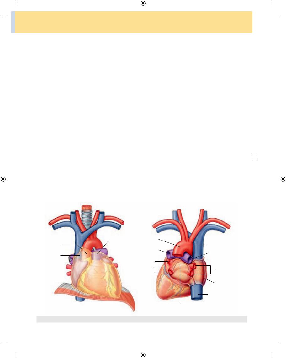
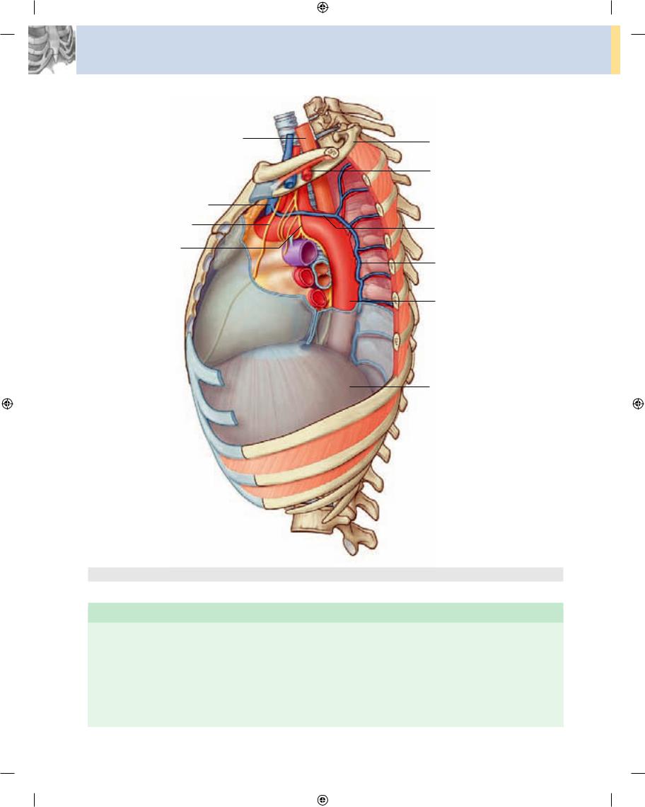
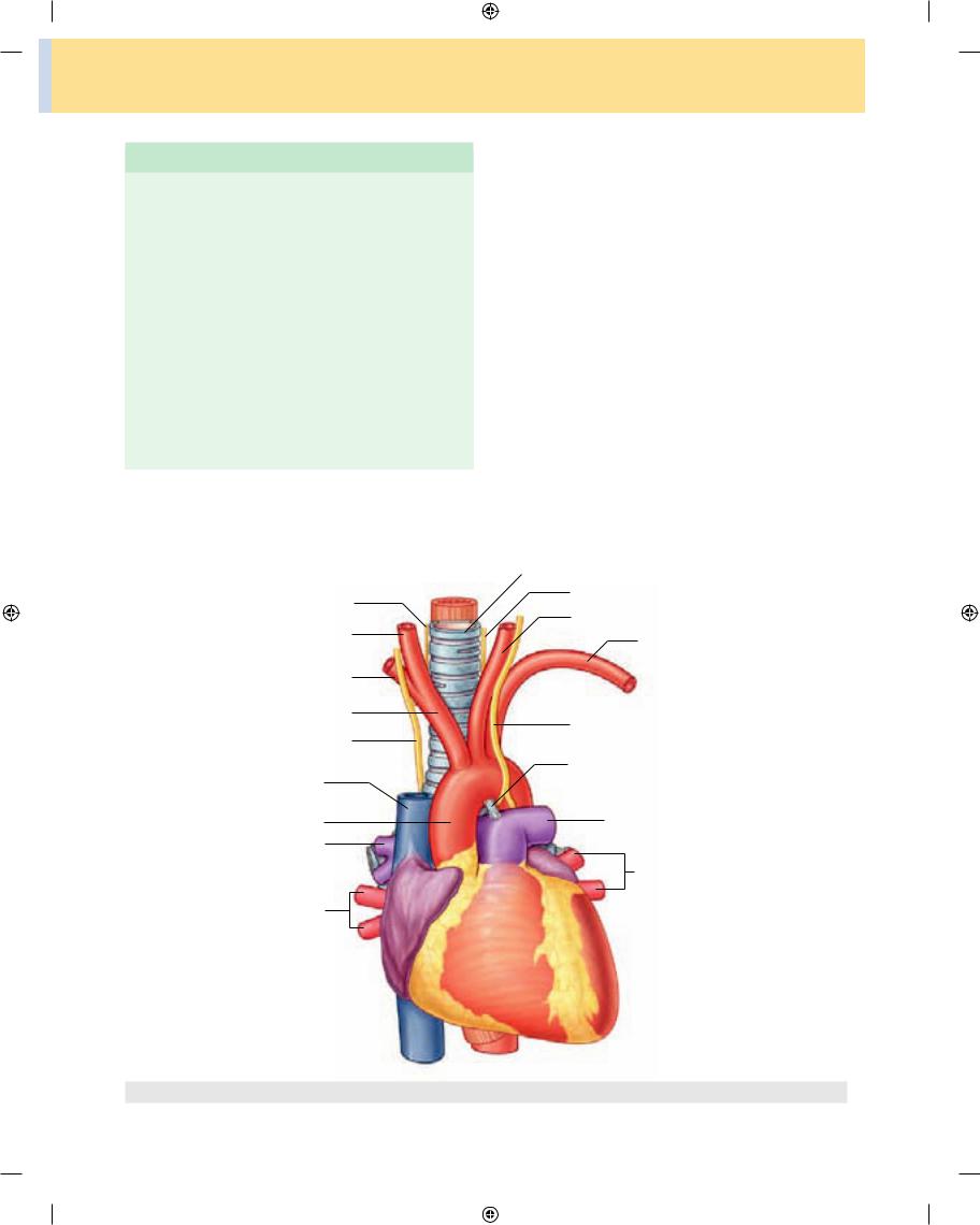
Pulmonary trunk
The pulmonary trunk is contained within the pericardial sac (Fig. 3.77), is covered by the visceral layer of serous
A B
Pulmonary trunk |
Arch of aorta |
Ascending |
|
aorta |
Left pulmonary |
Superior |
artery |
|
|
vena cava |
|
|
Left pulmonary |
|
veins |
Oblique pericardial sinus
Fig. 3.77 Major vessels within the middle mediastinum. A. Anterior view. B. Posterior view.
Superior vena cava
Right pulmonary artery
Right pulmonary veins
Right atrium
Inferior vena cava
3-83
Drake_ch03_main.indd 83 |
|
|
8/25/2008 4:13:42 PM |
|
|
||
|
|
|
|
Thorax
pericardium and is associated with the ascending aorta in a common sheath. It arises from the conus arteriosus of the right ventricle at the opening of the pulmonary trunk slightly anterior to the aortic orifice and ascends, moving posteriorly and to the left, lying initially anterior and then to the left of the ascending aorta. At approximately the level of the intervertebral disc between vertebrae TV and TVI, opposite the left border of the sternum and posterior to the third left costal cartilage, the pulmonary trunk divides into:
■the right pulmonary artery, which passes to the right, posterior to the ascending aorta and the superior vena cava, to enter the right lung; and
■the left pulmonary artery, which passes inferiorly to the arch of the aorta and anteriorly to the descending aorta to enter the left lung.
Ascending aorta
The ascending aorta is contained within the pericardial sac and is covered by a visceral layer of serous pericardium, which also surrounds the pulmonary trunk in a common sheath (Fig. 3.77A).
The origin of the ascending aorta is the aortic orifice at the base of the left ventricle, which is level with the lower edge of the third left costal cartilage, posterior to the left half of the sternum. Moving superiorly, slightly forward and to the right, the ascending aorta continues to the level of the second right costal cartilage. At this point, it enters the superior mediastinum and is then referred to as the arch of the aorta.
Immediately superior to the point where the ascending aorta arises from the left ventricle are three small outward bulges opposite the semilunar cusps of the aortic valve. These are the posterior, right, and left aortic sinuses. The right and left coronary arteries originate from the right and left aortic sinuses, respectively.
Other vasculature
The inferior half of the superior vena cava is located within the pericardial sac (Fig. 3.77B). It passes through the fibrous pericardium at approximately the level of the second costal cartilage and enters the right atrium at the lower level of the third costal cartilage. The portion within the pericardial sac is covered with serous pericar dium except for a small area on its posterior surface.
After passing through the diaphragm, at approximately the level of vertebra TVIII, the inferior vena cava enters the fibrous pericardium. A short portion of this vessel is within the pericardial sac before entering the right atrium.
3-84 While within the pericardial sac, it is covered by serous
pericardium except for a small portion of its posterior surface (Fig. 3.77B).
A very short segment of each of the pulmonary veins is also within the pericardial sac. These veins, usually two from each lung, pass through the fibrous pericardium and enter the superior region of the left atrium on its posterior surface. In the pericardial sac, all but a portion of the pos terior surface of these veins is covered by serous pericar dium. In addition, the oblique pericardial sinus is between the right and left pulmonary veins, within the pericardial sac (Fig. 3.77).
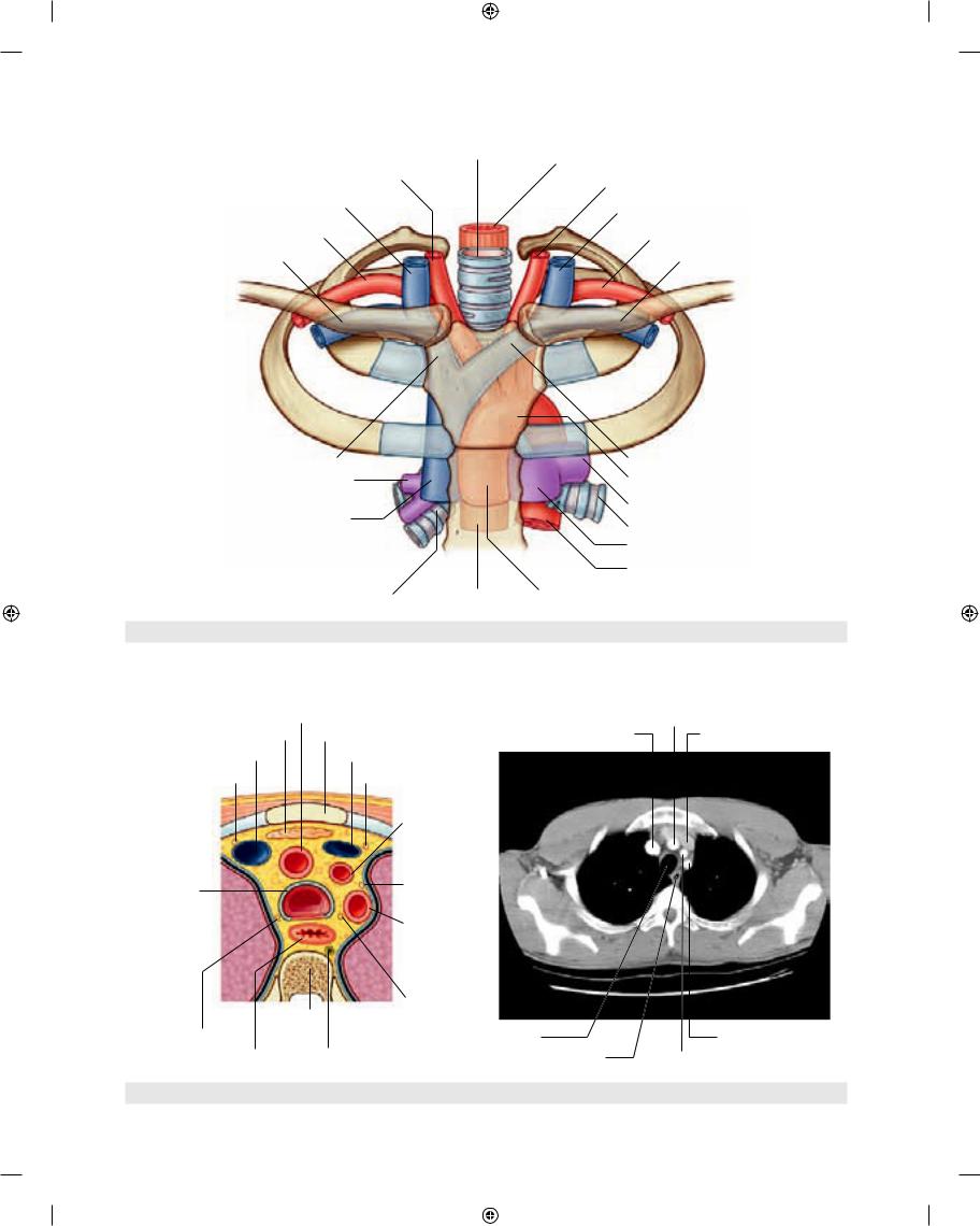
Superior mediastinum
The superior mediastinum is posterior to the manu brium of the sternum and anterior to the bodies of the first four thoracic vertebrae (see Fig. 3.52).
■Its superior boundary is an oblique plane passing from the jugular notch upward and posteriorly to the supe rior border of vertebra TI.
■Inferiorly, a transverse plane passing from the sternal angle to the intervertebral disc between vertebra TIV/V separates it from the inferior mediastinum.
■Laterally, it is bordered by the mediastinal part of the parietal pleura on either side.
The superior mediastinum is continuous with the neck superiorly and with the inferior mediastinum inferiorly.
The major structures found in the superior mediasti num include the:
■thymus;
■right and left brachiocephalic veins;
■left superior intercostal vein;
■superior vena cava;
■arch of the aorta with its three large branches;
■trachea;
■esophagus;
■phrenic nerves;
■vagus nerves;
■left recurrent laryngeal branch of the left vagus nerve;
■thoracic duct; and
■other small nerves, blood vessels, and lymphatics (Figs. 3.78 and 3.79).
Thymus
The thymus is the most anterior component of the superior mediastinum, lying immediately posterior to the manubrium of the sternum. It is an asymmetric, bilobed structure (Fig. 3.80).
Drake_ch03_main.indd 84 |
|
|
8/25/2008 4:13:43 PM |
|
|
||
|
|
|
|
|
Regional anatomy • Mediastinum |
3 |
|
|
|
Right common carotid artery
Right internal jugular vein
Right subclavian artery
Right subclavian vein
Right brachiocephalic vein
Right pulmonary artery
Superior vena cava
Right main bronchus
Fig. 3.78 Structures in the superior mediastinum.
Trachea
Esophagus
Left common carotid artery
Left internal jugular vein
Left subclavian artery
Left subclavian vein
Left brachiocephalic vein
Arch of aorta
Left pulmonary artery
Left main bronchus
Pulmonary trunk
Thoracic aorta
Esophagus |
Ascending aorta |
|
A |
Brachiocephalic trunk |
B |
Brachiocephalic trunk |
||
|
Thymus |
Manubrium of sternum |
Right brachiocephalic vein |
Left brachiocephalic vein |
|
Right brachiocephalic vein |
Left brachiocephalic vein |
|
|
||
Right phrenic nerve |
|
|
Left phrenic nerve |
|
|
|
|
|
Left common |
|
|
|
|
|
carotid artery |
|
|
Trachea |
|
|
Left vagus nerve |
|
|
|
|
|
|
|
|
|
|
|
Left subclavian |
|
|
|
|
|
artery |
|
|
|
|
|
Left recurrent |
|
|
|
|
TIII |
laryngeal nerve |
|
|
|
|
|
|
|
|
Right vagus nerve |
|
|
|
Trachea |
Left subclavian artery |
Esophagus |
Thoracic duct |
Esophagus |
Left common carotid artery |
||
Fig. 3.79 Cross-section through the superior mediastinum at the level of vertebra TIII. A. Diagram. B. Axial computed tomography image. |
3-85 |
|
Drake_ch03_main.indd 85 |
|
|
8/25/2008 4:13:46 PM |
|
|
||
|
|
|
|
Thorax
The upper extent of the thymus can reach into the neck as high as the thyroid gland; a lower portion typically extends into the anterior mediastinum over the pericardial sac.
Involved in the early development of the immune system, the thymus is a large structure in the child, begins to atrophy after puberty, and shows considerable size vari ation in the adult. In the elderly adult, it is barely identifi able as an organ, consisting mostly of fatty tissue that is sometimes arranged as two lobulated fatty structures.
Arteries to the thymus consist of small branches origi nating from the internal thoracic arteries. Venous drain age is usually into the left brachiocephalic vein and possibly into the internal thoracic veins.
Lymphatic drainage returns to multiple groups of nodes at one or more of the following locations:
■along the internal thoracic arteries (parasternal);
■at the tracheal bifurcation (tracheobronchial); and
■in the root of the neck.
Right and left brachiocephalic veins
The left and right brachiocephalic veins are located imme diately posterior to the thymus. They form on each side at the junction between the internal jugular and subclavian veins (see Fig. 3.78). The left brachiocephalic vein crosses the midline and joins with the right brachiocephalic vein to form the superior vena cava (Fig. 3.81).
■The right brachiocephalic vein begins posterior to the medial end of the right clavicle and passes vertically
In the clinic
Ectopic parathyroid glands in the thymus
The parathyroid glands develop from the third pharyngeal pouch, which also forms the thymus. The thymus is therefore a common site for ectopic parathyroid glands and, potentially, ectopic parathyroid hormone production.
Right internal thoracic artery |
Left internal thoracic artery |
|
|
|
TIV/V vertebral level |
Thymus
Pericardial sac
3-86 |
Fig. 3.80 Thymus. |
|
Drake_ch03_main.indd 86 |
|
|
8/25/2008 4:13:49 PM |
|
|
||
|
|
|
|

downward, forming the superior vena cava when it is joined by the left brachiocephalic vein. Venous tributar ies include the vertebral, first posterior intercostal, and internal thoracic veins. The inferior thyroid and thymic veins may also drain into it.
■ The left brachiocephalic vein begins posterior to the medial end of the left clavicle. It crosses to the right, moving in a slightly inferior direction, and joins with the right brachiocephalic vein to form the superior vena cava posterior to the lower edge of the right first costal cartilage close to the right sternal border. Venous tribu taries include the vertebral, first posterior intercostal, left superior intercostal, inferior thyroid, and internal thoracic veins. It may also receive thymic and pericar dial veins. The left brachiocephalic vein crosses the midline posterior to the manubrium in the adult. In infants and children the left brachiocephalic vein rises above the superior border of the manubrium and there fore is less protected.
Left superior intercostal vein
The left superior intercostal vein receives the second, third and sometimes the fourth posterior intercostal veins,
Regional anatomy • Mediastinum |
3 |
|
|
usually the left bronchial veins, and sometimes the left pericardiacophrenic vein. It passes over the left side of the aortic arch, lateral to the left vagus nerve and medial to the left phrenic nerve, before entering the left brachiocephalic vein (Fig. 3.82). Inferiorly, it may connect with the accessory hemiazygos vein (superior hemiazygos vein).
Superior vena cava
The vertically oriented superior vena cava begins posterior to the lower edge of the right first costal cartilage, where the right and left brachiocephalic veins join, and termi nates at the lower edge of the right third costal cartilage, where it joins the right atrium (see Fig. 3.78).
The lower half of the superior vena cava is within the pericardial sac and is therefore contained in the middle mediastinum.
The superior vena cava receives the azygos vein imme diately before entering the pericardial sac and may also receive pericardial and mediastinal veins.
The superior vena cava can be easily visualized forming part of the right superolateral border of the mediastinum on a chest radiograph (see Fig. 3.60A).
Left common carotid artery
Right vagus nerve |
|
Right brachiocephalic vein |
Left brachiocephalic vein |
|
Left vagus nerve |
Azygos vein |
Left pulmonary artery |
Superior vena cava |
|
|
Left pulmonary veins |
Fig. 3.81 Superior mediastinum with thymus removed. |
3-87 |
|
Drake_ch03_main.indd 87 |
|
|
8/25/2008 4:13:51 PM |
|
|
||
|
|
|
|
Thorax
Esophagus
Left brachiocephalic vein
Left phrenic nerve
Left vagus nerve
Fig. 3.82 Left superior intercostal vein.
Rib I
Left subclavian artery
Left superior intercostal vein
Accessory hemiazygos vein
Thoracic aorta
Diaphragm
In the clinic
Venous access for central and dialysis lines
Large systemic veins are used to establish central venous access for administering large amounts of fluid, drugs, and blood. Most of these lines (small bore tubes) are introduced through venous puncture into the axillary, subclavian, or internal jugular veins. The lines are then passed through the main veins of the superior
3-88
mediastinum, with the tips of the lines usually residing in the distal portion of the superior vena cava or in the right atrium.
Similar devices, such as dialysis lines, are inserted into patients who have renal failure, so that a large volume of blood can be aspirated through one channel and reinfused through a second channel.
Drake_ch03_main.indd 88 |
|
|
8/25/2008 4:13:56 PM |
|
|
||
|
|
|
|
In the clinic
Using the superior vena cava to access the inferior vena cava
Because the superior and inferior vena cava are oriented along the same vertical axis, a guide-wire, catheter, or line can be passed from the superior vena cava through the right atrium and into the inferior vena cava. This is a common route of access for such procedures as:
■transjugular liver biopsy;
■transjugular intrahepatic portosystemic shunts (TIPS); and
■insertion of an inferior vena cava filter to catch emboli dislodged from veins in the lower limb and pelvis (i.e., patients with deep vein thrombosis [DVT]).
Regional anatomy • Mediastinum |
3 |
|
|
Arch of aorta and its branches
The thoracic portion of the aorta can be divided into ascending aorta, arch of aorta, and thoracic(descending) aorta. Only the arch of the aorta is in the superior mediastinum. It begins when the ascending aorta emerges from the pericardial sac and courses upward, backward, and to the left as it passes through the superior mediasti num, ending on the left side at vertebral level TIV/V (see Fig. 3.78). Extending as high as the midlevel of the manu brium of sternum, the arch is initially anterior and finally lateral to the trachea.
Three branches arise from the superior border of the arch of the aorta; at their origins, all three are crossed anteriorly by the left brachiocephalic vein.
The first branch
Beginning on the right, the first branch of the arch of aorta is the brachiocephalic trunk (Fig. 3.83). It is the largest of the three branches and, at its point of origin
Right recurrent laryngeal nerve
Right common carotid artery
Right subclavian artery
Brachiocephalic trunk
Right vagus nerve
Superior vena cava
Ascending aorta
Right pulmonary artery
Right pulmonary veins
Fig. 3.83 Superior mediastinum with thymus and venous channels removed.
Trachea
Left recurrent laryngeal nerve
Left common carotid artery
Left subclavian artery
Left vagus nerve
Ligamentum arteriosum
Left pulmonary artery
Left pulmonary veins
3-89
Drake_ch03_main.indd 89 |
|
|
8/25/2008 4:13:58 PM |
|
|
||
|
|
|
|

Thorax
behind the manubrium of sternum, is slightly anterior to the other two branches. It ascends slightly posteriorly and to the right. At the level of the upper edge of the right sternoclavicular joint, the brachiocephalic trunk divides into:
■the right common carotid artery; and
■the right subclavian artery (see Fig. 3.78).
The arteries mainly supply the right side of the head and neck and the right upper limb, respectively.
Occasionally, the brachiocephalic trunk has a small branch, the thyroid ima artery, which contributes to the vascular supply of the thyroid gland.
The left subclavian artery is the major blood supply to the left upper limb.
Ligamentum arteriosum
The ligamentum arteriosum is also in the superior medi astinum and is important in embryonic circulation, when it is a patent vessel (the ductus arteriosus). It connects the pulmonary trunk with the arch of aorta and allows blood to bypass the lungs during development (Fig. 3.83). The vessel closes soon after birth and forms the ligamen tous connection observed in the adult.
The second branch
The second branch of the arch of aorta is the left common carotid artery (Fig. 3.83). It arises from the arch imme diately to the left and slightly posterior to the brachioce phalictrunkandascendsthroughthesuperiormediastinum along the left side of the trachea.
The left common carotid artery supplies the left side of the head and neck.
The third branch
The third branch of the arch of the aorta is the left subclavian artery (Fig. 3.83). It arises from the arch of aorta immediately to the left of, and slightly posterior to, the left common carotid artery and ascends through the superior mediastinum along the left side of the trachea.
In the clinic
In the clinic
Coarctation of the aorta
Coarctation of the aorta is a congenital abnormality in which the aortic lumen is constricted just distal to the origin of the left subclavian artery. At this point, the aorta becomes significantly narrowed and the blood supply to the lower limbs and abdomen is diminished. Over time, collateral vessels develop around the chest wall and abdomen to supply the lower body. The coarctation also affects the heart, which has to pump the blood at higher pressure to maintain peripheral perfusion. This in turn may produce cardiac failure.
Thoracic aorta
Diffuse atherosclerosis of the thoracic aorta may occur in patients with vascular disease, but this rarely produces symptoms. There are, however, two clinical situations in which aortic pathology can produce life-threatening situations.
Trauma
The aorta has three fixed points of attachment:
■the aortic valve;
■the ligamentum arteriosum;
■the point of entry behind the crura of the diaphragm.
The rest of the aorta is relatively free from attachment to other structures of the mediastinum. A serious
3-90
deceleration injury (e.g., in a road traffic accident) is most likely to cause aortic trauma at these fixed points.
Aortic dissection
In certain conditions, such as in severe arteriovascular disease, the wall of the aorta can split longitudinally, creating a false channel, which may or may not rejoin into the true lumen distally. This aortic dissection
occurs between the intima and media anywhere along its length. If it occurs in the ascending aorta or arch of the aorta, blood flow in the coronary and cerebral arteries may be disrupted, resulting in myocardial infarction or stroke. In the abdomen the visceral vessels may be disrupted, producing ischemia to the gut or kidneys.
Drake_ch03_main.indd 90 |
|
|
8/25/2008 4:13:59 PM |
|
|
||
|
|
|
|
|
Regional anatomy • Mediastinum |
3 |
|
|
|
In the clinic |
Trachea and esophagus |
Aortic arch and its anomalies
A right-sided arch of aorta occasionally occurs and may be asymptomatic. It can be associated with dextrocardia (right-sided heart) and, in some instances, with complete situs inversus (left-to-right inversion of the body’s organs). It can also be associated with abnormal branching of the great vessels.
In the clinic
Abnormal origin of great vessels
Great vessels occasionally have an abnormal origin, including:
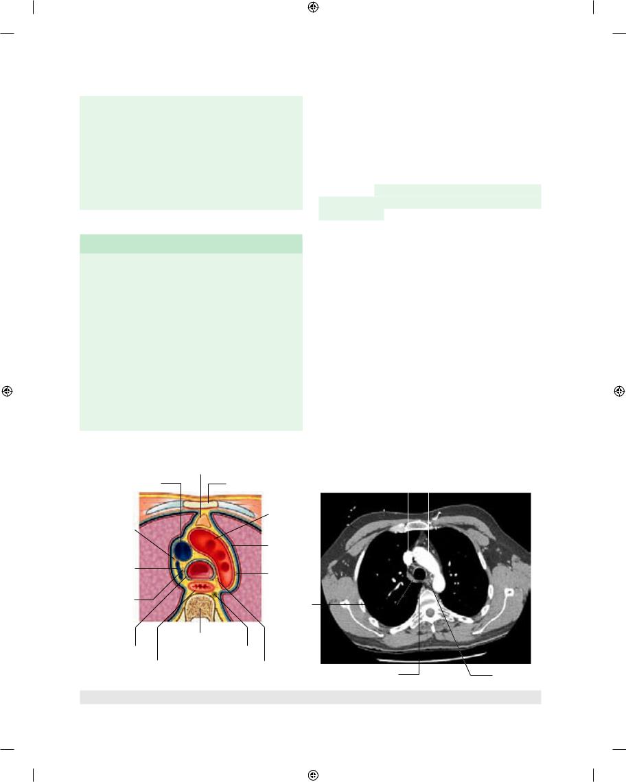
The trachea is a midline structure that is palpable in the jugular notch as it enters the superior mediastinum. Pos terior to it is the esophagus, which is immediately anterior to the vertebral column (Fig. 3.84, and see Figs. 3.78 and 3.79). Significant mobility exists in the vertical positioning of these structures as they pass through the superior mediastinum. Swallowing and breathing cause positional shifts, as may disease and the use of specialized instrumentation.
As the trachea and esophagus pass through the supe rior mediastinum, they are crossed laterally by the azygos vein on the right side and the arch of aorta on the left side.
The trachea divides into the right and left main bronchi at, or just inferior to, the transverse plane between the sternal angle and vertebral level TIV/V (Fig. 3.85), whereas
■a common origin of the brachiocephalic trunk and the esophagus continues into the posterior mediastinum. the left common carotid artery;
■the left vertebral artery originating from the aortic arch; and
■the right subclavian artery originating from the distal portion of the aortic arch and passing behind the esophagus to supply the right arm—as a result, the great vessels form a vascular ring around the trachea and the esophagus, which can potentially produce difficulty swallowing.
Nerves of the superior mediastinum
Vagus nerves
The vagus nerves [X] pass through the superior and posterior divisions of the mediastinum on their way to the abdominal cavity. As they pass through the thorax, they provide parasympathetic innervation to the thoracic viscera and carry visceral afferents from the thoracic viscera.
A |
Thymus |
B |
|
|
|
|
Superior vena cava |
Manubrium of sternum |
Superior vena cava |
|
|
|
Arch of aorta |
|
|
|
||||
|
Arch of aorta |
|
|
|
|
|
Right phrenic |
|
|
|
|
|
|
nerve |
Left phrenic |
|
|
|
|
|
|
|
|
|
|
|
|
Arch of |
nerve |
|
|
|
|
|
|
|
|
|
|
||
Left vagus |
|
|
|
|
|
|
azygos vein |
|
|
|
|
|
|
nerve |
|
|
|
|
|
|
|
|
|
|
|
|
|
Right vagus |
Arch of |
|
|
|
|
|
nerve |
|
|
|
|
|
|
azygos vein |
|
|
|
|
|
|
|
|
|
|
|
|
|
|
TIV |
|
|
|
|
|
Trachea |
Thoracic duct |
|
|
|
|
|
Esophagus |
Left recurrent |
|
|
|
|
|
|
laryngeal nerve |
Trachea |
|
|
Esophagus |
|
Fig. 3.84 Cross-section through the superior mediastinum at the level of vertebra TIV. A. Diagram. B. Axial computed tomography image. |
3-91 |
|
Drake_ch03_main.indd 91 |
|
|
8/25/2008 4:14:00 PM |
|
|
||
|
|
|
|