

Учебник по анатомии (для англ.яз)
.pdfThorax
TI
Sympathetic ganglion
Sympathetic trunk
TV
Gray and white rami communicantes
Thoracic spinal nerve
Gray and white rami communicantes
Greater splanchnic nerve
Lesser splanchnic nerve
Least splanchnic nerve
Fig. 3.95 Thoracic portion of sympathetic trunks.
This portion of the sympathetic trunks consists of two parallel cords punctuated by 11 or 12 ganglia (Fig. 3.95). The ganglia are connected to adjacent thoracic spinal nerves by white and gray rami communicantes and are numbered according to the thoracic spinal nerve with which they are associated.
In the superior portion of the posterior mediastinum, the trunks are anterior to the neck of the ribs. Inferiorly, they become more medial in position until they lie on the lateral aspect of the vertebral bodies. The sympathetic trunks leave the thorax by passing posterior to diaphragm
3-102
under the medial arcuate ligament or through the crura of the diaphragm. Throughout their course the trunks are covered by parietal pleura.
Branches from the ganglia
Two types of medial branches are given off by the ganglia:
■the first type includes branches from the upper five ganglia;
Drake_ch03_main.indd 102 |
|
|
8/25/2008 4:15:52 PM |
|
|
||
|
|
|
|

■the second type includes branches from the lower seven ganglia.
The first type, which includes branches from the upper five ganglia, consists mainly of postganglionic sympathetic fibers, which supply the various thoracic viscera. These branches are relatively small, and also contain visceral afferent fibers.
The second type, which includes branches from the lower seven ganglia, consists mainly of preganglionic sym pathetic fibers, which supply the various abdominal and pelvic viscera. These branches are large, also carry visceral afferent fibers, and form the three thoracic splanchnic nerves referred to as the greater, lesser, and least splanch nic nerves (Fig. 3.95).
■The greater splanchnic nerve on each side usually arises from the fifth to ninth or tenth thoracic ganglia. It descends across the vertebral bodies moving in a medial direction, passes into the abdomen through the crus of the diaphragm, and ends in the celiac ganglion.
■The lesser splanchnic nerve usually arises from the ninth and tenth, or tenth and eleventh thoracic ganglia. It descends across the vertebral bodies moving in a medial direction, and passes into the abdomen through the crus of the diaphragm to end in the aorticorenal ganglion.
Regional anatomy • Mediastinum |
3 |
|
|
■The least splanchnic nerve (lowest splanchnic nerve) usually arises from the twelfth thoracic gan glion. It descends and passes into the abdomen through the crus of the diaphragm to end in the renal plexus.
Anterior mediastinum
The anterior mediastinum is posterior to the body of the sternum and anterior to the pericardial sac (see Fig. 3.52).
■Its superior boundary is a transverse plane passing from the sternal angle to the intervertebral disc between vertebra TIV and TV, separating it from the superior mediastinum.
■Its inferior boundary is the diaphragm.
■Laterally, it is bordered by the mediastinal part of pari etal pleura on either side.
The major structure in the anterior mediastinum is a portion of thymus, described previously (see Fig. 3.80). Also present are fat, connective tissue, lymph nodes, mediastinal branches of the internal thoracic vessels, and sternopericardial ligaments, which pass from the posterior surface of the body of the sternum to the fibrous pericardium.
3-103
Drake_ch03_main.indd 103 |
|
|
8/25/2008 4:15:52 PM |
|
|
||
|
|
|
|

Thorax
Surface anatomy
Thorax surface anatomy
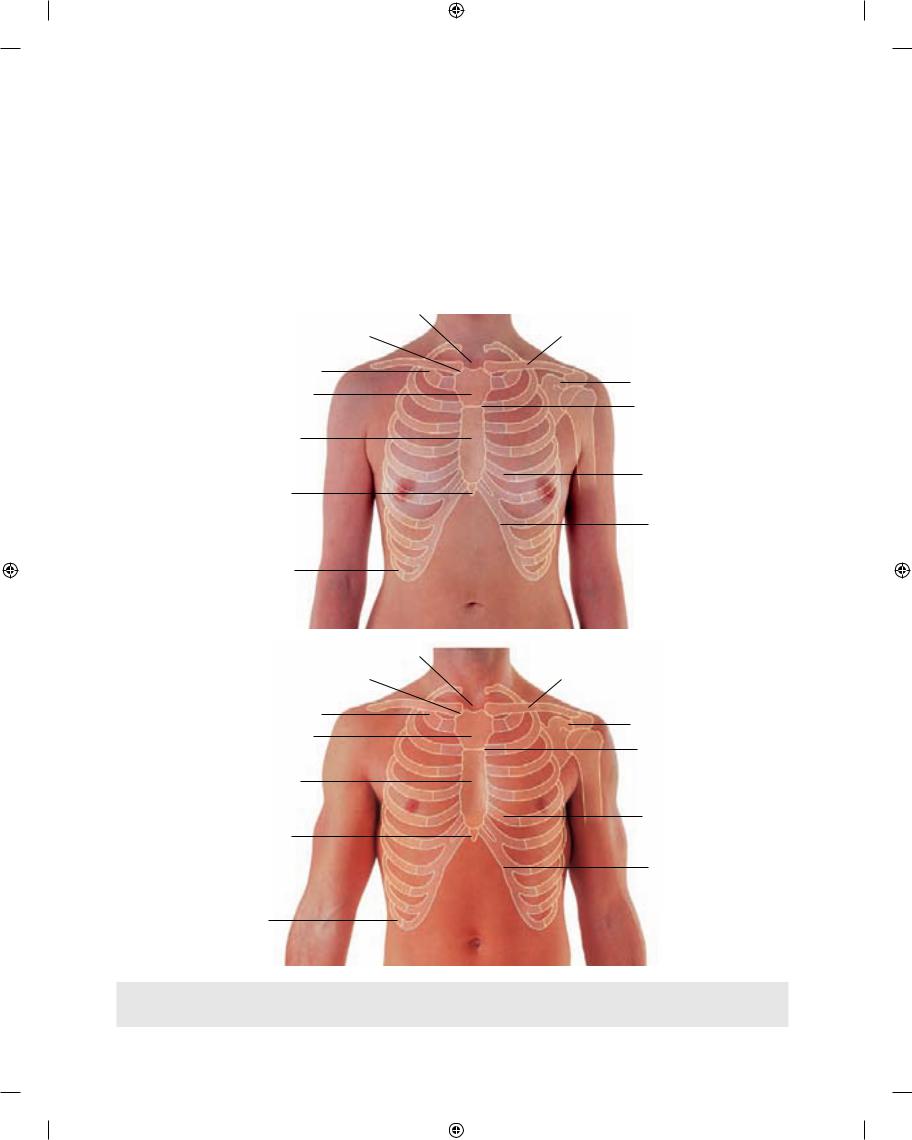
The ability to visualize how anatomical structures in the thorax are related to surface features is fundamental to a physical examination. Figs. 3.96 and 3.97 have been ren dered to show landmarks on the body’s surface that can be used to locate deep structures and to assess function by auscultation and percussion.
A
How to count ribs
Knowing how to count ribs is important because different ribs provide palpable landmarks for the positions of deeper structures. To determine the location of specific ribs, palpate the jugular notch at the superior extent of the manu brium of the sternum. Move down the sternum until a ridge is felt. This ridge is the sternal angle, which identi
B
Fig. 3.96 Anterior chest wall. A. In a woman. B. In a man.
A B
3-104 |
Fig. 3.97 Posterior chest wall. A. In a woman. B. In a man. |
|
Drake_ch03_main.indd 104 |
|
|
8/25/2008 4:16:34 PM |
|
|
||
|
|
|
|
|
Surface anatomy • Surface anatomy of the breast in women |
3 |
|
|
|
fies the articulation between the manubrium of sternum and the body of sternum. The costal cartilage of rib II artic ulates with the sternum at this location. Identify rib II. Then continue counting the ribs, moving in a downward and lateral direction (Fig. 3.98).
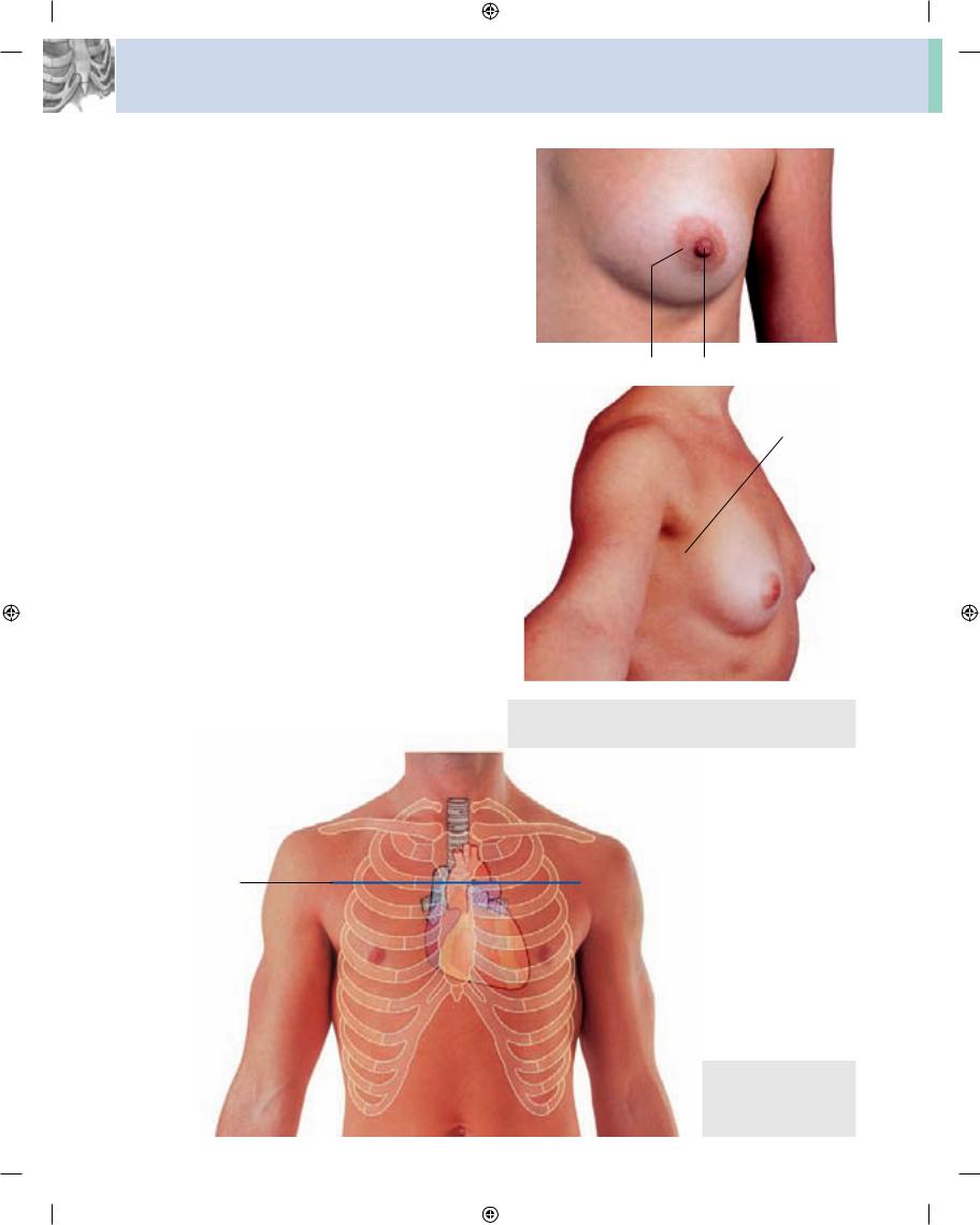
Surface anatomy of the breast in women
Although breasts vary in size, they are normally positioned on the thoracic wall between ribs II and VI and overlie the pectoralis major muscles. Each mammary gland extends
|
Jugular notch |
A |
Sternoclavicular joint |
Rib I
Manubrium of sternum
II
III
Body of the sternum
IV
V Xiphoid process VI
VII
VIII
IX
Rib X
|
Jugular notch |
B |
Sternoclavicular joint |
Rib I |
|
Manubrium of sternum |
II |
|
|
Body of the sternum |
III |
|
|
|
IV |
|
V |
Xiphoid process |
VI |
|
Clavicle
Coracoid process
Sternal angle
Costal cartilage
Costal margin
Clavicle
Coracoid process
Sternal angle
Costal cartilage
VII |
Costal margin |
VIII |
|
IX |
|
Rib X
Fig. 3.98 Anterior view of chest wall with the locations of skeletal structures shown. A. In women. The location of the nipple relative to a |
|
specific intercostal space varies depending on the size of the breasts, which may not be symmetric. B. In men. Note the location of the |
|
nipple in the fourth intercostal space. |
3-105 |
Drake_ch03_main.indd 105 |
|
|
8/25/2008 4:17:01 PM |
|
|
||
|
|
|
|
Thorax
superolaterally around the lower margin of the pectoralis major muscle and enters the axilla (Fig. 3.99). This portion of the gland is the axillary tail or axillary process. The posi tions of the nipple and areola vary relative to the chest wall depending on breast size.
Visualizing structures at the TIV/V vertebral level
The TIV/V vertebral level is a transverse plane that passes through the sternal angle on the anterior chest wall and the intervertebral disc between TIV and TV vertebrae pos teriorly. This plane can easily be located, because the joint between the manubrium of sternum and the body of sternum forms a distinct bony protuberance that can be palpated. At the TIV/V level (Fig. 3.100):
■the costal cartilage of rib II articulates with the sternum;
■the superior mediastinum is separated from the inferior mediastinum;
■the ascending aorta ends and the arch of aorta begins;
■the arch of aorta ends and the thoracic aorta begins;
■the trachea bifurcates.
A
Areola Nipple
B
Axillary process
Fig. 3.99 A. Close-up view of nipple and surrounding areola of the breast. B. Lateral view of the chest wall of a woman showing the axillary process of the breast.
TIV/V vertebral level
Fig. 3.100 Anterior view of the chest wall of a man
3-106 showing the locations of various structures related to the TIV/V level.
Drake_ch03_main.indd 106 |
|
|
8/25/2008 4:17:27 PM |
|
|
||
|
|
|
|
Surface anatomy • Visualizing the margins of the heart
Visualizing structures in the superior mediastinum
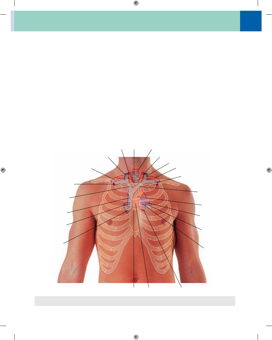
A number of structures in the superior mediastinum in adults can be visualized based on their positions relative to skeletal landmarks that can be palpated through the skin (Fig. 3.101).
■The arch of aorta begins and ends at the transverse plane between the sternal angle anteriorly and vertebral level TIV/V posteriorly. The arch may reach as high as the midlevel of the manubrium of sternum.
■On each side, the internal jugular and subclavian veins join to form the brachiocephalic veins behind the sternal ends of the clavicles near the sternoclavicular joints.
■The left brachiocephalic vein crosses from left to right behind the manubrium of sternum.
■The brachiocephalic veins unite to form the superior vena cava behind the lower border of the costal carti lage of the right first rib.
Visualizing the margins of the heart
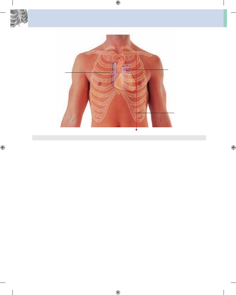
Surface landmarks can be palpated to visualize the outline of the heart (Fig. 3.102).
■The upper limit of the heart reaches as high as the third costal cartilage on the right side of the sternum and the second intercostal space on the left side of the sternum.
Right common carotid artery |
Trachea |
Esophagus |
Right internal jugular vein |
|
Left common carotid artery |
|
|
Left internal jugular vein |
Right subclavian artery |
|
Left subclavian artery |
|
|
Left subclavian vein |
Right subclavian vein |
|
|
|
|
Left brachiocephalic |
Right |
|
vein |
|
|
|
brachiocephalic vein |
|
Arch of aorta |
|
|
|
Superior |
|
Left pulmonary |
vena cava |
|
|
|
|
artery |
Right pulmonary |
|
|
artery |
|
Left main bronchus |
Right main |
|
|
bronchus |
|
Pulmonary trunk |
|
|
Esophagus |
Ascending aorta |
Thoracic aorta |
Fig. 3.101 Anterior view of the chest wall of a man showing the locations of different structures in the superior mediastinum as they relate
to the skeleton.
3
3-107
Drake_ch03_main.indd 107 |
|
|
8/25/2008 4:17:58 PM |
|
|
||
|
|
|
|
Thorax
Second intercostal space
Third costal cartilage
Sixth costal cartilage |
|
|
|
Fifth intercostal space |
|
|
|
Midclavicular line
Fig. 3.102 Anterior view of the chest wall of a man showing skeletal structures and the surface projection of the heart.
■The right margin of the heart extends from the right third costal cartilage to near the right sixth costal cartilage.
■The left margin of the heart descends laterally from the second intercostal space to the apex located near the midclavicular line in the fifth intercostal space.
■The lower margin of the heart extends from the sternal end of the right sixth costal cartilage to the apex in the fifth intercostal space near the midclavicular line.
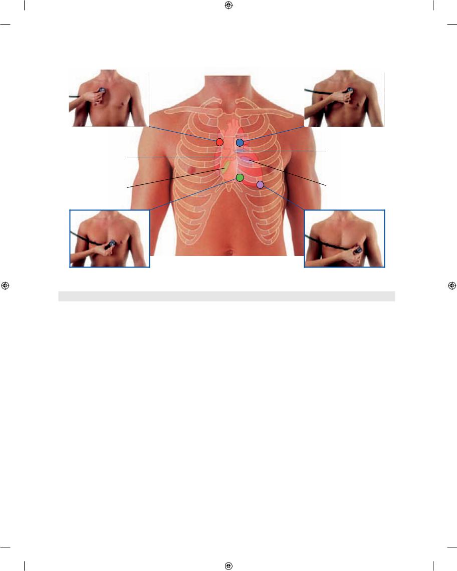
Where to listen for heart sounds
To listen for valve sounds, position the stethoscope down stream from the flow of blood through the valves (Fig. 3.103).
■The tricuspid valve is heard just to the left of the lower part of the sternum near the fifth intercostal space.
3-108
■The mitral valve is heard over the apex of the heart in the left fifth intercostal space at the midclavicular line.
■The pulmonary valve is heard over the medial end of the left second intercostal space.
■The aortic valve is heard over the medial end of the right second intercostal space.
Visualizing the pleural cavities and lungs, pleural recesses, and lung lobes and fissures
Palpable surface landmarks can be used to visualize the normal outlines of the pleural cavities and the lungs and to determine the positions of the pulmonary lobes and fissures.
Superiorly, the parietal pleura projects above the first costal cartilage. Anteriorly, the costal pleura approaches
Drake_ch03_main.indd 108 |
|
|
8/25/2008 4:18:20 PM |
|
|
||
|
|
|
|
|
|
|
Surface anatomy • Where to listen for lung sounds |
3 |
||
|
|
|
|
|
|
|
|
|
|
|
|
|
|
|
|
|
|
|
|
|
Auscultation position for aortic valve
Auscultation position for pulmonary valve
Pulmonary valve
Aortic valve
Tricuspid valve |
Mitral valve |
|
Auscultation position for tricuspid valve
Auscultation position for mitral valve
Fig. 3.103 Anterior view of the chest wall of a man showing skeletal structures, heart, location of the heart valves, and auscultation points.
the midline posterior to the upper portion of the sternum. Posterior to the lower portion of the sternum, the left pari etal pleura does not come as close to the midline as it does on the right side. This is because the heart bulges onto the left side (Fig. 3.104A).
Inferiorly, the pleura reflects onto the diaphragm above the costal margin and courses around the thoracic wall following an VIII, X, XII contour (i.e., rib VIII in the mid clavicular line, rib X in the midaxillary line, and vertebra TXII posteriorly).
The lungs do not completely fill the area surrounded by the pleural cavities, particularly anteriorly and inferiorly.
■Costomediastinal recesses occur anteriorly, particularly on the left side in relationship to the heart bulge.
■Costodiaphragmatic recesses occur inferiorly between the lower lung margin and the lower margin of the pleural cavity.
In quiet respiration, the inferior margin of the lungs travels around the thoracic wall following a VI, VIII, X contour (i.e., rib VI in the midclavicular line, rib VIII in the midaxillary line, and vertebra TX posteriorly).
In the posterior view, the oblique fissure on both sides is located in the midline near the spine of vertebra TIV (Figs. 3.104B and 3.105A). It moves laterally in a downward direction, crossing the fourth and fifth intercostal spaces and reaches rib VI laterally.
In the anterior view, the horizontal fissure on the right side follows the contour of rib IV and its costal cartilage and the oblique fissures on both sides follow the contour of rib VI and its costal cartilage (Fig. 3.105B).
Where to listen for lung sounds
The stethoscope placements for listening for lung sounds are shown in Fig. 3.106.
3-109
Drake_ch03_main.indd 109 |
|
|
8/25/2008 4:18:41 PM |
|
|
||
|
|
|
|

Thorax
A
Superior lobe
Horizontal fissure
Middle lobe
Rib VI
Inferior lobe
Rib VIII
Rib X
Parietal pleura
B
TIV
Rib V
Rib VI
TX
Rib X
TXII
Superior lobe
Costomediastinal recess
Inferior lobe
Costodiaphragmatic recess
Upper lobe
Oblique fissure
Lower lobe
Parietal pleura
Fig. 3.104 Views of the chest wall showing the surface projections of the lobes and the fissures of the lungs. A. Anterior view in a woman. On the right side, the superior, middle, and inferior lobes are illustrated. On the left side, the superior and inferior lobes are illustrated.
B. Posterior view in a woman. On both sides, the superior and inferior lobes are illustrated. The middle lobe on the right side is not visible in this view.
3-110
Drake_ch03_main.indd 110 |
|
|
8/25/2008 4:18:58 PM |
|
|
||
|
|
|
|

A
TIV spine
B
Superior lobe
TIV spine
Rib V
Rib VI
Middle lobe
Inferior lobe
Rib VIII
Rib X
Midaxillary line
Surface anatomy • Where to listen for lung sounds |
3 |
|
|
Superior lobe
Oblique fissure
Medial margin of scapula
Inferior lobe
Horizontal fissure
Oblique fissure
Parietal pleura
Costodiaphragmatic recess
Fig. 3.105 Views of the chest wall. A. Posterior view in a woman with arms abducted and hands positioned behind her head. On both sides, the superior and inferior lobes of the lungs are illustrated. When the scapula is rotated into this position, the medial border of the scapula parallels the position of the oblique fissure and can be used as a guide for determining the surface projection of the superior and inferior lobes of the lungs. B. Lateral view in a man with his right arm abducted. The superior, middle, and inferior lobes of the right lung are illustrated. The oblique fissure begins posteriorly at the level of the spine of vertebra TIV, passes inferiorly crossing rib IV, the fourth intercostal space, and rib V. It crosses the fifth intercostal space at the midaxillary line and continues anteriorly along the contour of rib VI. The horizontal fissure crosses rib V in the midaxillary space and continues anteriorly, crossing the fourth intercostal space and following the contour of rib IV and its costal cartilage to the sternum.
3-111
Drake_ch03_main.indd 111 |
|
|
8/25/2008 4:19:12 PM |
|
|
||
|
|
|
|