


cough of younger children. Chest radiographs in this group of patients are almost always normal, despite the intensity of the cough illness.
Although infection with H. capsulatum is usually asymptomatic in older children and adolescents, infants and young children are at risk for symptomatic infection, which may cause respiratory distress and hypoxemia.
Pneumonia is the most common cause of acute chest syndrome, which occurs in 15–43 % of patients with sickle cell disease. This syndrome is characterized by fever, chest pain, dyspnea, cough, tachypnea, crackles.
Alternative diagnoses and missed diagnosis. There are a few other conditions that should be considered in children with this presentation. Bronchiolitis in babies manifests with rhinorrhoea, fever and tachypnoea. Bilateral crackles and/or wheeze may be evident. Children with upper respiratory tract infections have normal saturations and a clear chest on auscultation. Babies with cardiac failure often have a known history of congenital heart disease and may have bilateral chest signs without fever. Urinary tract infection and bacteraemia should be considered in children with fever who have minimal respiratory symptoms or signs. Tachypnoea alone may be a sign of underlying metabolic acidosis e.g. diabetic ketoacidosis. Occasionally, lower lobe pneumonia may present with abdominal pain and fever. In these patients, the increased respiratory rate and low saturations may aid the diagnosis, however these signs can be absent or minimal. Children with pneumonia may also present with fever alone.
DIAGNOSIS
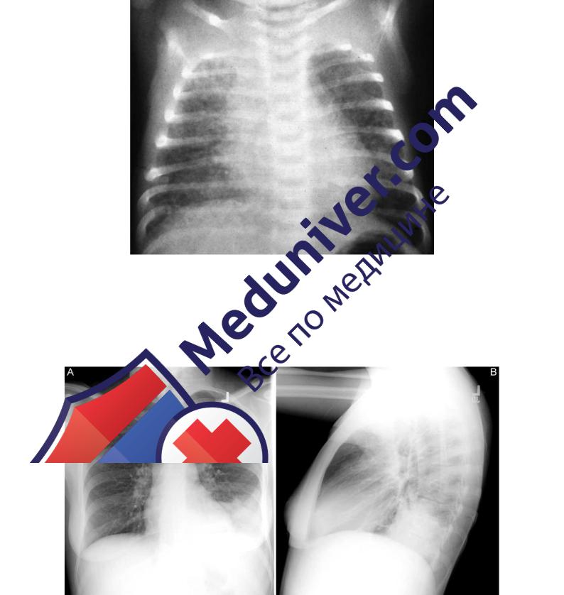
The chest radiograph confirms the diagnosis of pneumonia and may indicate a complication such as a pleural effusion or empyema. Viral pneumonia is usually characterized by hyperinflation with bilateral interstitial infiltrates and peribronchial cuffing (fig. 2).
А |
|
В |
|
|
|
Figure 2. Anteroposterior (A) and lateral (B) radiographs from a child with presumptive viral pneumonia
13
Chest X-ray shows hyperinflation with bilateral, symmetrical interstitial infiltrates in infants with pneumonia caused by Chlamydia trachomatis (fig. 3).
Figure 3. Pneumonia caused by Chlamydia trachomatis in a 3-month-old infant with inclusion conjunctivitis
Confluent lobar consolidation is typically seen with pneumococcal pneumonia (fig. 4). The radiographic appearance alone is not diagnostic and other clinical features must be considered. Repeat chest x-rays are not required for proof of cure for patients with uncomplicated pneumonia.
А |
|
В |
|
|
|
Figure 4. Anteroposterior (A) and lateral (B) radiographs from a child with a left lower lobe infiltrate
14
The peripheral white blood cell (WBC) count can be useful in differentiating viral from bacterial pneumonia. In viral pneumonia, the WBC count can be normal or elevated but is usually not higher than 20,000/mm3, with a lymphocyte predominance. Bacterial pneumonia (occasionally, adenovirus pneumonia) is often associated with an elevated WBC count in the range of 15,000–40,000/mm3 and a predominance of granulocytes. A large pleural effusion, lobar consolidation, and a high fever at the onset of the illness are also suggestive of a bacterial etiology.
Atypical pneumonia due to C. pneumoniae or M. pneumoniae is difficult to distinguish from pneumococcal pneumonia by x-ray and other labs, and although pneumococcal pneumonia is associated with a higher WBC count, erythrocyte sedimentation rate (ESR), and C-reactive protein (CRP), there is considerable overlap.
The definitive diagnosis of a viral infection rests on the isolation of a virus or detection of the viral genome or antigen in respiratory tract secretions. Growth of respiratory viruses in tissue culture usually requires 5–10 days. Reliable DNA or RNA tests for the rapid detection of RSV, parainfluenza, influenza, and adenoviruses are available and accurate. Serologic techniques can also be used to diagnose a recent respiratory viral infection but generally require testing of acute and convalescent serum samples for a rise in antibodies to a specific viral agent. This diagnostic technique is laborious, slow, and not generally clinically useful because the infection usually resolves by the time it is confirmed serologically. Serologic testing may be valuable as an epidemiologic tool to define the incidence and prevalence of the various respiratory viral pathogens.
The definitive diagnosis of a bacterial infection requires isolation of an organism from the blood, pleural fluid, or lung. Culture of sputum is of little value in the diagnosis of pneumonia in young children.
Blood culture remains the non-invasive gold standard for determining the precise etiology of pneumonia. However the sensitivity of this test is very low. Positive blood cultures are found only in 10 % to 30 % of patients with pneumonia. Blood culture should be performed in severe pneumonia or when there is poor response to the first line antibiotics.
In M. pneumoniae infections, cold agglutinins at titers > 1 : 64 are found in the blood in ≈ 50 % of patients. Cold agglutinins are nonspecific, however, because other pathogens such as influenza viruses may also cause increases. Acute infection caused by M. pneumoniae can be diagnosed on the basis of a positive PCR test or seroconversion in an IgG assay. Serologic evidence such as the antistreptolysin O (ASO) titer may be useful in the diagnosis of group A streptococcal pneumonia.
SEVERITY ASSESSMENT
Children with CAP may present with a range of symptoms and signs: fever, tachypnoea, breathlessness, difficulty in breathing, cough, wheeze, headache, abdominal pain and chest pain. The spectrum of severity of CAP can be mild to severe (table 2).
15

Table 2
Severity assessment
|
Mild to moderate |
Severe |
Infants |
Temperature < 38.5 °C |
Temperature > 38.5 °C |
|
Respiratory rate < 50 breaths/min |
Respiratory rate > 70 breaths/min |
|
Mild recession |
Moderate to severe recession |
|
Taking full feeds |
Nasal flaring |
|
|
Cyanosis |
|
|
Intermittent apnoea |
|
|
Grunting respiration |
|
|
Not feeding |
|
|
Tachycardia* |
|
|
Capillary refill time ≥ 2 s |
|
|
Chronic conditions |
Older |
Temperature < 38.5 °C |
Temperature > 38.5 °C |
children |
Respiratory rate < 50 breaths/min |
Respiratory rate > 50 breaths/min |
|
Mild breathlessness |
Severe difficulty in breathing |
|
No vomiting |
Nasal flaring |
|
|
Cyanosis |
|
|
Grunting respiration |
|
|
Signs Tachycardia* |
|
|
Capillary refill time ≥ 2 s |
|
|
Signs of dehydration |
|
|
Chronic conditions** |
* Values to define tachycardia vary with age and with temperature. Thorax 2011; ** Congenital heart disease, chronic lung disease of prematurity, chronic respiratory conditions leading to infection such as cystic fibrosis, bronchiectasis, immune deficiency.
Infants and children with mild to moderate respiratory symptoms can be managed safely in the community. The most important decision in the management of CAP is whether to treat the child in the community or refer and admit for hospital-based care. This decision is best informed by an accurate assessment of severity of illness at presentation and an assessment of likely prognosis. In previously well children there is a low risk of complications and treatment in the community is preferable. This has the potential to reduce inappropriate hospital admissions and the associated morbidity and costs. Management in these environments is dependent on an assessment of severity. Severity assessment will influence microbiological investigations, initial antimicrobial therapy, route of administration, duration of treatment and level of nursing and medical care.
A referral to hospital will usually take place when a general practitioner assesses a child and feels the clinical severity requires admission. In addition to assessing severity, the decision whether to refer to hospital or not should take account of any underlying risk factors the child may have together with the ability of the parents/carers to manage the illness in the community. This decision may be influenced by the level of parental anxiety (table 3).
16

Table 3
Factors suggesting need for hospitalization of children with pneumonia
Age < 6 mo
Sickle cell anemia with acute chest syndrome
Multiple lobe involvement
Immunocompromised state
Toxic appearance
Severe respiratory distress
Requirement for supplemental oxygen
Dehydration
Vomiting
No response to appropriate oral antibiotic therapy
Noncompliant parents
Pneumonia / ed. by H. B. Jenson, R. S. Baltimore // Pediatric Infectious Diseases: Principles and Practice. Philadelphia, WB Saunders, 2002. 801 p.
Children with CAP may also access hospital services when the parents/carers bring the child directly to a hospital emergency department. In these circumstances hospital doctors may come across children with mild disease that can be managed in the community. Some with severe disease will require hospital admission for treatment. One key indication for admission to hospital is hypoxaemia. In a study carried out in the developing world, children with low oxygen saturations were shown to be at greater risk of death than adequately oxygenated children. The same study showed that a respiratory rate of > 70 breaths/ min in infants aged < 1 year was a significant predictor of hypoxaemia.
There is no single validated severity scoring system to guide the decision on when to refer for hospital care. An emergency care-based study assessed vital signs as a tool for identifying children at risk from a severe infection. Features including a temperature > 39 °C, saturations < 94 %, tachycardia and capillary refill time > 2 s were more likely to occur in severe infections. Auscultation
revealing absent |
breath sounds |
with a dull percussion note should raise |
the possibility of |
a pneumonia |
complication by effusion and should trigger |
a referral to hospital. There is some evidence that an additional useful assessment is the quality of a child’s cry and response to their parent’s stimulation; if these are felt to be abnormal and present with other worrying features, they may also strengthen the case for referral for admission to hospital. A global assessment of clinical severity and risk factors is crucial in identifying the child likely to require hospital admission.
Transfer to a pediatric intensive care unit is warranted when the child cannot maintain an oxygen saturation level greater than 92 percent despite a fraction of inspired oxygen greater than 0.6, the patient is in shock, the respiratory rate and pulse rate are rising, and the child shows evidence of severe respiratory distress and exhaustion (with or without a rise in partial arterial carbon dioxide tension), or when the child has recurrent episodes of apnea or slow, irregular breathing.
17