

4 курс / Лучевая диагностика / ЛУЧЕВАЯ ДИАГНОСТИКА
.pdfМИНИСТЕРСТВО ЗДРАВООХРАНЕНИЯ РЕСПУБЛИКИ БЕЛАРУСЬ
УЧРЕЖДЕНИЕ ОБРАЗОВАНИЯ «ГОМЕЛЬСКИЙГОСУДАРСТВЕННЫЙМЕДИЦИНСКИЙУНИВЕРСИТЕТ»
Кафедра онкологии с курсом лучевой диагностики и лучевой терапии
Н. М. ЕРМОЛИЦКИЙ
ЛУЧЕВАЯ ДИАГНОСТИКА
Учебно-методическое пособие для студентов 3 курса факультета по подготовке специалистов
для зарубежных стран учреждений высшего медицинского образования
В двух частях
Часть 1
2-е издание
RADIOLOGY
Teaching workbook
for 3rd year students of the Faculty of preparation of experts for foreign countries of medical higher educational institutions
In two parts
Part 1
Гомель
ГомГМУ
2018

УДК 615.849(072)=111 ББК 53.6я73
Е 74
Рецензенты:
кандидат медицинских наук, доцент, директор Институт радиобиологии Национальной академии наук Беларуси
И. А. Чешик;
кандидат медицинских наук, доцент, заведующий клинико-диагностической лабораторией Республиканского научно-практического центра радиационной медицины и экологии человека
Ю. И. Ярец
Ермолицкий, Н. М.
Е 74 Лучевая диагностика: учеб.-метод. пособие для студентов 3 курса факультета по подготовке специалистов для зарубежных стран меди-
цинских вузов: в 2 ч. = Radiology: Teaching workbook work for 3rd year students of the Faculty of preparation of experts for foreign countries of medical higher educational institutions: in two parts / Н. М. Ермолицкий. 2-е изд. — Гомель: ГомГМУ, 2018. Ч. 1. 108 с.
ISBN 978-985-588-089-0
Учебно-методическое пособие подготовлено в соответствии с типовой программой по специальности «Лечебное дело», включены темы, изучаемые в начальном семестре, и в связи с этим содержит необходимые для усвоения разделы дисциплины: принципы и методы диагностической радиологии; основы защиты от ионизирующих излучений; радиология опорно-двигательной системы, грудной клетки и сердечно-сосудистой системы.
Материалы адаптированы к русскоязычному варианту изложения лучевой диагностики и лучевой терапии с учетом объема часов, могут быть использованы студентами старших курсов факультета по подготовке специалистов для зарубежных стран при изучении терапевтических и хирургических дисциплин.
Предназначено для студентов 3 курса факультета по подготовке специалистов для зарубежных стран учрежденийвысшегомедицинскогообразования.
Утверждено и рекомендовано к изданию научно-методическим советом учреждения образования «Гомельский государственный медицинский университет» 20 июня 2018 г., протокол № 4.
|
УДК 615.849(072)=111 |
|
ББК 53.6я73 |
ISBN 978-985-588-088-3 |
© Учреждение образования |
ISBN 978-985-588-089-0 (Ч. 1) |
«Гомельский государственный |
|
медицинский университет», 2016 |
2
|
CONTENTS |
|
1. Overview and principles of diagnostic radiology imaging. |
|
|
Contrast radiologic examinations. An approach to image interpretation. |
|
|
Protection in radiological practice......................................................................... |
4 |
|
2. |
Radiology of musculoskeletal system..................................................... |
38 |
3. |
Chest imaging.......................................................................................... |
60 |
4. |
Cardiovascular system imaging.............................................................. |
95 |
References................................................................................................. |
105 |
|
3

1. OVERVIEW AND PRINCIPLES OF DIAGNOSTIC RADIOLOGY IMAGING
Introduction
The realm of diagnostic radiology encompasses various modalities of imaging that may be used individually or, more commonly, in combination to provide the clinician with enough information to aid in making a diagnosis.
Of high importance in today's managed care environment, diagnostic imaging can also be used for the early identification of potential medical problems, to help prevent their occurrence.
Radiologists and other physicians interpret the resulting images to diagnose various medical illness or injury so that patient treatment and therapy can be specifically planned and implemented. Diagnostic imaging is also used to guide surgical planning and is often used to follow surgery and/or monitor the outcomes of therapeutic procedures.
The newest frontier in diagnostic imaging, molecular imaging, makes possible the identification of certain molecules within cell structures.
Main areas of diagnostic radiology imaging
The specialty of radiology includes conventional techniques that use ionizing radiation: X-ray examinations, computed tomography (CT); and nuclear medicine (NM).
High quality diagnostic radiology images also may be obtained via the techniques of magnetic resonance imaging (MRI) and ultrasound (US), thereby avoiding the risks of radiation.
Three modes of image production
There are three basic means by which radiologic images are produced: transmission of energy, reflection of energy, and emission of energy. Physicians have understood this, and they also know that the three methods are complementary, each providing information at a different level.
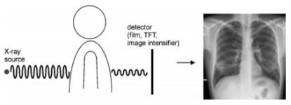
Transmission imaging
Radiograph is produced by the transmission of energy. A beam of highenergy photons is passed through the body, some of which are attenuated or blocked when they strike subatomic particles. The principal transmission modalities include plain radiography (such as chest radiographs and abdominal radiographs), fluoroscopy, and CT.
Fluoroscopy represents a kind of «movie», or moving picture, in which continuous detection and display of the pattern of photon transmission enables the visu-
4

alization of dynamic processes in real time. CT gathers transmission data from multiple perspectives and employs a mathematical algorithm (the Fourier transform) to reconstruct an image of the slice of tissue through which the x-ray beams passed.
Reflection imaging
The radiologic modality that exemplifies reflection imaging is ultrasound. As we shall see, ultrasound creates images not according to the density differences between various tissues, but by their acoustic differences. A very lowdensity structure such as fat, which appears black on a radiograph, may be very «echogenic» acoustically, and hence appear bright on a sonogram.
Emission imaging
The emission modalities include MRI and nuclear medicine. MRI creates images by distinguishing between the nuclear magnetic properties of various tissues. MRI also utilizes no ionizing radiation, generating images using a magnetic field and radio waves.
The other emission modality, nuclear medicine, creates images by introducing radioisotopes into the human body and then detecting their emission of gamma rays (like x-rays, except from a different source). In contrast to MRI, nuclear medicine involves ionizing radiation.
Different types of ionizing radiation
The gamma (γ) ray corresponds to the emission of short wavelength and variable energy photons (figure 1.1–1.2). This radiation translates the loss of excessive energy in the nucleus and its transformation into a more stable state, as opposed to the production of X-rays, obtained after excitation and ionization of electrons. X- and gamma rays are ideal medical diagnosis tools. As they are very penetrating, they can go through large thicknesses of matter and travel for hundreds of meters in the air. Dense materials such as lead, very thick concrete or deep water are able to stop them or, at least, strongly attenuate them.
Figure 1.1 Different types of radiations to which man is daily exposed
5

An X-ray is an electromagnetic wave of the same type as light waves, but with a higher energy level, thus capable to some extent of travelling through matter. The technique is based on matter’s capacity to attenuate an external beam of X-rays depending on whether it is solid, liquid or gaseous. Therefore, organs will allow radiation to pass through them according to their density, thickness and constitution; the intensity of the rays is measured on photographic film or using a specific detector. Initial applications distributed on organs and tissues presenting a different coefficient of absorption.
Definition
X-rays, or roentgen rays, are a form of electromagnetic radiation or energy of extremely short wavelength. X-rays in the diagnostic range are near the end of the spectrum of short wavelengths. The shorter the wavelength of an electromagnetic radiation form, the greater its energy and, as a rule, the greater its ability to penetrate various materials.
Figure 1.2 Electromagnetic spectrum extending over several orders
of magnitude listing values of wavelengths, frequency, and identifying values in some of the more common regions of the spectrum. Ionizing radiation
has sufficient energy to penetrate human tissue.
X-rays are described in terms of particles or packets of energy called quanta or photons, which travel at the speed of light. The amount of energy carried by each photon depends on the wavelength of the radiation. This is mea-sured in electron volts. An atom is ionized when it loses an electron. Any photon with approximately 15 or more electron volts of energy is capable of producing ionization in atoms and molecules (ionizing radiation). X-rays, γ-rays, and certain types of ultraviolet radiation are all typical ionizing radiation forms.
Production of X-Rays
X-rays used in diagnostic radiology require a vacuum and the presence of a high potential difference between a cathode and an anode. Moving these elec-
6
trons toward the anode at an energy level sufficient to produce x-rays requires a high potential — up to 125,000 V (125 kV). When the accelerated electrons strike the tungsten anode, x-rays are produced.
X-rays follow the same physical laws as light, and as such, will darken photographic (or, in the past x-ray) film. For the purpose of this discussion, the word «film» is used interchangeably with any type of recording medium (digital receptor plates).
Production of images
Image production by x-rays results from attenuation of those x-rays by the material through which they pass. Attenuation is the process by which x-rays are removed from a beam through absorption and scatter. In general, the greater the material’s density — that is, the number of grams per cubic centimeter — the greater its ability to absorb or scatter x-rays. Absorption is also influenced by the atomic number of the structure. The denser the structure, the greater the attenuation, which results in less blackening of the «film» (fewer x-rays strike the «film»). Less-dense structures attenuate the beam to a lesser degree and result in more blackening of the «film» (more x-rays strike the «film», figure 1.3). A small percentage of the incident radiation beam exits the patient and strikes a detector. An image receptor is required to convert the radiation into an image after it has passed through the area of interest.
Figure 1.3 X-rays penetrate the human body and produce an image that shows of tissue-specific absorption along a path from the X-ray source to a detector
Radiographic density
It is important to differentiate between two types of densities that you will hear mentioned when discussing radiographs with radiologists or other colleagues: physical density and radiographic density. Physical density is the type of density just described. Radiographic density refers to the degree of blackness of a film. Radiographic contrast is the difference in radiographic densities on a film. The radiographic density of a substance is related to its physical density. The effect on film or other recording media occurs paradoxically: structures of high physical density produce less radiodensity and vice versa. Structures that produce more blackening on film are referred to as being radiolucent; those that produce less blackening are called radiopaque or radiodense. There are four
7

types of radiographic densities; in increasing order of physical density, these are gas (air), fat, soft tissue (water), and bone (metal). Radiographically, these appear as black, gray-black, gray, and white, respectively.
Both density and thickness tissues, then, are factors to take into account in assessing the degree of opacification noted on a radiograph (figure 1.4). Radiographs were initially referred to as «skiagraphs», from the Greek for «shadow pictures», because they represent recordings of the shadows cast by anatomic structures as photons pass through the body.
Plain radiography may be conceptualized as a snapshot: it provides a static view of anatomic structure obtained in a fraction of a second.
A 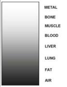 B
B 
Figure 1.4 (A) Diagram of radiographic densities on NEGATIVE image.
(B) Note how the four basic densities are visible on negative frontal chest radiograph
Recording media (detector)
X-ray film
The most common type of recording medium used to be x-ray film. X-ray film is still used in many parts of the world. However, state-of-the-art radiology departments have replaced x-ray film with electronic recording media.
X-ray film consists of a plastic sheet coated with a thin emulsion that contains silver bromide and a small amount of silver iodide. This emulsion is sensitive to light and radiation. A protective coating covers the emulsion. When the film is exposed to light or to ionizing radiation and then developed, chemical changes take place within the emulsion, resulting in the deposition of metallic silver, which is black. The amount of blackening on the film depends entirely on the amount of radiation reaching it and therefore on the amount attenuated or removed from the beam by the subject.
The classic imaging receptor is a film/screen combination into cassette. The x-ray beam strikes a fluorescent screen (into cassette), which produces light that exposes the film, and then the film is developed.
Fluoroscopic screen (for fluoroscopy)
A fluoroscopic screen is coated with a substance (phosphor) that gives off visible light (or fluoresces) when it is irradiated. The brightness of the light is
8
proportional to the intensity of the x-ray beam striking the plate and depends on the amount of radiation removed from the beam by the object being irradiated. Today, the fluoroscopic screen is combined with an electronic device that converts the visible light into an electron stream that amplifies the image (makes it brighter) by converting the electron pattern back into visible light.
This system allows the radiologist to see the image clearly without requiring dark adaptation of the eyes, as was necessary in non-image-enhanced fluoroscopy. The technology of image intensification was originally developed around 1950 for military use at night. Intensifying screens, variants of fluoroscopic screens, were used in most film cassettes to reduce the amount of radiation needed to produce an acceptable exposure.
A little bit of history
In 1895, Dutch physicist Wilhelm Conrad Roentgen developed equipment that could generate an unknown radiation, unknown because indefinable and original, which he called X. Wilhelm Roentgen succeeded in creating the first image of an X-rayed hand and demonstrated that human tissues behave differently depending on their density.
This first radiography of Mrs Roentgen’s hand, dating from 1896, became famous and opened the way for a new discipline of medicine, diagnostic and therapeutic radiology (figure 1.5).
The recognition of the value and advantages of this technology was such that special radiology services were created within five years.
Plain film (x-ray, projection, conventional) radiography
The x-rays are directed in a limited beam towards the patient (figure 1.5). The radiograph itself is a two-dimensional representation of the threedimensional structures of the patient's body. These structures are visible because of the differences in attenuation of the x-ray beam. Attenuation refers to the process by which x-rays are removed.
Plain radiographs are often called «plain X-rays» — but you can't see the X-rays, only the images created by them. Radiographs can be produced using a variety of imaging methods. The image or picture is basically a shadow of the parts of the patient that absorb or block the X-rays.
The radiographic image is a «photographic negative» of the object — the «shadows» are white regions (where the X-rays were blocked by the object). The image is black in the regions that did not stop the X-rays, and they passed through to expose the film or sensor.
Plain radiographs («plain films») are usually taken by a trained registered radiologic technologist. The resulting films or images are then interpreted by the radiologist to make a diagnosis or suggest further tests.
9

Radiography is more particularly used in orthopaedics, rheumatology and orthodontics. It is also very informative in pneumology (preventive radiology of the lungs or where there is a suspicion of lung cancer) and in oncology (mammography, preventive examination for the detection of breast cancer).
Principle radiography: the rays pass through the area under investigation, and depending on the tissues, a variable amount is absorbed — this is known as differential attenuation. The exact amount of whiteness depends on how much calcium or other heavy metal is present. Bone containing calcium — absorbs almost all of the rays and so shows up as white, fat absorbs much less, and air absorbs none so it appears black. The rays can then be imaged either using film, to get a conventional x-ray, or can be detected using special equipment to get digital radiographs. The routine x-ray is a shadow picture.
A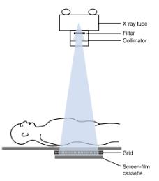 B
B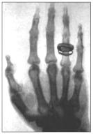
Figure 1.5 (A) Scematic representation of film screen radiography.
(B) Radiograph of Mrs Roentgen’s hand, taken in 1896
A patient that has just been subject to a radiological examination is not radioactive, even if X-rays are a form of radioactivity. Outside the field, the effect of the radiation disappears. Progress in these last years has effectively aimed at reducing both the time taken to acquire the image and the dose necessary for this acquisition, while still maintaining, even improving, quality.
Projection radiography terminology
Typical x-ray projections (figure 1.6)
X-ray projections are typically listed as AP or PA. This depends on whether the x-ray beam passed to the patient from anterior to posterior (AP) or the reverse. Lateral (LAT) and oblique (OBL) views also are commonly obtained.
Note: the word 'view' is often used erroneously to describe a radiographic projection.
10