Добавил:
Опубликованный материал нарушает ваши авторские права? Сообщите нам.
Вуз: Предмет: Файл:

KAPLAN_USMLE_STEP_1_LECTURE_NOTES_2018_BIOCHEMISTRY_and_GENETICS

.pdf
Скачиваний:
7
Добавлен:
29.01.2024
Размер:
10.81 Mб
Скачать

Part I Biochemistry

Vitamin D Toxicity

A 45-year-old man had a 3-week history of weakness, excessive urination, intense thirst, and staggering walk. For most of his adult life, he took excessive amounts of vitamin C because he was told it would help prevent the common cold. The past month, he took excessive amounts of vitamin D and calcium every day because he learned that he was developing osteoporosis. Recent lab tests revealed greatly elevated serum calcium, and vitamin D toxicity was diagnosed.

Vitamin D is highly toxic at consumption levels that continuously exceed 10× RDA, resulting in hypercalcemia. Unlike water-soluble vitamins, which are excreted in excess amounts, vitamin D can be stored in the liver as 25-hydroxycholecalciferol. The excess vitamin D can promote intestinal absorption of calcium and phosphate.

The direct effect of excessive vitamin D on bone is resorption similar to that seen in vitamin D deficiency. Therefore, the increased intestinal absorption of calcium in vitamin D toxicity contributes to hypercalcemia. Rather than help

the man’s osteoporosis, a large amount of vitamin D can contribute to it. Hypercalcemia can impair renal function, and early signs include polyuria, polydipsia, and nocturia. Prolonged hypercalcemia can result in calcium deposition in soft tissues, notably the kidney, producing irreversible kidney damage.

Clinical Correlate

Isotretinoin, a form of retinoic acid, is used in the treatment of acne. It is teratogenic (malformations of the craniofacial, cardiac, thymic, and CNS structures) and is therefore absolutely contraindicated in pregnant women. Use with caution in women of childbearing age.

Note

What to Know for the Exam

Vitamins:

Clinical manifestations of deficiencies

Enzymes that accumulate

Pathways

Preventions of deficiencies

Treatments of deficiencies

Vitamin D Deficiency

High-Yield


Deficiency of vitamin D in childhood produces rickets, a constellation of skeletal abnormalities most strikingly seen as deformities of the legs (although many other developing bones are affected). Muscle weakness is common.

Deficiency of vitamin D after epiphyseal fusion causes osteomalacia, which produces less deformity than rickets. Osteomalacia may present as bone pain and muscle weakness.

VITAMIN A

Vitamin A (carotene) is converted to several active forms in the body associated with two important functions, maintenance of healthy epithelium and vision. Biochemically, there are 3 vitamin A structures that differ on the basis of the functional group on C-1: hydroxyl (retinol), carboxyl (retinoic acid), and aldehyde (retinal).

Maintenance of Epithelium

Retinol and retinoic acid are required for the growth, differentiation, and maintenance of epithelial cells. In this capacity they bind intracellular receptors, which are in the family of Zn-finger proteins, and they regulate transcription through specific response elements.

154

Chapter 10 Vitamins

Vision

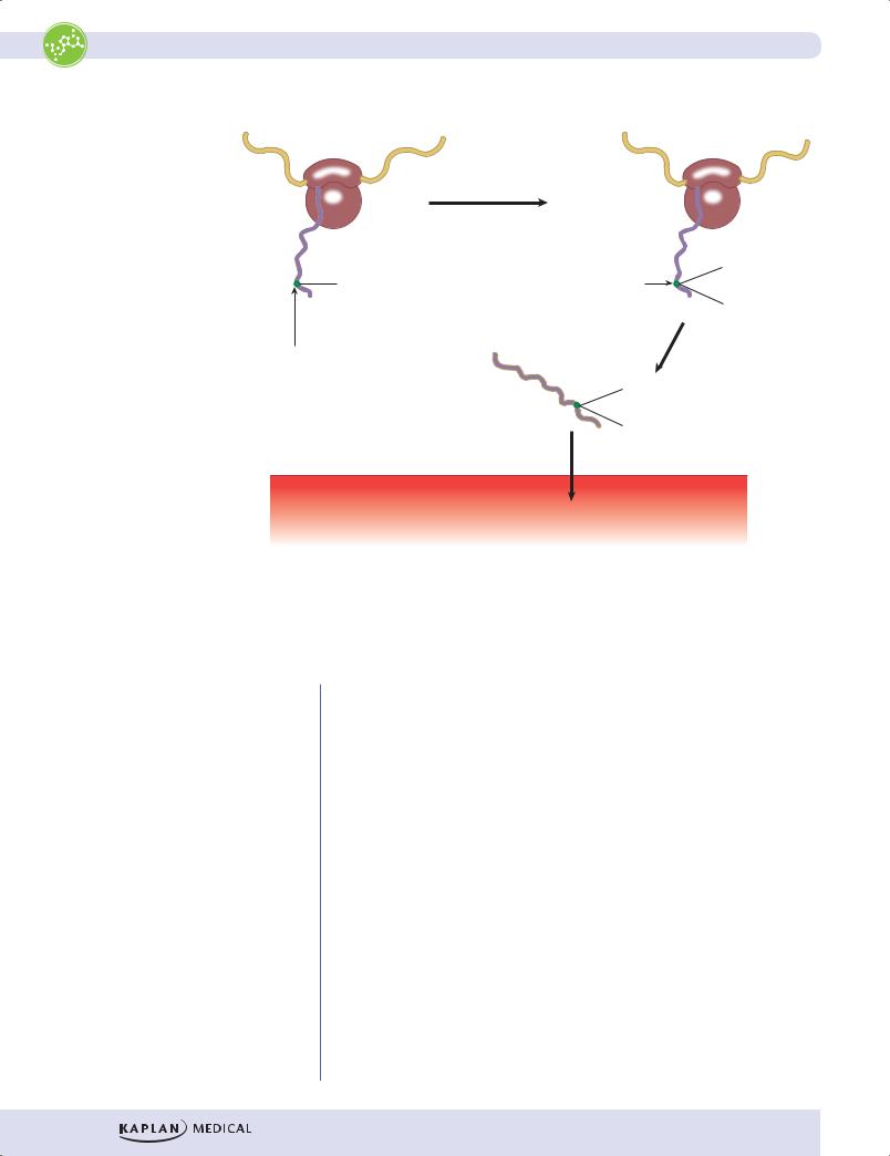
When first formed, all the double bonds in the conjugated double bond system in retinal are in the trans configuration. This form, all-trans retinal is not active. The conversion of all-trans retinal to the active form cis-retinal takes place in the pigmented epithelial cells. Cis-retinal is then transferred to opsin in the rod cells forming the light receptor rhodopsin. It functions similarly in rod and cone cells. When exposed to light, cis-retinal is converted all-trans retinal. A diagram of the signal transduction pathway for light-activated rhodopsin in the rod cell is shown in Figure I-10-2, along with the relationship of this pathway to rod cell anatomy and changes in the membrane potential. Note the following points:

Rhodopsin is a 7-pass receptor coupled to the trimeric G protein transducin (Gt).

When light is present, the pathway activates cGMP phosphodiesterase, which lowers cGMP.

Rhodopsin and transducin are embedded in the disk membranes in the outer rod segment.

cGMP-gated Na+ channels in the cell membrane of the outer rod segment respond to the decrease in cGMP by closing and hyperpolarizing the membrane.

The rod cell is unusual for an excitable cell in that the membrane is partially depolarized (~ –30 mV) at rest (in darkness) and hyperpolarizes on stimulation.

Because the membrane is partially depolarized in the dark, its neurotransmitter glutamate is continuously released. Glutamate inhibits the optic nerve bipolar cells with which the rod cells synapse. By hyperpolarizing the rod cell membrane, light stops the release of glutamate, relieving inhibition of the optic nerve bipolar cell and thus initiating a signal into the brain.

155

Part I Biochemistry

Outer

Rhodopsin

Intradisk space


rod


segment


α


β

cGMP


γ

PDE

Light


Cytoplasm


Gt


Light

cGMP


Dark


5´GMP

+


(inactive)

cGMP


Light


Na+


–30


Membrane


potential


Inner

(meV)


Cell membrane

–35


rod

3 sec


segment


Bipolar

cell

Light

Figure I-10-2. Light-Activated Signal Transduction in the Retinal Rod Cell

156

Chapter 10 Vitamins

Vitamin A Deficiency

A severe drought in portions of Kenya wiped out a family’s yam crop, their primary food staple. Within several months, a 3-year-old child in the family began to complain of being unable to see very well, especially at dusk or at night. Also, the child’s eyes were red due to constant rubbing because of dryness.

Due to the ability of the liver to store vitamin A, deficiencies which are severe enough to result in clinical manifestations are unlikely to be observed, unless there is an extreme lack of dietary vitamin A over several months. Vitamin A deficiency is the most common cause of blindness and is a serious problem in developing countries. It has a peak incidence at age 3–5. In the United States, vitamin A deficiency is most often due to fat malabsorption or liver cirrhosis.

Vitamin A deficiency results in night blindness (rod cells are responsible for vision in low light), metaplasia of the corneal epithelium, xerophthalmia (dry eyes), bronchitis, pneumonia, and follicular hyperkeratosis. The spots or patches noted in the eyes of patients with vitamin A deficiency are known as Bitot spots. Because vitamin A is important for differentiation of immune cells, deficiencies can result in frequent infections.

β-carotene is the orange pigment in yams, sweet potatoes, carrots, and yellow squash. Upon ingestion, it can be cleaved relatively slowly to two molecules of retinal by an intestinal enzyme, and each retinal molecule is then converted to all-trans-retinol and then absorbed by interstitial cells. Therefore, it is an excellent source of vitamin A.

Recall Question

Resection of the terminal ileum in Crohn’s disease leads to deficiency of which of the following vitamins?

A.Biotin

B.Cyanocobalamin

C.Pyridoxine

D.Riboflavin

E.Thiamine

Answer: B

Note

If vitamin A is continuously ingested at levels greater than 15× RDA, toxicity develops; symptoms include excessive sweating, brittle nails, diarrhea, hypercalcemia, hepatotoxicity, vertigo, nausea, and vomiting. Unlike vitamin A, β-carotene is not toxic at high levels.

VITAMIN K

Vitamin K is required to introduce Ca2+ binding sites on several calciumdependent proteins. The modification which introduces the Ca2+ binding site is a γ-carboxylation of glutamyl residue(s) in these proteins, often identified simply as the γ-carboxylation of glutamic acid. Nevertheless, this vitamin K- dependent carboxylation is a cotranslational modification occurring as the proteins are synthesized on ribosomes associated with the rough endoplasmic reticulum (RER) during translation.

157

Part I Biochemistry


HEPATOCYTE


mRNA

mRNA


Ribosome

Vitamin K

Prothrombin

γ-Carboxylation

on RER

by γ-Glutamyl


Carboxylase


COO


NH2


Glutamic


COO

acid


Prothrombin

γ-Carboxy


Glutamic Acid


(binds Ca2+)

NH2


COO


Ribosome

on RER

COO

COO

Prothrombin

COO


NH2

Secretion by exocytosis

Blood

Prothrombin

Figure I-10-3. Vitamin K–Dependent γ-Carboxylation of Prothrombin during Translation on the Rough Endoplasmic Reticulum (RER)

Examples of proteins undergoing this vitamin K–dependent carboxylation include the coagulation factors II (prothrombin), VII, IX, and X, as well as the anticoagulant proteins C and S. All these proteins require Ca2+ for their function.

Vitamin K deficiency produces prolonged bleeding, easy bruising, and potentially fatal hemorrhagic disease. Conditions predisposing to a vitamin K deficiency include:

Fat malabsorption (bile duct occlusion)

Prolonged treatment with broad-spectrum antibiotics (eliminate intestinal bacteria that supply vitamin K)

Breast-fed newborns (little intestinal flora, breast milk very low in vitamin K), especially in a home-birth where a postnatal injection of vitamin K may not be given

Infants whose mothers have been treated with certain anticonvulsants during pregnancy such as phenytoin (Dilantin)

158

Chapter 10 Vitamins

Vitamin K Deficiency

A 79-year-old man living alone called his 72-year-old sister and then arrived at the hospital by ambulance complaining of weakness and having a rapid heartbeat. His sister said that he takes no medications and has a history of poor nutrition and poor hygiene. Physical examination confirmed malnourishment and dehydration. A stool specimen was positive for occult blood. He had a prolonged prothrombin time (PT), but his liver function tests (LFTs) were within normal range. He was given an injection of a vitamin that corrected his PT in 2 days.

Poor nutrition and malnourishment, lack of medications, occult blood in the stool specimen, prolonged PT, and normal LFTs are all consistent with vitamin K deficiency. Without vitamin K, several blood clotting factors (prothrombin, X, IX, VII) are not γ-carboxylated on glutamate residues by the γ-glutamyl carboxylase during their synthesis (cotranslational modification) in hepatocytes. The PT returned to normal 2 days after a vitamin K injection.

Vitamin K deficiency should be distinguished from vitamin C deficiency.

Table I-10-3. Vitamin K versus Vitamin C Deficiency


Vitamin K Deficiency


Vitamin C Deficiency


Easy bruising, bleeding


Easy bruising, bleeding


Normal bleeding time


Increased bleeding time


Increased PT


Normal PT


Hemorrhagic disease with no


Gum hyperplasia, inflammation,


connective tissue problems


loss of teeth


• Skeletal deformity in children


Poor wound healing


Anemia


Associated with:


Associated with:


• Fat malabsorption


• Diet deficient in citrus fruit, green


• Long-term antibiotic therapy


vegetables


Breast-fed newborns

Infant whose mother was taking anticonvulsant therapy during pregnancy

159

Part I Biochemistry

Clinical Correlate

Relative to the other proteins that undergo γ-carboxylation, protein C has a short half-life. Thus, initiation of warfarin therapy may cause a transient hypercoagulable state.

Clinical Correlate

Vitamin K (SC, IM, oral, or IV) is used to reverse bleeding from hypothrombinemia caused by excess warfarin.

Anticoagulant Therapy

High-Yield


Warfarin and dicumarol antagonize the γ-carboxylation activity of vitamin K and thus act as anticoagulants. They interfere with the cotranslational modification during synthesis of the precoagulation factors.

Once these proteins have been released into the bloodstream, vitamin K is no longer important for their subsequent activation and function.

Related to this are 2 important points:

Warfarin and dicoumarol prevent coagulation only in vivo and cannot prevent coagulation of blood in vitro (drawn from a patient into a test tube).

When warfarin and dicumarol are given to a patient, 2–3 days are required to see their full anticoagulant activity. Heparin or low- molecular-weight heparin is often given to provide short-term anticoagulant activity. Heparin is an activator of antithrombin III.

VITAMIN E

Vitamin E (α-tocopherol) is an antioxidant. As a lipid-soluble compound, it is especially important for protecting other lipids from oxidative damage. It prevents peroxidation of fatty acids in cell membranes, helping to maintain their normal fluidity.

Vitamin E deficiency can lead to hemolysis, neurologic problems, and retinitis pigmentosa.

High blood levels of vitamin E can cause hemorrhage in patients given warfarin.

160

Chapter 10 Vitamins

Review Questions

Select the ONE best answer.

1.Retinitis pigmentosa (RP) is a genetically heterogeneous disease characterized by progressive photoreceptor degeneration and ultimately blindness. Mutations in more than 20 different genes have been identified in clinically affected patients. Recent studies have mapped an RP locus to the chromosomal location of a new candidate gene at 5q31. One might expect this gene to encode a polypeptide required for the activity of a(n)

A.receptor tyrosine kinase

B.cGMP phosphodiesterase

C.phospholipase C

D.adenyl cyclase

E.protein kinase C

2.A 27-year-old woman with epilepsy has been taking phenytoin to control her seizures. She is now pregnant, and her physician is considering changing her medication to prevent potential bleeding episodes in the infant. What biochemical activity might be deficient in the infant if her medication is continued?

A.Hydroxylation of proline

B.Glucuronidation of bilirubin

C.Reduction of glutathione

D.γ-Carboxylation of glutamate

E.Oxidation of lysine

3.A 75-year-old woman is seen in the emergency room with a fractured arm. Physical examination revealed multiple bruises and perifollicular hemorrhages, periodontitis, and painful gums. Her diet consists predominately of weak coffee, bouillon, rolls, and plain pasta. Lab results indicated mild microcytic anemia. Which of the following enzymes should be less active than normal in this patient?

A.Homocysteine methyltransferase

B.γ-Glutamyl carboxylase

C.Dihydrofolate reductase

D.ALA synthase

E.Prolyl hydroxylase

161

Part I Biochemistry

Answers

1.Answer: B. Only phosphodiesterase participates as a signaling molecule in the visual cycle of photoreceptor cells.

2.Answer: D. Phenyl hydantoins decrease the activity of vitamin K, which is required for the γ-carboxylation of coagulation factors (II, VII, IX, X), as well as proteins C and S.

3.Answer: E. The patient has many signs of scurvy from a vitamin C deficiency. The diet, which contains no fruits or vegetables, provides little vitamin C. Prolyl hydroxylase requires vitamin C, and in the absence of hydroxylation, the collagen α-chains do not form stable, mature collagen. The anemia may be due to poor iron absorption in the absence of ascorbate.

162

Energy Metabolism 11

Learning Objectives

Explain information related to metabolic sources of energy

Interpret scenarios about metabolic energy storage and fuel metabolism

Answer questions about patterns of fuel metabolism in tissues

METABOLIC SOURCES OF ENERGY

Energy is extracted from food via oxidation, resulting in the end products carbon dioxide and water. This process occurs in 4 stages.

In stage 1, metabolic fuels are hydrolyzed in the gastrointestinal (GI) tract to a diverse set of monomeric building blocks (glucose, amino acids, and fatty acids) and absorbed.

In stage 2, the building blocks are degraded by various pathways in tissues to a common metabolic intermediate, acetyl-CoA.

Most of the energy contained in metabolic fuels is conserved in the chemical bonds (electrons) of acetyl-CoA.

A smaller portion is conserved in reducing nicotinamide adenine dinucleotide (NAD) to NADH or flavin adenine dinucleotide (FAD) to FADH2.

Reduction indicates the addition of electrons that may be free, part of a hydrogen atom (H), or a hydride ion (H).

In stage 3, the citric acid (Krebs, or tricarboxylic acid [TCA]) cycle oxidizes acetyl-CoA to CO2. The energy released in this process is primarily conserved by reducing NAD to NADH or FAD to FADH2.

The final stage is oxidative phosphorylation, in which the energy of NADH and FADH2 is released via the electron transport chain (ETC) and used by an ATP synthase to produce ATP. This process requires O2.

163