


32 2 Local Anaesthesia
2.7 Troubleshooting
The goal of injecting local anaesthesia is to prevent the patient from perceiving pain when tissues are manipulated. There are a number of reasons why anaesthetic may fail, although the majority of cases are due to poor technique:
●●Anxiety and dental fear by the patient.
●●Local inflammatory conditions or the pulp or surrounding tissues.
●●Variations in local anatomy.
●●Accessory innervation of the teeth or surrounding structures.
●●Thick cortical bone preventing diffusion.
●●Inadequate dose for the procedure.
●●Inadequate time for onset of anaesthetic effect.
A common misconception amongst patients undergoing procedures with local anaesthesia is that they will feel nothing. If patients are not sufficiently informed that anaesthesia only inhibits local pain signalling, they may perceive the sensation of pushing forces on the jaws as failed anaesthesia. Anxiety and dental fear can also heighten the experience of pain and otherwise innocuous stimuli. Patients should be informed that whilst profound anaesthesia can inhibit local pain signalling, this does not extend to reducing pressure on the entire mandible and maxilla. If, despite forewarning, the patient still does not tolerate the procedure, additional forms of anaesthesia may be indicated.
Inadequate anaesthesia may also be related to improper technique or variations in anatomy. When administering an inferior alveolar nerve block, there are a number of anatomical variants which can lead to failed anaesthesia. A flared ramus, accessory mandibular foramen, or abnormal position of the mandibular foramen may prevent the block from being effective. Failure of an infiltration anaesthetic may also be due to local anatomical factors, such as a thick cortical plate or exostosis limiting the diffusion of the anaesthetic agent.
In some cases, mandibular teeth may receive accessory sensory innervation by the nerve to mylohyoid. This nerve branches from the inferior alveolar nerve before it enters the mandibular foramen. Its primary function is as a motor nerve to the mylohyoid muscle, but it may also carry sensory fibres, which can provide innervation to mandibular molars. As this nerve branches higher in the pterygomandibular space, a traditional inferior alveolar nerve block may not adequately anaesthetise the mandibular teeth. In such cases – where the operator is assured that the lower lip and chin are anaesthetised, and adequate buccal anaesthesia has been obtained – additional local anaesthesia in the form of intraligamental injections may be used to account for this anatomic variant.
Localised infections, dental abscesses, and inflammatory oedema can significantly reduce the efficacy of local anaesthetic. In solution, local anaesthetic molecules exist in both ionised form, which cannot cross nerve membranes, and unionised form, which can. Tissues undergoing an inflammatory or infectious process may be more acidic, increasing the proportion of injected anaesthetic molecules that are ionised versus unionised. Less of the solution will effectively block the voltage-gated sodium channels, and its anaesthetic effect will thus be reduced. Use of buffered anaesthetic agents containing bicarbonate allows for less ionisation and therefore more effective anaesthesia.
Inflammatory conditions of the pulpal tissues, or ‘hot pulp’, can also lead to a failure to adequately anaesthetise the tooth, through hyperalgesia and nerve sensitisation due to the presence of inflammatory mediators. In these situations, additional forms of anaesthesia may be required for patient comfort.
https://t.me/DentalBooksWorld

2.7 Troubleshootin 33
An inadequate dose is another common reason for failure. For general and restorative dentistry, an approach of using as little anaesthesia as possible is generally favoured. The advantage from the patient’s perspective is that the lack of sensation is shorter in duration, there is less chance of producing tachycardia, and fewer injections are required. However, when using local anaesthesia for dentoalveolar extractions, more profound anaesthesia is required, and the clinician should bear this in mind when planning dose and delivery. The anaesthetic technique must ensure that the tissues are sufficiently anaesthetised and that the patient has sufficient time after the procedure to commence analgesics before the local anaesthetic effect wears off.
https://t.me/DentalBooksWorld
https://t.me/DentalBooksWorld

35
3
Basic Surgical Instruments
A complete surgical instrument tray is essential to safely and effectively perform dentoalveolar extractions. Every piece of equipment on the surgical setup serves a specific role in the removal of a tooth from the oral cavity. An understanding of the correct indications, uses, and limitations of each is therefore paramount in reducing the risk of failed extraction or prolonged operating time. Whilst a great range of instruments are available, a basic set is usually sufficient, awareness of which can save the surgeon significant practice costs. This chapter introduces the various tools one should be familiar with in an oral surgical practice, outlining their names, indications, and instructions for use.
As with any surgical procedure, adequate pre-planning is paramount when undertaking dentoalveolar extractions to ensure that the surgeon has all equipment ready and within easy reach, and to reduce delays in completion. This is particularly important for extractions under local anaesthetic, where both appropriate use of instruments and minimisation of operative time can significantly improve patient comfort and tolerance.
Broadly, surgical instruments for dental extraction can be categorised as follows:
●●Retractors. Instruments used to displace tissues and improve surgical access.
●●Elevators, Luxators, and Periotomes. Shanked instruments used to disrupt the periodontal ligament and mobilise teeth against the bony socket.
●●Extraction Forceps. Pincer-type instruments used to grasp, mobilise, and deliver teeth or roots.
●●Ancillary Soft Tissue Instruments. Additional instruments used during and after dental extraction, to manipulate the soft tissues.
●●Suturing Equipment. Specialised equipment necessary for the handling of a suture needle and soft tissues, for wound closure.
●●Surgical Suction and Irrigation. Suction and irrigation devices that can be safely and effectively used in a surgical site.
●●Surgical Handpieces and Burs. Drills designed specifically for use in dentoalveolar surgery.
3.1Retractors
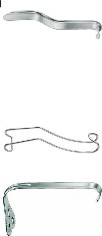
The Cawood-Minnesota retractor is a versatile instrument used by the surgeon during surgical extraction procedures (Figure 3.1). It is broad and flat, with one curved end, which can be used as a lip retractor, and one flat, pointed end, which is usually used to retract mucoperiosteal flaps. It is held in the nondominant hand, with the index and middle fingers on the front of the instrument
Principles of Dentoalveolar Extractions, First Edition. Seth Delpachitra, Anton Sklavos and Ricky Kumar. © 2021 John Wiley & Sons Ltd. Published 2021 by John Wiley & Sons Ltd.
Companion website: www.wiley.com/go/delpachitradentoalveolarextractions
https://t.me/DentalBooksWorld
36 3 Basic Surgical Instruments
Figure 3.1 Cawood-Minnesota retractor. Only a gentle finger grip is required in its use. Excessive grip strength reduces tactile feedback from the instrument, can strain the operator’s hand, and can lead to unnecessary soft tissue injury from the sharp tip of the retractor. Source: KLS Martin.
and the thumb positioned directly behind the fingers on the back. The sharp end should only be pressed against hard tissues, as excessive downward force against soft tissues can lead to mucosal or gingival lacerations.
A wire cheek retractor is used by the surgical assistant during oral surgical procedures, to retract the lips and improve surgical access (Figure 3.2). It is named for its construction from a heavy-gauge, stainless-steel wire that has been twisted to produce a curved retractor on either end. It is particularly useful during suturing, when the primary operator’s hands are both in use.
A tongue retractor is used only during general anaesthetic cases (Figure 3.3). It has a large, broad blade attached at 90° to the handle. It is excellent for displacement of the lingual soft tissues to improve surgical access, but is very uncomfortable for the awake patient, and can initiate a gag reflex.
Figure 3.2 Wire cheek retractor. This instrument is most commonly held by the surgeon’s assistant. One major limitation is its propensity to slip away from the oral cavity whilst in use. Holding it in the palm of the hand, with a gentle grip, and an outward and downward direction, helps to engage the lip and cheek and reduce retractor slippage. Source: KLS Martin.
Figure 3.3 Tongue retractor. This instrument is large and can damage the anterior teeth if used incorrectly. It should be inserted such that the blade is parallel to the occlusal plane; once inside the oral cavity, it may be rotated 90° to rest against the tongue. Source: KLS Martin.
https://t.me/DentalBooksWorld