

NEUROPHYSIOLOGY |
Synaptic Transmission: Temporal and Spatial Summation |
Temporal and Spatial Summation
of Excitation and Inhibition
Excitatory fibers |
mV |
|
–70 |
|
Axon |
Inhibitory fibers |
|
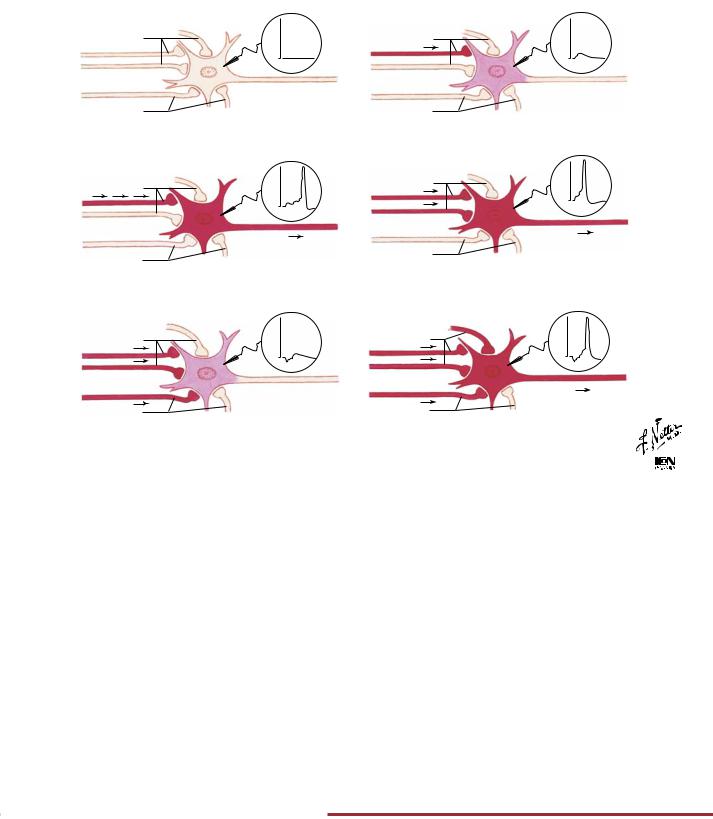
A. Resting state: motor nerve cell shown with synaptic boutons of excitatory and inhibitory nerve fibers ending close to it
Excitatory fibers |
mV |
|
–70 |
Axon
Inhibitory fibers
C. Temporal excitatory summation: a series of impulses in one excitatory fiber together produce a suprathreshold depolarization that triggers an action potential
Excitatory fibers |
mV |
|
–70 |
Axon
Inhibitory fibers
B. Partial depolarization: impulse from one excitatory fiber has caused partial (below firing threshold) depolarization of motor neuron
Excitatory fibers |
mV |
|
–70 |
Axon
Inhibitory fibers
D. Spatial excitatory summation: impulses in two excitatory fibers cause two synaptic depolarizations that together reach firing threshold triggering an action potential
Excitatory fibers |
mV |
|
–70 |
Axon
Inhibitory fibers
E. Spatial excitatory summation with inhibition: impulses from two excitatory fibers reach motor neuron but impulses from inhibitory fiber prevent depolarization from reaching threshold
Excitatory fibers |
mV |
|
–70 |
|
Axon |
Inhibitory fibers |
|
E. (continued): motor neuron now receives additional excitatory |
|
impulses and reaches firing threshold despite a simultaneous |
|
inhibitory impulse; additional inhibitory impulses might still |
|
prevent firing |
© |
CHART 2.1 SUMMARY OF SOME NEUROTRANSMITTERS AND WHERE WITHIN THE CENTRAL AND PERIPHERAL NERVOUS SYSTEM THEY ARE FOUND
Transmitter |
Location |
Transmitter |
Location |
Acetylcholine |
Neuromuscular junction, autonomic end- |
Gas |
|
|
ings and ganglia, CNS |
Nitric oxide |
CNS, GI tract |
Biogenic amines |
|
Peptides |
|
Norepinephrine |
Sympathetic endings, CNS |
-Endorphins |
CNS, GI tract |
Dopamine |
CNS |
Enkephalins |
CNS |
Serotonin |
CNS, GI tract |
Antidiuretic |
CNS (hypothalamus/posterior |
Amino acids |
|
hormone |
pituitary) |
-Aminobutyric |
CNS |
Pituitary-releasing |
CNS (hypothalamus/anterior |
acid (GABA) |
|
hormones |
pituitary) |
Glutamate |
CNS |
Somatostatin |
CNS, GI tract |
|
|
Neuropeptide Y |
CNS |
Purines |
|
||
Adenosine |
CNS |
Vasoactive |
|
Adenosine |
CNS |
intestinal peptide |
CNS, GI tract |
triphosphate (ATP) |
|
|
|
|
|
|
|
CNS, Central nervous system; GI, gastrointestinal.
FIGURE 2.9 TEMPORAL AND SPATIAL SUMMATION •
Neurons receive multiple excitatory and inhibitory inputs. Temporal summation occurs when a series of subthreshold impulses in one excitatory fiber produces an action potential in the postsynaptic cell (panel C). Spatial summation occurs when subthreshold impulses from two or more different fibers trigger an action poten-
tial (panel D). Both temporal and spatial summation can be modulated by simultaneous inhibitory input (panel E). Inhibitory and excitatory neurons use a wide variety of neurotransmitters, some of which are summarized here.
60

Cerebrospinal Fluid (CSF): Brain Ventricles and CSF Composition |
NEUROPHYSIOLOGY |
Left lateral phantom view
Right lateral ventricle
Left interventricular foramen (Monro)
3rd ventricle
CHART 2.2 CSF COMPOSITION
Frontal (anterior) horn |
|
|
Central part |
Left |
|
Temporal (inferior) horn |
lateral |
|
ventricle |
||
Occipital (posterior) horn |
||
|
Cerebral aqueduct (Sylvius)
4th ventricle
Left lateral aperture (foramen of Luschka)
Left lateral recess
Median aperture (foramen of Magendie)
Central canal of spinal cord
©

|
CSF |
Blood Plasma |
Na (mEq/L) |
140–145 |
135–147 |
K (mEq/L) |
3 |
3.5–5.0 |
Cl− (mEq/L) |
115–120 |
95–105 |
HCO3− (mEq/L) |
20 |
22–28 |
Glucose (mg/dL) |
50–75 |
70–110 |
Protein (g/dL) |
0.05–0.07 |
6.0–7.8 |
pH |
7.3 |
7.35–7.45 |
|
|
|
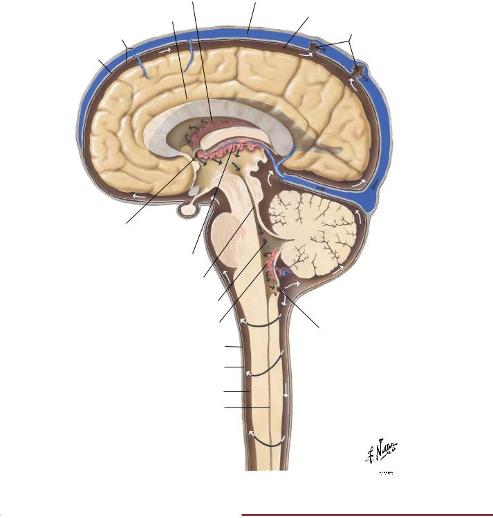
FIGURE 2.10 BRAIN VENTRICLES AND CSF COMPOSITION•
CSF circulates through the four brain ventricles (two lateral ventricles and a third and fourth ventricle) and in the subarachnoid space surrounding the brain and spinal cord. The electrolyte composition of the CSF is regulated by the choroid plexus, which
secretes the CSF. Importantly, the CSF has a lower [HCO3 ] than plasma and therefore a lower pH. This allows small changes in blood PCO2 to cause changes in CSF pH, which in turn regulates the rate of respiration (see Chapter 5).
61
NEUROPHYSIOLOGY |
Cerebrospinal Fluid (CSF): Circulation of CSF |
Choroid plexus of lateral ventricle (phantom)
Cistern of corpus callosum
Dura mater
Arachnoid
Interventricular foramen (Monro)
Choroid plexus of 3rd ventricle
Cerebral aqueduct (Sylvius)
Lateral aperture (foramen of Luschka)
Choroid plexus of 4th ventricle
Dura mater
Arachnoid
Subarachnoid space
Central canal of spinal cord
Superior sagittal sinus
Subarachnoid space
Arachnoid granulations
Median aperture (foramen of Magendie)
©

FIGURE 2.11 CIRCULATION OF CEREBROSPINAL FLUID•
CSF circulates through the four brain ventricles (two lateral ventricles and a third and fourth ventricle) and in the subarachnoid space surrounding the brain and spinal cord. Most of the CSF is
reabsorbed into the venous system through the arachnoid granulations and through the walls of the capillaries of the central nervous system and pia mater.
62
Spinal Cord: Ventral Rami |
NEUROPHYSIOLOGY |
Base of skull
C1 spinal nerve
C8 spinal nerve
T1 spinal nerve
1st rib
Intercostal nerves
T12 spinal nerve
12th rib
Conus medullaris
L1 spinal nerve
Cauda equina
S1 spinal nerve
Sacrum (cut away)
Termination of
dural sac
Coccygeal nerve
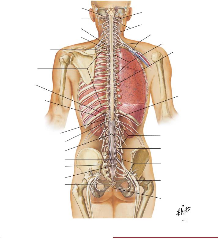
FIGURE 2.12 SPINAL CORD AND VENTRAL RAMI IN SITU•
C1 vertebra (atlas)
Cervical plexus
Brachial plexus
Spinal dura mater
Filaments of spinal nerve roots (T7 and T8)
L1 vertebra
Lumbar plexus
L5 vertebra
Sacral plexus
Sciatic nerve
Coccyx
©

The spinal cord gives rise to 31 pairs of spinal nerves that distribute segmentally to the body. These nerves are organized into plexuses that distribute to the neck (cervical plexus), upper limb (brachial plexus), and pelvis and lower limb (lumbosacral plexus). Motor
fibers of these spinal nerves innervate skeletal muscle, and sensory fibers convey information back to the central nervous system from the skin, skeletal muscles, and joints.
63