

6 курс / Судебная медицина / Морфологические_особенности_волос_человека_в_аспекте_судебно_медицинской
.pdfSUMMARY
T he -chapter "Structure of human hair" illustrates the structure of the hair cortical substance, core, root and peripheral ends.
The section "Deposition of pigment" describes hair with variable amounts,of
granular (figs 1-6) and diffuse (fig 7) pigments of various |
colours |
deposited |
centrally |
(fig 7), uniformly (figs 1-5) or peripherally (fig 6). |
|
|
|
The section "Colour and amount of pigment" contains illustrations of hair of |
|||
different colour: blonde (fig 8), light fair (fig 9), fair (figs |
10-12, |
22), dark |
fair (figs |
13-16), black (figs 17-19) and red (figs 20, 21) containing pigment in the form of: fine granules (figs 8, 21), fine and middle-sized granules (figs 9, 22, 23), middlesized granules (figs 10, 11, 15), middle-sized and large granules (figs 12-19), large granules (fig 14). The colour of pigment varies: light brown (fig 8), light brown with reddish tint (fig 21), brown (figs 9-11, 13, 14, 22), brown with reddish tint (fig 12), dark brown (figs 15, 16, 18, 19), black (figs 17-19), red (figs 20, 22, 23). No pigment was found in gray hair (fig 24). The illustrations show pigmentophores of fine, middle and large size and of various forms: rounded oval, fusiform, with or without processes, distributed evely all over the thickness of the hair or predominently in its periphery (figs 25, 26).
The section "Core of the hair" shows that some hairs are devoid of the core (fig 27); in the others it constitutes from V7-V8 to '/г-'/з of the hair thickness (figs 27-32). In individual hairs the core looks as an intermittent irregular cord of uneven contour (figs 27-30), or as uninterrupted cord (figs 28-32). The core is located mostly centrally (figs 27-32), sometimes it appears as a double cord (fig 32).
The sections "Peripherla ends" and "Root ends" show that the surface of hair cutting may be: a) even (figst 33, 38), with fine tubercles (fig 34), with large tubercles (figs 35, 36), enlarged in its diameter (fig 37), stepped (fig 39); the hair end may be split broom-like (fig 40) or needle-thinned (fig 41). b) The bulb may be dead (fig 42), damaged and pick-shaped (fig 43), may have sheaths (fig 43), be devoid of sheaths (figs 45, 46) or have sheath remnants (figs 45, 46).
The chaptes "Hairs of different parts of the body" contains illustrations of all kinds of human hair.
Long hair of the head: red (figs 47, 48), fair (figs 49-52), dark fair (figs 53, 54), getting gray (figs 55, 56). In some of the hairs the core amounts to '/s-1/? of their thickness. The cortical substance contains diffuse (figs 47, 48) and granular (figs 49-56) pigments of various colour and amount, deposited centrally (figs 47, 48), uniformly (figs 49-55) or peripheralle (fig 56). The root ends are dry cylindrical and flask-shaped bulbs. The peripheral ends have transversal and oblique surfaces of separation (fig 57), may acquire the form of truncated sone; they are thinned and polished (fig 58).
Long hair of the face
Hair of the beard: black (figs 60, 61), red (fig 62), fair (fig 63), dark fair (figs 64, 65). In the majority of hairs the core is of broken controur and constitutes '/з-'/б °f their thickness, located centrally (figs 60-64) and excentrally (figs 65, 66). The cortical substance contains various quantitites of pigment, the colour of which varies from light brown to black, distributed uniformly (figs 60-62, 65, 66) and in some hairs predominantly centrally (figs 63, 64). In individual hairs the amount of pigment in one half of the hain exceeds that in the other (figs 60, 61, 64). The root end is a rich hooked bulb (fig 60); the peripheral ends are polished (fig 62) (traces of old hair-cutting).
Hairs of the moustache: red (figs 67, 68), fair (figs 69, 74, 75), light fair (fig 70). In the majority of hairs the core is situated centrally (figs 68, 70, -74, 75) or excentrally (figs 67, 69, 71): in some hairs it appears as a double cord (figs 68, 70, 71) of broken contour. The cortical substance contains different amounts of diffuse (fig 69) and granular (figs 67, 69-75) pigments deposited uniformly (figs 69-75) or centrally (figs 67, 68, 70, 73). In some cases there is more pigment in one half of the hair than in the other (figs 69, 71, 73, 74). Along the core of some hairs there are many cavities and
136
cracks |
in |
the cortical substance. The peripheral ends are polished (traces of |
old |
hair-cutting). |
|
||
Hairs |
of the whiskers: fair (figs 78, 79, 83, 84), dark fair (figs 80, 85), light fair |
||
(fig 86), |
black (fig 82) and getting gray (fig 81). In the majority of hairs the core |
||
equals |
to |
'Ar'/e OI their thickness and is located both centrally (figs 78-82. 84-86) |
and |
excentrally (fig 83). The cortical substance contains various amounts of granular pigment deposited uniformly (figs 78-85) or predominently peripherally (fig 86). In some cases the amonut of pigment in one half of the hair exceeds that in the other (figs 78, 80, 84). In the cortical substance of some hairs there are many cavities and cracks. The peripheral ends are thinned (fig 79) or rounded and polished (fig 85).
Short thick hairs of the face
Hairs of the eyebrows: the core of the hairs constitutes from 1/3 to Ve of their thickness. The cortical substance contains diffuse (fig 87) and granular (figs 88, 91-96) pigments deposited uniformly (figs 88, 91-96) and centrally (fig 87). The amount of pigment varies along the length of the hair, being the greatest in the middle part of the shaft. The cortical substance in some hairs exhibits many cavities and cracks. The peripheral ends are thinned (figs 88, 90-96) with markedly pronounced dentation of the optical margin; the root ends are dry flask-shaped bulbs (figs 88-91).
Hairs of the eyelids (eyelashes) the core of the hairs constitutes from '/з to '/7 of their thickness. The cortical substance contains the granular pgiment deposited uniformly (figs 97-101) and, in various amounts, along the length of the hairs, being the greatest in the middle parts of the latter. The hair apices are thinned, optical margins are clearly dentated; the root ends are dry bulbs of cylindrical forms (fig 97).
Hairs of the nostrils: the cortical substance (fig 102) is yellowish, individual pigmental granules are poorly discernible; sometimes there is more pigment in one half of the hair than in the other. The cuticle is damaged and shows some deposits; the root ends (fig 103) are dry cylindrical bulbs; the apices are thinned.
Long hairs of the torso
Hairs of the armpit: the core of some hairs equals to V6-V7 of their thickness (figs 105-108). The cortical substance contains various of the granular pigment evenly distributed (figs 105-107), or with predominent peripheral arrengement (fig 108). In one case (fig 108) the amount of pigment in one half of the hair exceeds that in the other. The root ends are cylindrical and piriform bulbs (figs 104, 109, 111); the peripheral ends are thinned. On the hair cuticle there are extensive homogenous
(structureless) deposits (figs 112, 113); the cuticle is split. |
|
|
|
|
||||
Hairs |
of the perenium and pubis: the core of the |
hair shafts (figs |
114, |
115, |
||||
119-123) |
equals to |
' / 3 - V 7 |
of the |
hair thickness. The |
cortical |
substance contains |
||
different |
amounts of |
the |
granular |
pigment distributed |
uniformly |
(figs |
114, |
115, |
117-119) or mostly peripherally (fig |
116). In individual hairs the amount of pigment in |
|||||||
one half of the hair exceeds that in the other (figs 119, 120). In some hairs (figs |
118, |
|||||||
121) pigmental granules are poorly discernible (urine action). On the cuticle there are homogenous (structurelless) deposits (fig 122). The root ends are bulbs of hooked (fig 116) and cylindrical (fig 119) forms; the peripheral ends are thinned (figs 117, 119).
Hairs of the breast: the core of the hair shafts (figs 125, 126) constitutes V6-V7 of the hair thickness. The cortical substance contains various amounts of the granular pigment distributed uniformly (figs 125-127), gray hair contains no pigment (fig 131). The root end is a dry flask-shaped bulb (fig 129); the peripheral ends are thinned and split (figs 130, 132).
Hairs of the abdomen: the core of the hair shafts constitutes '/V'/s of the hair
thickness (fig 128). The cortical |
substance contains various amounts of the granular |
Pigment distributed uniformly (figs |
128, 131, 132) and peripherally (fig 130); gray hair |
ls devoid of pigment (fig 131); the root ends are dry flask-shaped bulbs (fig 129); the |
|
Peripheral ends are thinned and |
split (figs 130, 132). |
137
Short hairs of the body
Hairs of the arm: the core constitutes V5 - V7 of the hair thickness (figs 133, 135 136, 138). In the cortical substance there are various amounts of the granular pigment uniformly distributed (figs 133-139). No pigment is in the gray hair. In some cases,(fig 134) the amount of pigment in one half of the hair exceeds that in the other. Th e root ends are piriform bulbs with defects at the site of the papilla attachment; the apices are thinned (figs 133, 136, 138) and polished.
Hairs of the leg: the core constitutes Vs-'/e of the hair thickness. Th e cortical substance contains various amounts of the granular pigment deposited uniformly (figs 143, a, b, 144) and peripherally (figs 141, 143, b); in some sases (fig 141) there is more pigment in one half of the hair than in the other. In gray hair pigment is absent. The root end (fig 145) is a bulb of cylindrical form.
Hairs of the back: the core of the hair shafts amounts to Vs-'/e of the hair thickness (fig 146). Th e cortical substance contains the granular pigment distributed uniformly and peripherally (figs 146; 147, 148). Th e hair apices are thinned (figs 147 149); the root ends are dry cylindrical bilbs (fig 150).
The downy hair (lanugo): the core of the hair shaft consists of islets (fig 152). The cortical substance has granular uniformly distributed pigment (figs 153, 157) the amount of which markedly diminishes towards the apex. Th e peripheral ends are thinned, the optical margin is clearly dentated; the root ends are dry cylindrical (fig 154) or fusiform (fig 155) bulbs.
Imprints of hair cuticles of: long hair of the head (figs 205-210), moustache (figs 211-213), beard (figs 215, 216), eyelids (figs 217, 218), eyebrows (figs 219, 220), armpit (figs 221-223), pubis (figs 224, 225), leg (figs 226-228).
Cross sections of human hair: long hair of the head (figs 158-163), eyebrows (164-167), eyelids (figs 168, 169), moustache (figs 170-176), beard figs 177-181), pubis (figs 182-190), armpit (figs 191-195), leg (figs 196, 197), breast (figs 198-201) arm (fig 202) and back (figs 203-204).
Changes in hair peripheral ends depending upon the time of haircutting: 3 days (fig 205), 8 days (fig 206), 9 days (figs 207, 208), 16 days (fig 209), 21 days (fig 210), one month (fig 211), two months (fig 212), six months (fig 213),
1 38

Глава 2
ПОВРЕЖДЕНИЯ ВОЛОС (рис. 241—362)
МЕХАНИЧЕСКАЯ ТРАВМА
Отделение волос острым предметом
Рис. 241
Отделение волос бритвой: поперечная ровная поверхность сечения с острыми краями.
Рис. 241 ( в а р и а н т ) .
139

Рис. 242
Отделение волос скальпелем: косая и поперечная крупно- и мелкозернистая поверхность сечения; концы некото рых волос слегка расширены в попе речнике; края отделения острые.
Рис. 242 ( в а р и а н т ) .
Рис. 243
Отделение волос острым ножом: косая среднебугристая поверхность сечения с острыми краями, конец одного волоса слегка расширен в поперечнике.
110
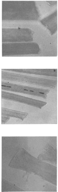
Рис. 243 ( в а р и а н т ) .
Рис. 244
Отделение волос ножом: концы расши рены в поперечнике, имеют попереч ную и косую крупнобугристую повер хность сечения, а местами — с выступа ми коркового вещества.
Рис. 244 (в а р и а н т ) .
I l l
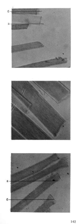
Рис. 245
Отделение волос в тонком пучке острыми ножницами: косая и попереч ная мелкобугристая поверхность сече ния с острыми краями. У волос (а) и
(б) видны клиновидные выступы кор кового вещества; у одного волоса на поверхности отделения виден дефект в корковом веществе.
Рис. 245 ( в а р и а н т ) .
Рис. 246
Отделение волос в тонком пучке тупы ми ножницами: концы расширены в поперечнике с небольшими продоль ными трещинами в корковом веществе. Поверхность отделения поперечная, мелко- и крупнобугристая с выступами коркового вещества; кутикула у волос
(а) и (б) разволокнена и отщеплена от стержня волоса.

Рис. 246 ( в а р и а н т ) .
Рис. 247
Отделение волос острым скальпелем: поперечная мелкобугристая повер хность сечения с острыми краями; у волоса (а) поверхность отделения поч ти ровная.
143

Отделение волос тупым предметом
3 Рис. 248
Отделение волос ребром тупогранного предмета: концы расширены в попе речнике, в корковом веществе видны продольные трещины, поверхность от деления крупнобугристая, косая, попе речная, с острыми краями. В стержне волоса (а) видно веретенообразное расширение с небольшими поперечны ми трещинами в корковом веществе.
Рис. 248 ( в а р и а н т ) .
Рис. 249
Отделение волос колуном: поперечни ки волос расширены; в корковом веще с т в е — продольные трещины; в стержнях видно веретенообразное рас ширение с частичным разрушением коркового вещества, а также продоль ные и поперечные трещины.
144

с. 249 ( в а р и а н т ) .
с. 250
тделение волос колуном: концы волос езко расширены в поперечнике, поерхность отделения неровная с проольными трещинами в корковом веестве; стержни тоже расширены в оперечнике и имеют трещины в кор овой веществе.
с. 250 ( в а р и а н т ) .
)* 3699