

5 курс / Пульмонология и фтизиатрия / Pulmonary vascular disease
.pdfNOTES
NOTES
PULMONARY VASCULAR
DISEASE
GENERALLY, WHAT IS IT?
PATHOLOGY & CAUSES |
DIAGNOSIS |
▪ Diseases affecting blood fl ow through |
X-ray, chest CT scan, spirometry, ultra- |
pulmonary vasculature, or fl uid fl ow from |
sound, echocardiogram, ECG |
vasculature |
|
▪ Can be caused by process within lungs/ |
TREATMENT |
elsewhere in body |
|
SIGNS & SYMPTOMS |
▪ Supportive, treat underlying disease, |
optimize organ function (heart, lungs) |
▪Dyspnea, poor effort tolerance, chest pain, tachypnea
Figure 129.1 Chronic thromboembolic pulmonary hypertension is an example of a pulmonary vascular disease that originates outside the lungs. In this case, an embolism blocks the pulmonary vessels, causing pulmonary blood pressure to rise beyond normal levels.
912 OSMOSIS.ORG

Chapter 129 Pulmonary Vascular Disease
PULMONARY EDEMA
osms.it/pulmonary-edema
PATHOLOGY & CAUSES
▪Alteration in Starling forces → build up of fl uid within interstitial space, air spaces of
lung
CAUSES
Cardiogenic (heart disease)
▪Left sided heart failure → ineffi cient pumping of blood from heart by left ventricle → blood backs up into left atrium
→pulmonary circulation → pulmonary hypertension (raised hydrostatic pressure)
→more fl uid in lung interstitium → pulmonary edema
▫Severe systemic hypertension
(> 180/110mmHg) → left ventricle cannot pump effectively against extreme afterload → blood backs up into left atrium → pulmonary circulation → pulmonary edema
Non-cardiogenic (damage to pulmonary capillaries or alveoli)
▪Direct damage to alveoli/vasculature → infl ammatory response → leaky capillaries
▫Pulmonary infection, toxin inhalation, chest trauma, pulmonary vein occlusion, burns
▫Sepsis → systemic infl ammation → global edema
▫Insuffi cient circulation of osmotically active proteins, e.g. albumin → low oncotic pressure in capillaries
▪Malnutrition
▪Liver failure
▪Excessive protein loss (nephrotic syndrome, protein losing enteropathies)
COMPLICATIONS
▪Impaired gas exchange: oxygen/carbon dioxide must diffuse through wide layer of fl uid → blood unable to fully saturate
▪Free fl uid predisposes to secondary infection
SIGNS & SYMPTOMS
▪Dyspnea, productive cough (pink frothy sputum), excessive sweating, anxiety, tachycardia, end-inspiratory crackles, dullness to percussion, cyanosis (decreased hemoglobin saturation)
▪Pulmonary edema in heart failure may also include
▫Orthopnea (shortness of breath worse when lying fl at)
▫Paroxysmal nocturnal dyspnea (episodes of severe sudden breathlessness at night)
▫Peripheral pitting edema
▫Raised jugular venous pressure
▫Hepatomegaly
DIAGNOSIS
DIAGNOSTIC IMAGING
Chest X-ray
▪Kerley B lines (thickened subpleural interlobular septa, usually seen at base of lung)
▪Increased vascular shadowing → batwing perihilar pattern
▪Upper lobe diversion (prominent upper lobe pulmonary veins)
▪Pleural effusion (if edema severe)
Non-contrast high resolution chest CT scan
▪Airspace opacity
▪Smooth thickening of interlobular septae
Chest ultrasound
▪Detection of small amounts of fl uid
▪Echo-free space between visceral and parietal pleura
▪Septations in pleural fl uid → underlying
OSMOSIS.ORG 913
infection, chylothorax/hemothorax
Echocardiograph
▪Evaluation of cardiac function, can demonstrate left ventricular failure
LAB RESULTS
▪Serum electrolytes
▪Renal function
▪Infl ammatory markers
▪Low oxygen saturation
▪Increased carbon dioxide
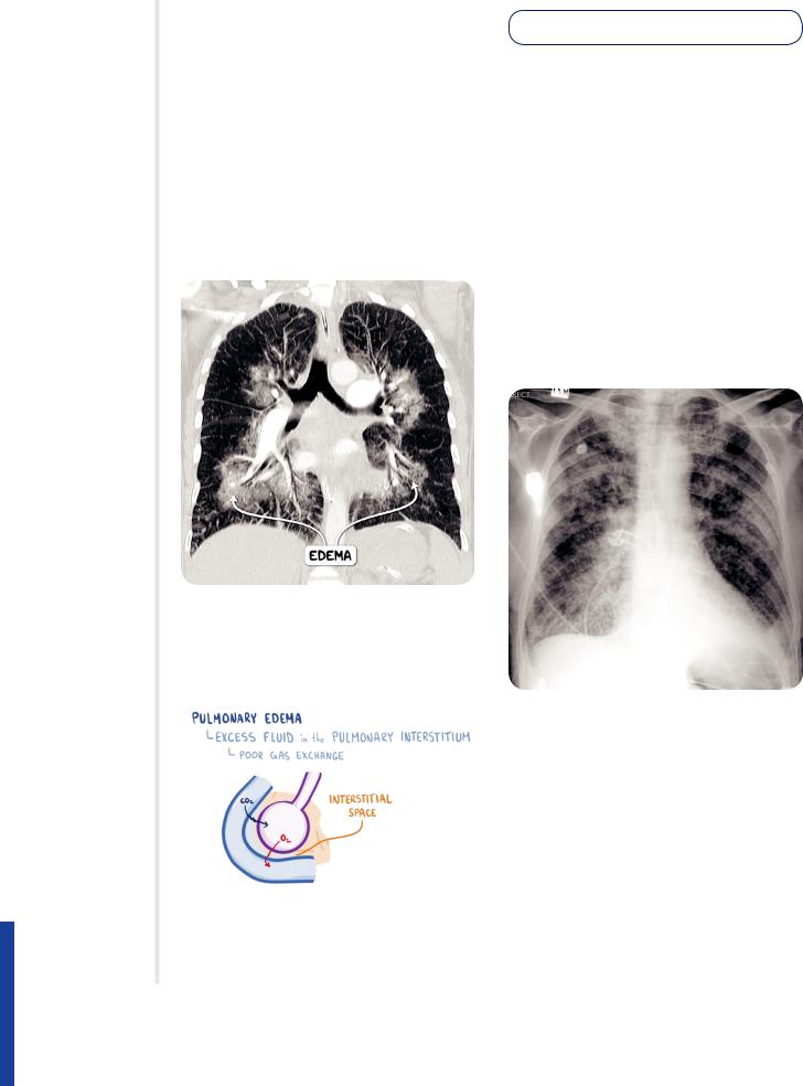
Figure 129.2 A CT scan of the chest in the coronal plane demonstrating the
peribronchovascular distribution of acute pulmonary edema.
Figure 129.4 Illustration depicting pulmonary edema.
TREATMENT
MEDICATIONS
▪If cardiogenic
▫Preload reduction: nitroglycerin, diuretics, morphine sulphate
▫Afterload reduction: ACE inhibitors, angiotensin II receptor blockers, nitroprusside
▪If non-cardiogenic
▫Manage illness (e.g. treat infection)
OTHER INTERVENTIONS
▪Continuous positive airway pressure (CPAP)
▪Intubation: mechanical ventilation if level of consciousness compromised
Figure 129.3 A plain chest radiograph demonstrating pulmonary edema. There is interstitial edema, represented by fi ne stranded opacities known as Kerley B lines, as well as alveolar edema, represented by confl uent nodular opacities.
914 OSMOSIS.ORG

Chapter 129 Pulmonary Vascular Disease
Figure 129.5 The histological appearance of pulmonary edema.
PULMONARY EMBOLISM
osms.it/pulmonary-embolism
PATHOLOGY & CAUSES
▪Blockage of pulmonary artery by a substance brought there via bloodstream
▪Thrombus in remote site embolizes → lodges in pulmonary vascular tree → “pulmonary embolism”
▪Obstruction of blood fl ow distal to embolism → increased pulmonary vascular resistance → increased pulmonary artery pressure → increased right ventricular pressure → cor pulmonale (if severe obstruction)
▪Regional decrease in lung perfusion → dead space (ventilation, but no perfusion) → hypoxemia → tachypnea
Source of embolus
▪Lower extremity deep vein thrombosis
▫Most arise from deep veins above knee, iliofemoral deep vein thrombosis
▫Can arise from pelvic deep veins
▫Pelvic thrombi tend to advance to more proximal veins before embolizing
▪Upper extremity deep veins (rarely)
▪Uncommon embolic material: air, fat, amniotic fl uid
RISK FACTORS
▪Virchow’s triad: endothelial injury, stasis of blood fl ow, blood hypercoagulability
▪> 60 years old, malignancy, history of deep vein thrombosis/pulmonary embolism, hypercoagulable states, genetic disorders (e.g. Factor V Leiden thrombophilia), dehydration, prolonged immobilization (bed rest, travel), cardiac disease, obesity, nephrotic syndrome, major surgery, trauma, pregnancy, estrogen-based medication (e.g. oral contraceptives)
▪Increased risk of fat embolism with bone fractures (e.g. hip, femur)
SIGNS & SYMPTOMS
▪Dyspnea, pleuritic chest pain, cough, hemoptysis
▪Signs, symptoms of deep vein thrombosis
▫Tender, swollen, erythematous extremity
▪Syncope
▪Often asymptomatic (in the case of small emboli)
OSMOSIS.ORG 915
MNEMONIC: TOM
SCHREPFER
Risk factors for Pulmonary embolism
Trauma
Obesity
Malignancy
Surgery Cardiac disease Hospitalization Rest (bed-ridden) Elderly
Past history Fracture
Estrogen (pregnancy, postpartum)
Road trip
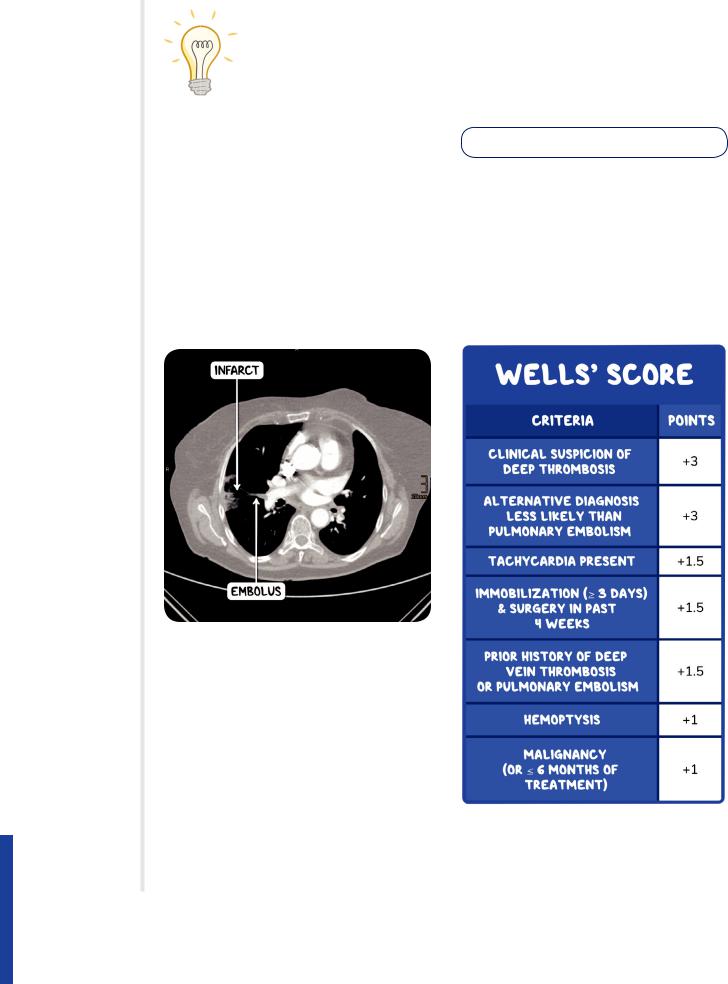
Figure 129.6 A CT pulmonary angiogram demonstrating a pulmonary embolus and subsequent right middle lobe infarct.
▪Low SpO2, tachypnea, rales, tachycardia, S4 heart sound, increased P2 (closure of pulmonary valve), shock, low-grade fever, decreased breath sounds, percussion dullness, pleural friction rub, sudden death (pulmonary saddle embolism)
DIAGNOSIS
Wells’ score
▪Used to assess probability of pulmonary embolism (multiple different probability tests available)
▫Score > 4: pulmonary embolism likely, consider diagnostic imaging
▫Score ≤ 4: pulmonary embolism unlikely, consider D-dimer test to rule out
916 OSMOSIS.ORG

DIAGNOSTIC IMAGING
Chest X-ray
▪ Typically normal
CT pulmonary angiography
▪Defi nitive test
▪Visualize decreased blood supply
Venous duplex ultrasound
▪Of lower extremities
▫May reveal origin of pulmonary embolism
▫Negative result does not exclude pulmonary embolism
Ventilation-perfusion scan
▪ Normal scan rules out pulmonary embolism
LAB RESULTS
▪D-dimer (high negative predictive value)
▫Positive result does not prove pulmonary embolism
▫Negative result rules out pulmonary embolism
▪Arterial blood gas
▫↓ PaO2 → hypoxemia
▫Hyperventilation → ↑ PaCO2 → ↑ pH → respiratory alkalosis
▫A-a gradient elevated (indicated V/Q mismatch)
▪Tests for causes of secondary pulmonary embolism
▫Full blood count, clotting profi le, erythrocyte sedimentation rate, renal function, liver function, electrolytes
OTHER DIAGNOSTICS
ECG
▪Excludes other causes of chest pain
▪ECG features of pulmonary embolism (or any pulmonary hypertension) include
▫Sinus tachycardia
▫Right bundle branch block
▫Right ventricular strain pattern: T wave
inversion in right precordial (V1–V4), and inferior leads (II, III, aVF)
▫Right atrial enlargement (P pulmonale)
▫Right atrial dilatation → right axis deviation
Chapter 129 Pulmonary Vascular Disease
▫Dominant R wave in V1
▫S1Q3T3 pattern: Deep S wave in lead I, Q wave in lead III, negative wave in lead
III
▪Nonspecifi c ST segment, T wave changes
▪Pulmonary embolism can be excluded if
▫SaO2 exceeds 95%
▫Age < 50
▫No unilateral leg swelling, hemoptysis, history of deep vein thrombosis/ pulmonary embolism, recent surgery/ trauma, hormone use (or estrogenbased medications), tachycardia
TREATMENT
MEDICATIONS
Anticoagulation
▪Acute phase (days–weeks)
▫Prevent further thromboembolic events
▫Unfractionated heparin, low molecular weight heparin, fondaparinux
▪Long-term (vitamin K antagonists)
▫Warfarin, acenocoumarol, phenprocoumon
Thrombolysis
▪Used for massive pulmonary embolism causing hemodynamic instability
▪Carries risk of secondary hemorrhage
▪Thrombolytics used to break up clots
▫Streptokinase, staphylokinase, urokinase, anistreplase
▫Recombinant tissue plasminogen activators (alteplase, reteplase, tenecteplase)
SURGERY
Pulmonary thromboendarterectomy
▪Surgical removal of a chronic thromboembolism
▪Rare
Inferior vena cava fi lter
▪Vascular fi lter inserted into inferior vena cava to prevent life-threatening pulmonary emboli
▪Indications: anticoagulant therapy contraindicated, major embolic event
OSMOSIS.ORG 917

despite anticoagulation
OTHER INTERVENTIONS
Preventative measures
▪Unfractionated heparin, low molecular weight heparin
▪Factor Xa inhibitor
▪Long-term low-dose aspirin
▪Anti-thrombosis compression stockings/ intermittent pneumatic compression
Figure 129.7 A plant chest radiograph of the same individual, demonstrating the pulmonary infarct which is visible as a wedge shaped opacity in the lateral art of the right lung fi eld.
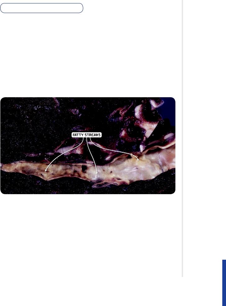
Figure 129.8 The gross pathological appearance of a pulmonary embolus.
Figure 129.9 The ECG changes associated with a pulmonary embolism. There is a right bundle branch block, sinus tachycardia and T-wave inversions in leads V1-3 and III.
918 OSMOSIS.ORG

Chapter 129 Pulmonary Vascular Disease
PULMONARY HYPERTENSION
osms.it/pulmonary-hypertension
PATHOLOGY & CAUSES
▪Increased blood pressure in pulmonary circulation
▪Mean pulmonary arterial pressure > 25mmHg (normal ~15mmHg)
▪Pulmonary hypertension → excess fl uid in pulmonary interstitium (pulmonary edema)
→impaired gas exchange
▪Pulmonary hypertension → strain on right heart → hypertrophy → right heart oxygen demand eventually exceeds supply → right-sided heart failure
▫Right heart failure caused by lung disease → cor pulmonale → backup of blood in venous system → signs, symptoms of right heart failure
▪Raised jugular venous pressure
▪Fluid build up in liver → hepatomegaly
▪Fluid build up in legs → leg edema
▪Left ventricle receives less blood → compensation → pumps harder, faster (tachycardia)
TYPES
Group I
▪Pulmonary arterial hypertension, pulmonary veno-occlusive disease, pulmonary capillary hemangiomatosis
▪Abnormal increase in pulmonary arteriolar resistance → increased strain on right heart (pumping fl uid through narrower pipe)
▪Damage to endothelial cells lining pulmonary arteries → release of endothelin-1 serotonin, thromboxane, produce less nitric oxide and prostacyclin → constriction of arterioles, hypertrophy of smooth muscle → pulmonary hypertension
▪Over time affected vessels become stiffer, thicker (fi brosed) due to vasoconstriction, thrombosis, vascular remodeling → greater increase in blood pressure in lungs, more strain on right heart
▪Idiopathic, inherited, drug/toxin associated causes connective tissue disease, HIV infection, portal hypertension congenital heart disease (shunting)
Group II
▪Pulmonary hypertension secondary to left heart disease
▪Pulmonary hypertension due to left heart disease (heart failure, valvular dysfunction)
→left heart fails to pump blood effi ciently
→backup of blood in pulmonary veins, capillary beds → increased pressure in pulmonary artery → pulmonary edema, pleural effusion
▪Raised back pressure may trigger secondary vasoconstriction → increased right heart strain
▪Common causes include
▫Left ventricular systolic/diastolic dysfunction
▫Valvular heart disease
▫Congenital/acquired in/out-fl ow tract obstruction
▫Congenital cardiomyopathy
▫Pulmonary venous stenosis
Group III
▪Pulmonary hypertension due to lung disease/chronic hypoxia
▪Low oxygen levels in alveoli pulmonary arteries constrict
▪Chronic lung disease → region of diseased lung → ineffi cient/total lack of gas exchange → hypoxic vasoconstriction (pulmonary arterioles) → shunting of blood away from damaged areas
▪Prolonged alveolar hypoxia across wide portion of pulmonary vascular bed → increase in pulmonary arterial pressure → thickening of pulmonary vessel walls → greater effort required from right heart → sustained pulmonary hypertension
▪Causes include
▫COPD
OSMOSIS.ORG 919

▫Interstitial lung disease
▫Mixed restrictive/obstructive pattern disease
▫Sleep-disordered breathing
▫Alveolar hypoventilation
▫Chronic exposure to high altitude
Group IV
▪Chronic arterial obstruction/ thromboembolic disease
▪Recurrent blood clots in pulmonary vasculature
▪Blockage/narrowing of pulmonary vessel with unresolved obstruction (e.g. clot)
→increased pressure, shear stress (turbulence) in pulmonary circulation
→vessel wall remodelling → sustained pulmonary hypertension
▪Causes endothelium to release histamine, serotonin → constriction of pulmonary arterioles → rise in pulmonary blood pressure → chronic thromboembolic pulmonary hypertension
▪Other causes of arterial obstruction
▫Angiosarcoma, arteritis, congenital pulmonary artery stenosis, parasitic infection
Group V
▪Unclear/multifactor mechanisms
▪Hematologic disease (e.g. hemolytic anemia)
▪Systemic disease (e.g. sarcoidosis, vasculitis)
▪Metabolic disorders (e.g. glycogen storage disease, thyroid disease)
▪Other (e.g. microangiopathy, chronic kidney disease)
RISK FACTORS
▪Family history, prior pulmonary embolic events, HIV/AIDS, sickle cells disease, cocaine use, COPD, sleep apnea, living at high altitude, mitral valve pathology
SIGNS & SYMPTOMS
▪Dyspnea, syncope, fatigue, chest pain, poor effort tolerance, loss of appetite, lightheadedness, orthopnea (left-sided heart failure)
▪Tachycardia, cyanosis, parasternal heave
▪Signs of systemic congestion/right heart failure:
▫Loud pulmonic component of second heart sound (P2)
▫Jugular venous distension
▫Ascites
▫Hepatojugular refl ux
▫Lower limb edema
DIAGNOSIS
DIAGNOSTIC IMAGING
Chest X-ray
▪Enlarged pulmonary arteries
▪Lung fi elds may or may not be clear, dependent on underlying cause
Echocardiogram
▪Increased pressure in pulmonary arteries, right ventricles → dilated pulmonary artery
▪Dilatation/hypertrophy of right atrium, right ventricle
▪Large right ventricle → bulging septum
Ventilation/perfusion scan
▪Identity / exclude ventilation-perfusion mismatches
OTHER DIAGNOSTICS
Right heart catheterisation (gold standard)
▪Catheter into right heart → most accurate measure of pressures
ECG
▪Right heart strain pattern: T wave inversion
in right precordial (V1–V4), and inferior leads (II, III, aVF)
Spirometry
▪ Unidentifi ed underlying cause
920 OSMOSIS.ORG
TREATMENT
MEDICATIONS
▪Pulmonary hypertension secondary to left ventricular failure → optimize left ventricular function
▫Diuretics (cautiously—individuals may be preload dependent)
▫Digoxin
▫Anticoagulants
▪Cardiogenic pulmonary arterial hypertension
▫Relax smooth muscle (promote vasodilation), reduce vascular remodelling, improve exercise capacity
Chapter 129 Pulmonary Vascular Disease
with prostanoids, phosphodiesterase inhibitors, endothelin antagonists
▪Pulmonary arterial hypertension
▫Endothelin receptor antagonists
▫Prostanoids
SURGERY
▪Lung transplant
▪Repair/replace damaged valves to optimize left ventricular function
Figure 129.10 The gross pathological appearance of the pulmonary arteries in a case of pulmonary hypertension. The underlying pathological process is similar to atherosclerosis found elsewhere in the cardiovascular system.
OSMOSIS.ORG 921