

4 курс / Лучевая диагностика / ЛУЧЕВАЯ_ДИАГНОСТИКА_И_ЛУЧЕВАЯ_ТЕРАПИЯ
.pdfA B
B  C
C  D
D 
Figure 1.3 Inflammatory polyp (A, B arrows) and oesophageal carcinoma (C, D).
(A) Spot radiograph of the esophagus performed while the patient stands and drinks high-density barium shows a column of barium (with an air-barium level, open arrow) in the mid-esophagus. A region of focal circumferential narrowing, 3 cm in length, is present. The narrowing appears smooth on one wall (white arrow) and has an irregular contour on the opposite wall (black arrows).
(B) Spot radiograph performed seconds later after the barium column has passed through the esophagus shows the same circumferential narrowing with a smooth contour on one side and an irregular contour on the other. The mucosal surface is now seen en face as well, however, revealing focal mucosal nodularity (open arrow) and bariumcoated lines (arrows) that disrupt the normal smooth surface of the esophagus and out-
line the outer margin of a mass.
(C) Early oesophageal carcinoma. En face there is a central ulcer (asterisk) with a nodular margin (arrows).
(D) Esophageal carcinoma, malignant stricture. A squamous cell carcinoma of the midesophagus causes an abrupt narrowing with irregular mucosa. Note the irregularity and overhanging edges (arrows)
A B
B C
C D
D
Figure 1.4 Schemes of esophagograms variants of esophageal tumours.
(A) Infiltrative, (B) polypoid, (C) annular stenotic, (D) ulcerative
On contrast studies, esophageal cancer may appear as an infiltrating, polypoid, ulcerative. Infiltrating cancers show irregular narrowing of the lumen with an associated nodular or ulcerated mucosa with well-defined borders. Polypoid lesions are usually greater than 3,5 cm in diameter and appear as lobulated or fungating intraluminal masses with possible areas of ulceration. Ulcerative carcinomas appear as well-defined meniscoid ulcers with a radiolucent rim of tumor surrounding the ulcer.
11

2. RADIOLOGY OF STOMACH
Introduction
The stomach are not well seen on plain x-ray films. In order to examine well it is necessary to swallow a barium fluid that can be seen on a fluoroscope screen and on the x-ray films. To X-ray anatomy of stomach: fornix, cardial part, small curvature, large curvature, body of the stomach, angle of the stomach, antral part, pyloric canal.
Single-contrast radiographic study: the stomach is filled and distended with dilute barium or a water-soluble contrast agent. Water-soluble contrast should be used when perforation or post-operative anastomotic failure is suspected. The stomach is compressed either manually or by positioning to allow for adequate x-ray penetration in the evaluation of each anatomical segment. Single-contrast technique assesses thickness of the gastric folds and evaluation of gastric emptying. Large luminal defects can be detected.
Double-contrast radiographic study: the stomach is temporarily paralyzed by administration of glucagon, filled with dense barium, and distended with gas using effervescent granules. Hence both barium and air are used for contrast. Images are obtained as the patient rolls in various positions to coat the gastric mucosa with contrast. Double-contrast technique provides improved visualization of the gastric mucosa. Several films may be made during this time (figure 2.1).
Limitation: requires fluoroscopy and requires follow up endoscopy to biopsy lesions.
Patient preparation: the patient should have nothing orally after midnight or the next morning preceding the radiographic study (no smoking or chewing gum). Fluid and food in the stomach degrade the examination by interfering with good mucosal visualization and causing artifacts that may mimic disease.
A  B
B 
Figure2.1 (A)Normalstomach.1—gastricfundus,2—gastricbody,3—gastricantrum,
4—pylorus(pyloricchannel),5—duodenalbulb,and6—secondportionofduodenum.
(B)isadouble-contrastimageofthedistalstomach.Rugalfoldsareseenasradiolucentfilling defectsinthebariumpool(whitearrow)andasparallelbariumetchedlines(blackarrows). Rugalfoldsarecomposedofmucosaandsubmucosaandaremostprominentalongthe greatercurvatureofthestomach.Thenormalgastricantrum(A) hasfew,ifany,rugalfolds inmostpatients
12

Magnetic resonance and CT imaging
Magnetic resonance (MR) imaging is the newest modality developed for cross-sectional imaging of the body and nearly all organ systems can be evaluated with this technique. MR imaging of the hollow organs of the gastrointestinal tract is increasingly being used to evaluate a wide assortment of gastrointestinal tract disorders. As with CT imaging, mild diseases and small focal lesions are not well detected with this technique; however, malignancies can be similarly evaluated and staged.
Inflammatory disorders
Gastritis
The appearance of a gastritis can vary. There may be oedema around small ulcers, thickened hyperaemic mucosa, or areas of atrophic mucosa. Islands of more 'normal' mucosa may stand proud from a sea of atrophic thinned mucosa, giving the polyps reported in the fundal type 'A' gastritis.
Perhaps as diverse as the underlying causes are the varied manifestations including erosions, rugal hypertrophy, rugal atrophy, and mucosal nodularity. Given that no single finding or combination thereof is specific for a particular etiology, gastritis is most commonly categorized as either acute or chronic.
Acute gastritis
Radiographic findings: thickened gastric rugae (> 5mm) secondary to edema mucosal nodularity, antral narrowing (indicative of h. pylori) (figure 2.2).
Erosions: manifest by small mucosal defects that collect contrast.
A 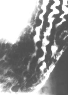 B
B
Figure 2.2 (A) Acute gastritis. Diffuse erosive gastritis with thick nodular folds. Erosions are scattered along the folds.
(B) Acute erosive gastritis. There are numerous erosions in the stomach (arrows). Each erosion consists of a small central collection of barium surrounded by a translucent ring (a small 'target' lesion)
13

Chronic gastritis
Radiographic findings: thinning or loss of rugal folds (however, thickening may be seen in early chronic disease); widening or loss of the area gastricae; as in acute gastritis, erosive lesions may be appreciated; mucosal nodularity; antral narrowing.
Gastric polyps
Gastric polyps are the most common benign gastric tumor with an incidence of 1,5–5 %. They are typically solitary, but may be multiple or diffuse. Gastric classification is similar to other GI polyps:
1. Hyperplastic: most common; usually small in size and have no malignant potential. Radiographic findings:
Shape: sessile or pedunculated. “Mexican hat sign” represents the stalk seen overlying the head of polyp.
Size: usually < 2cm in diameter.
Surface: sharply marginated polyp with smooth circular border.
Location: variable.
Often multiple.
2.Adenomatous: tend to be larger and papillary in appearance. Are a malignant precursor. Radiographic findings:
Shape: broad-based elliptical polyp +/- a pedicle.
Size: usually > 2cm in diameter.
Surface: contour is generally lobulated, but may be smooth.
Location: antrum is most common.
Gastric ulcers
Gastric ulcers are disruptions of the mucosa that extend into or through the submucosa. The radiologic imaging study of choice in the clinical evaluation of suspected gastric ulcers is a double-contrast upper GI series. Features to evaluate include location, shape, penetration, mucosal folds, and the ulcer mound. Ulcers are generally classified as either benign or malignant depending upon their radiographic features and underlying etiology.
Radiographic findings: the distortion and uninterrupted mucosal folds that radiate from (pulled into) the ulcer crater.
Ulcer crater seen en face: distinct collection of barium that persists on different views; the collection is most often round but can be linear. Ulcer crater seen in profile: barium collection extends outside the projected margin of the gastric or duodenal wall. Double-contrast studies: the crater has a white center with surrounding black “collar.”
Benign ulcers
Benign ulcers constitute approximately 95 % of all gastric ulcers. Most benign ulcers heal in 3–4 weeks following treatment with complete resolution by 6 weeks. All gastric ulcers should be followed by repeat examination in 6 weeks to confirm healing. Surgery is rarely performed for benign gastric ulcers but may be indicated if outlet obstruction occurs.
14
Radiographic findings:
Location: most commonly occur on the lesser curvature or posterior wall of the stomach.
Shape: ulcer crater is smooth and round or oval (figure 2.3).
Penetration: the ulcer projects beyond the normal margin of the gastric lumen.
Mucosal folds: extension of smooth gastric folds to the ulcer crater margin.
Ulcer collar: an edematous mucosal band across the ulcer neck.
A  B
B 
C  D
D 
Figure 2.3 Variants of gastric ulcer crater at double-contrast radiographic examination.
(A)A 6-mm focal barium collection (arrow) is seen en face. No mass effect is seen.
(B)When the barium collection is viewed in profile, the collection protrudes from the expected luminal contour (large arrow). A thin lucency (small arrow) crosses the collec-
tion at its interface with the luminal contour. These are findings of a benign gastric ulcer. The ulcer niche (crater) is filled with barium (large arrow) and protrudes outside the expected contour of the stomach. No mass or mucosal nodularity is seen to indicate that a tumor is present. As the ulcer extends into the soft submucosal fat, it spreads laterally, burrowing under the mucosa. The undermined mucosa is manifested as the lu-
cency (small arrow) crossing the edge of the ulcer, termed a Hampton line.
(C) Coarsely lobulated, nodular folds radiate toward a central barium collection (u). Compare a normal-sized fold (open arrow) with an enlarged, lobulated fold (black arrow).
(D) Benign gastric ulceration. Small posterior wall ulcer demonstrated en face. Radiating mucosal folds extend to the edge of the crater
Malignant ulcers
Approximately 5 % of gastric ulcers are found to be associated with malignancy. Of those, 90 % are due to adenocarcinoma, with the remaining 10 % di-
15

vided among lymphoma, sarcoma, and metastases. Radiographic imaging suggestive of malignancy warrants tissue biopsy.
Radiographic features:
The interrupted mucosal folds and irregular raised edge of the tumour indicate carcinoma of stomach.
Shape: the ulcer crater is irregular and nodular.
Non-penetrating: The ulcer does not project beyond the expected normal stomach margin.
Mucosal folds: do not radiate to the ulcer margin and are often nodular.
Ulcer mound: irregular and mass-like with eccentric crater.
Benign tumors
Benign tumors of the stomach are usually submucosal (lipoma, fibroma, schwannoma, hemangioma, lymphangioma, carcinoid (malignant transformation in 20 %)). Leiomyoma: most common benign tumor; may ulcerate, 10 % malignant.
Gastric carcinoma
Gastric carcinoma is the third most common GI malignancy after colorectal and pancreatic cancer, and represents the 6th leading cause of cancer deaths. Although they may present anywhere within the stomach, gastric carcinomas are most commonly located in the antrum or pylorus (50 %); 60 % are on the lesser curvature and 30 % occur at the GE junction.
The diagnosis is usually suggested by endoscopic biopsy. CT is useful in defining the extent of disease as hepatic or peritoneal metastases may be present.
Radiographic findings:
Gastric mass producing an irregular filling defect (figure 2.4)
Possible tumor ulceration. If an ulcer is present, radiating folds may be blunted or fused.
A  B
B  C
C  D
D
Figure 2.4 (A) 4-cm, smooth-surfaced mass (large arrows) is protruding into the stomach. The mass has sharp angles to the luminal contour (arrowheads). There is a sharp line between the obliquely oriented tumor and mucosal surface (thin arrows). These are the classic findings of a mass arising in the submucosa or muscularis propria, variably termed a submucosal or extramucosal mass.
(B) Adenomatous polyp. A long, thin pedicle (arrows) extends from the head of the polyp to the stomach wall.
(C) Polypoid gastric carcinoma. Single-contrast technique upper GI series reveals a lobulated filling defect (arrows) in the antrum of the stomach.
(D) Single contrast study from the same patient showing the apple core appearance (arrows) of the stomach due to the invasive gastric adenocarcinoma
16
3. RADIOLOGY OF INTESTINE
Introduction
Decreased natural contrast between adjacent structures of roughly similar radiographic density mandates the use of contrast material. Barium suspensions, high-density compounds mixed with water, are commonly used in the examination of the gastrointestinal tract. Barium enema is useful to diagnose problems in the intestine and to identify abnormal growths, ulcers, polyps, diverticula, and intestinal cancer. A satisfactory barium examination depends on the colon being carefully cleansed and empty.
Limitation: discomfort to patient, requires fluoroscopy, time consuming. Patient preparation: for small-bowel examination, the patient should have
nothing orally after midnight or the next morning preceding the radiographic study. Fluid and food in the stomach and small intestine degrade the examination by interfering with good mucosal visualization and causing artifacts that may mimic disease.
Preparation for the barium enema examination must be performed properly to obtain an accurate evaluation of the colon by this method. Various colonic preparations have been recommended and usually combine the use of dietary changes, oral fluids, and several cathartics the day preceding the barium enema examination. The presence of even small amounts of residual stool in the bowel may mimic colonic disease, or a filling defect in the colon may be passed off as stool but be a neoplasm.
CT Imaging
CT imaging of the abdomen can portray the various organs of the gastrointestinal tract. Mucosal disease, such as ulcers, and small neoplasms will not be shown with this imaging modality. Larger intestinal neoplasms, thickening of the walls of the hollow organs, and extrinsic processes can be detected with CT imaging. A major role of CT scanning, especially in the esophagus and colon, is staging malignancy of these organs. In the colon, for example, CT examination is used for initial staging, especially of distant metastases, and for evaluation of recurrence following surgery.
Magnetic resonance imaging
MR imaging of the hollow organs of the gastrointestinal tract is being used to evaluate and stage malignancies, especially of the esophagus and rectum, and also to assess inflammatory and obstructive bowel disease.
Radiography small intestine
The normal dimensions of the small bowel can be remembered by the “Rule of 3’s”: 1) bowel wall<3mm thick, 2) bowel folds<3mm thick, 3) bowel diameter<3cm wide. Patterns that should be assessed when evaluating the small
17

bowel include the location of the abnormality, the caliber of the lumen, the mucosal contour, the fold pattern (figure 3.1).
The small bowel generally lies centrally within the abdomen, “framed” by the large bowel. On imaging, the small bowel can be differentiated from the large bowel based on the presence of plicae circulares (circular folds), which traverse the entire diameter of the lumen. The large bowel does not possess these circular folds, but rather has saccular dividers called haustra.
Inflammatory diseases
Duodenal ulcer
Duodenal ulcers are two to three times more common than gastric ulcers. All bulbar duodenal ulcers are considered benign. Postbulbar or multiple ulcers raise the suspicion for Zollinger-Ellison syndrome.
Radiographic features of duodenal ulcers: persistent round or elliptical collection of the barium, radiating folds, spasm (figure 3.1).
A  B
B 
C 
Figure 3.1 (A) Spot radiograph of the duodenal bulb (B) and second portion of the duodenum (2) obtained during a double-contrast upper GI series. The folds of the duodenum (folds of Kerckring) (arrow) cross the duodenum except at the level of the papilla of Vater.
(B) Close-up from an overhead view obtained during a normal small bowel followthrough examination demonstrates the normal anatomy. Many loops of small intestine are visible. Two loops are well distended (thin arrows) and show that the valvulae conniventes lie perpendicular to the longitudinal axis of the small bowel.
(C) Scheme variants of duodenal ulcers
18

Ulcer largely replaces the duodenal bulb. A large ulcer crater may be mistaken for a deformed bulb but does not change shape during fluoroscopy. Duodenal ulcers often heal with a scar; this can lead to deformity and contraction of the duodenal bulb: cloverleaf deformity, or hourglass deformity. Postbulbar ulcers: any ulcer distal to the first portion of the duodenum should be considered to have underlying malignancy until proven otherwise (only 5 % are benign ulcers, mostly secondary to Zollinger-Ellison syndrome).
Crohn’s disease (regional enteritis)
Early findings represent active inflammation, while late findings represent chronic inflammation and/or fibrosis. Fluoroscopic findings include:
mucosal ulcers (punctate collections of barium surrounded by radiolucent mounds of edema)
luminal narrowing from edema, spasm.
fold thickening.
strictures, manifest by the “string sign”.
Ulcers
Fluoroscopy, using either single or double contrast, is the imaging study of choice for evaluating small bowel ulcerations. Barium pools in the ulcer base, seen as a radiopaque collection outside the confines of the small bowel. Ulcers can have a variety of appearances, ranging from aphthoid to linear to “punchedout” to “bull’s-eye,” depending on the etiology. In addition, pancreatic pseudocysts and diverticula may mimic the appearance of ulcers.
A 
B  C
C 
Figure 3.2 (A) Schemes variants Crohn’s disease of the small bowel.
(B) Spot radiograph of the terminal ileum shows numerous aphthoid ulcers en face as 2- to 5 mm punctate or slightly elongated collections of barium surrounded by radiolucent halos (thin arrows). In profile, the aphthoid ulcers appear as 2- to 4mm punctate barium collections (open arrow) protruding outside the expected luminal contour.
(C) Crohn disease of intestine (luminal narrowing from edema, spasm, fold thickening) with stomach involvement
19

Solitary mass
Masses projecting into or occurring with the small intestine appear as filling defects on radiographic studies. These filling defects may represent intraluminal, mucosal, intramural, or extrinsic masses (figure 3.3).
A
B
C
Figure 3.3 (A-C) Schemes of variants lumen, mucosal and fold patterns of small bowel
Radiography colon Inflammatory diseases
Crohn's disease
Crohn's disease is an idiopathic inflammatory disease of the GI tract that is characterized by ulcerations, erosions, noncaseating granulomas, and fullthickness bowel wall inflammation. The disease may affect any portion of the gastrointestinal system from the esophagus to the anus. The course is progressive in nature, with frequent remissions and relapses. The radiographic hallmarks of Crohn's disease are aphthous erosions, thickened and distorted folds due to bowel wall edema, "cobblestone" pattern of deep ulcerations (figure 3.2– 3.3), fibrosis with thickened walls, contractures, and stenosis resulting in the "string sign," fistula and sinus tract formations, stranding in mesenteric fat due to inflammation, and "skip" lesions with intermittent areas of normal bowel between diseased segments (figure 3.4).
20