

5 курс / Инфекционные болезни / Доп. материалы / Lower respiratory tract infection
.pdfNOTES
NOTES
LOWER RESPIRATORY TRACT
INFECTION
GENERALLY, WHAT IS IT?
PATHOLOGY & CAUSES |
DIAGNOSIS |
|
▪ Infections involving trachea, bronchi, |
LAB RESULTS |
|
bronchioles, lungs |
▪ Complete blood count (CBC) |
|
RISK FACTORS |
Microbe identifi cation |
|
▪ Blood culture, sputum culture; Gram stain, |
||
▪ Smoking, compromised immunity, age |
||
polymerase chain reaction (PCR) |
||
(children, elderly), comorbidities |
|
|
COMPLICATIONS |
TREATMENT |
|
▪ Respiratory compromise, infection spread, |
MEDICATIONS |
|
sepsis |
||
|
▪ Antimicrobials |
|
SIGNS & SYMPTOMS |
OTHER INTERVENTIONS |
|
▪ Cough, dyspnea, fatigue, fever |
▪ Ventilatory support |
|
|
BACTERIAL TRACHEITIS
osms.it/bacterial_tracheitis
PATHOLOGY & CAUSES |
RISK FACTORS |
|
▪ Antecedent viral infections, especially croup |
||
|
||
▪ Rare, potentially life-threatening exudative |
▪ Commonly affects children |
|
|
||
infection |
COMPLICATIONS |
|
▫ Characterized by mucosal ulceration, |
||
pseudomembrane formation, airway |
▪ Pneumonia, septicemia, pneumothorax, |
|
obstruction risk (due to edema, |
pneumomediastinum, hypoxia (secondary |
|
exudative sloughing) |
to airway obstruction), cardiorespiratory |
|
▪ Common infective agents: Staphylococcus |
arrest |
|
aureus, Moraxella catarrhalis, Streptococcus |
|
|
pneumoniae, H. infl uenzae |
|
878 OSMOSIS.ORG

SIGNS & SYMPTOMS
▪Prodromal respiratory viral infection presentation → acute onset of fever, hoarseness, sore throat, stridor
▪Productive, barky cough with copious tracheal secretions, retrosternal pain
▪Progressive respiratory distress
▫Dyspnea, retractions, fatigue, ↓ level of consciousness
DIAGNOSIS
DIAGNOSTIC IMAGING
Chest X-ray
▪Upper tracheal narrowing (“steeple sign”)
▪Tracheal pseudomembranes (irregular shadows)
LAB RESULTS
▪ CBC: leukocytosis, left shift
Microbe identifi cation
▪ Positive tracheal culture, Gram stain
OTHER DIAGNOSTICS
▪Laryngoscopy: subglottic edema; tracheal lumen narrowing; presence grayish exudate; slough, pus; friable tracheal mucosa
Chapter 125 Lower Respiratory Tract Infections
TREATMENT
MEDICATIONS
▪ Broad antibiotic coverage
OTHER INTERVENTIONS
▪Ventilatory support
▫Humidifi ed supplemental oxygen, intubation, endoscopic tracheal debridement
▪Fluid management
Figure 125.1 The endoscopic appearance of bacterial tracheitis in a nine-year-old boy.
BRONCHIOLITIS
osms.it/bronchiolitis
PATHOLOGY & CAUSES
▪Viral small airway respiratory infection
▪Viral spread through respiratory secretions, contaminated hands → infects lower respiratory tract cells → natural killer cells attack → cytokines released → epithelial cells produce mucus, vessels vasodilate →
fl uid leaks, walls swell → airway narrows (more severe in children)
▪Dead cells, mucus slide into airway → form mucus plugs → trap air → airways collapse (atelectasis)
CAUSES
▪Respiratory syncytial virus (RSV): most common, especially during winter months
▪Adenovirus, human bocavirus, human metapneumovirus
▪Mycoplasma pneumoniae
OSMOSIS.ORG 879
RISK FACTORS
▪Young age (children < two years old), previous infection, daycare attendance, decreased immunity, neuromuscular disorders, premature birth, cardiovascular malformations, airway malformations, exposure to smoking
COMPLICATIONS
▪ Hypoxemia, sepsis
SIGNS & SYMPTOMS
▪Congestion, pharyngitis, sore throat, cough
▪Hypoxia → tachycardia, tachypnea, exhaustion
▪If severe: dyspnea, wheezing, central apnea (brief periodic breathing arrest), nasal
fl aring, retractions, cyanosis, fever, poor feeding, ↓ activity
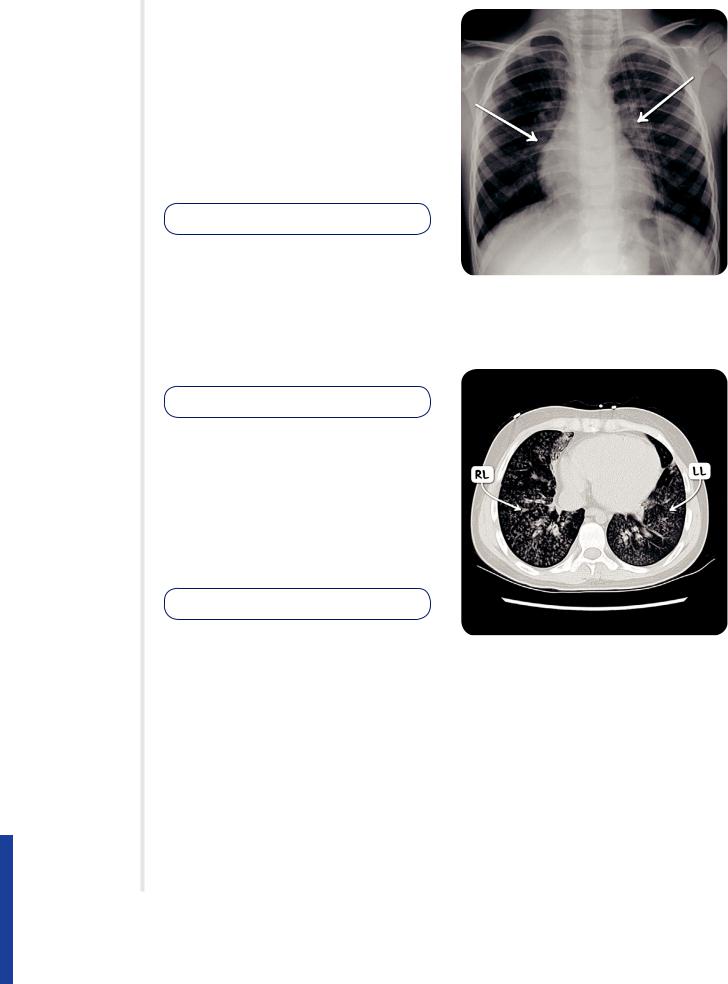
Figure 125.2 A plain chest radiograph in a child with bronchiolitis demonstrating bilateral hilar fullness.
DIAGNOSIS
DIAGNOSTIC IMAGING
X-ray
▪ Patchy infi ltrates, atelectasis
LAB RESULTS
▪Positive rapid viral testing (RT-PCR): suggests viral infection
TREATMENT
OTHER INTERVENTIONS
Immunoprophylaxis
▪Palivizumab: monoclonal antibody against RSV given monthly throughout RSV season for prematurely-born infants, chronic lung disease, congenital heart disease
▪Heated, humidifi ed supplemental oxygen (high-fl ow nasal cannula/continuous positive airway pressure (CPAP)), fl uids, nasal suctioning
▪Intubation (if hypoxia continues despite intervention)
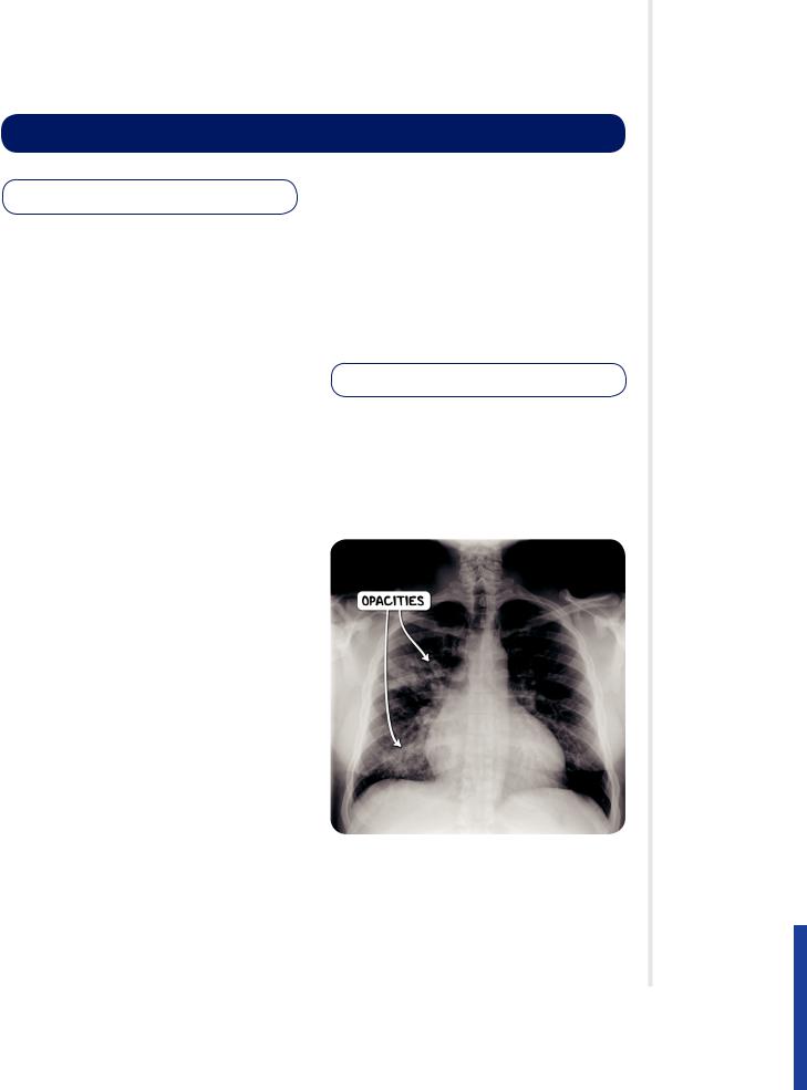
Figure 125.3 A CT scan of the chest in the axial plane in an individual with severe bronchiolitis. Both lung fi elds demonstrate the tree-in-bud pattern.
880 OSMOSIS.ORG
Chapter 125 Lower Respiratory Tract Infections
COMMUNITY–ACQUIRED
PNEUMONIA
osms.it/community-acquired_pneumonia
PATHOLOGY & CAUSES
▪Pneumonia acquired outside hospital/ healthcare setting
▪Viral pneumonia may → superimposed bacterial infection
Spread
▪Respiratory: from host to host
▪Hematogenous: from another infection with same pathogen (e.g. cellulitis)
Causative organisms
▪S. pneumoniae, S. aureus, H. infl uenzae, group A streptococci, infl uenza virus, respiratory syncytial virus (RSV), parainfl uenza
Resolution
▪Approx. day 8, can continue for three weeks
▫Exudate digested by enzymes, ingested by macrophages, coughed up
COMPLICATIONS
▪ Meningitis, sepsis, pleural effusions
SIGNS & SYMPTOMS
▪High fever, cough, hemoptysis, pleuritic chest pain, tachypnea, tachycardia, dyspnea, muscle pain, fatigue
▪Crepitation on palpation, dullness on percussion
RISK FACTORS
▪Advanced age, lowered immunity, smoking, alcohol abuse, malnutrition, chronic lung disease
STAGING
Congestion
▪Between days 1–2
▫Blood vessels, alveoli start fi lling with excess fl uid
Red hepatization
▪Between days 3–4
▫Exudate (contains red blood cells, neutrophils, fi brin) starts fi lling airspaces
→ solidifi es them → lungs develop liverlike appearance
Gray hepatization
▪Approx. days 5–7
▫Lungs remain fi rm but color changes → red blood cells in exudate start to break down
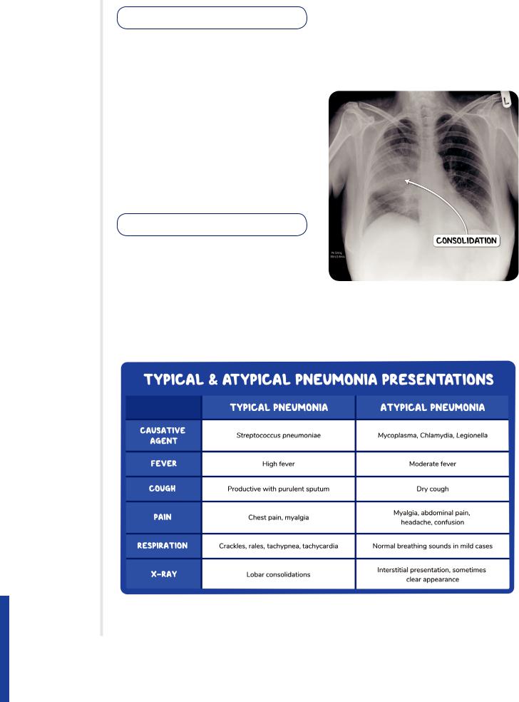
Figure 125.4 A plain chest radiograph demonstrating patchy peri-bronchial shadowing in an individual with bronchopneumonia.
OSMOSIS.ORG 881
DIAGNOSIS
DIAGNOSTIC IMAGING
X-ray
▪Interstitial infi ltrates; consolidation; may show pleural effusion
LAB RESULTS
▪↓ oxygen saturation
▪CBC: leukocytosis
▪Organism identifi cation: sputum Gram stain, culture; C-reactive protein test (CRP), PCR for typical viruses
▪Positive urine for S. pneumoniae
TREATMENT
MEDICATIONS
▪ Antibiotics
OTHER INTERVENTIONS
▪ Supplemental oxygen, fl uids
Prevention
▪23-valent vaccine (Pneumovax) available against pneumococcus
▫Recommended in splenectomised, immunocompromised individuals
Figure 125.5 A plain chest radiograph demonstrating consolidation of the right middle lobe in an individal with lobar pneumonia.
882 OSMOSIS.ORG
Chapter 125 Lower Respiratory Tract Infections
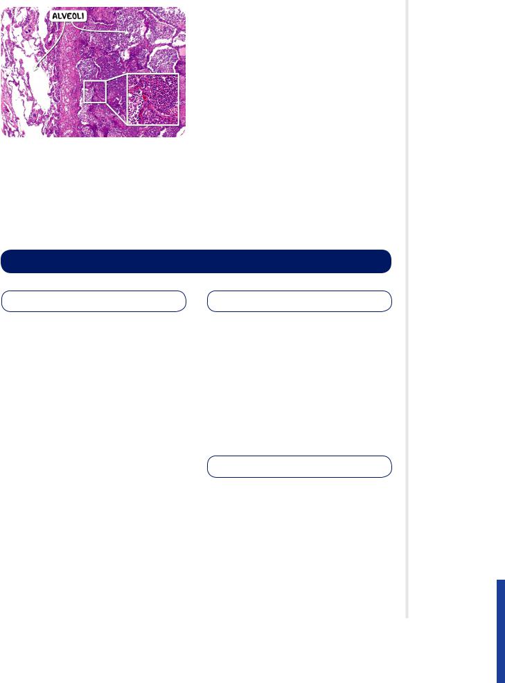
Figure 125.6 The histological appearance of acute pneumonia. In the affected part of the lung (right) the alveoli are fi lled with neutrophils.
CROUP
osms.it/croup
PATHOLOGY & CAUSES
▪Acute respiratory condition
▫Characterized by laryngotracheitis
▪Immune response to epithelial viral infection
▫Upper bronchi: larynx, trachea narrow due to swelling
▫Lower bronchi: terminal bronchioles, viral pneumonia
CAUSES
▪RSV, parainfl uenza, adenoviruses
▪Historically: Corynebacterium diphtheriae
(vaccine development → ↓ incidence)
RISK FACTORS
▪ Most common in children < six years old
COMPLICATIONS
▪Hypoxia, respiratory failure
▪Secondary bacterial infections → ↑ mortality
SIGNS & SYMPTOMS
▪Progressive respiratory symptoms; sore throat, hoarse voice (due to laryngeal involvement)
▪Respiratory symptoms
▫“barking” cough
▫Tachypnea
▫Grunting (attempt to increase endexpiratory pressure)
▫Prominent inhalation, inspiratory stridor, apnea
DIAGNOSIS
DIAGNOSTIC IMAGING
X-ray
▪ “Steeple sign,” narrowing below epiglottis
LAB RESULTS
▪ CBC: normal ↑ with left shift, or ↓
OSMOSIS.ORG 883

OTHER DIAGNOSTICS
▪Severity: Westley scale 0–17
▫3-7: moderate
▫8-11: severe
▫12 and above: indicates respiratory failure
TREATMENT
MEDICATIONS
▪ Dexamethasone, epinephrine (nebulized)
OTHER INTERVENTIONS
▪Humidifi ed supplemental oxygen, fl uids, antipyretics
▪Intubation (if impending respiratory failure)
Figure 125.7 A plain X-ray image demonstrating the steeple sign in an infant with croup.
884 OSMOSIS.ORG

Chapter 125 Lower Respiratory Tract Infections
NOSOCOMIAL PNEUMONIA
osms.it/nosocomial-pneumonia
PATHOLOGY & CAUSES
▪Hospital-acquired pneumonia
▫AKA healthcare-associated pneumonia
▫Includes ventilator-associated pneumonia
▪Involves microaspiration of organisms from oropharyngeal tract/sometimes from gastrointestinal tract
▪Severity varies depending on offending organism, individual’s immune system status
SIGNS & SYMPTOMS
▪Nonspecifi c symptoms (malaise, lethargy), fever, productive cough
DIAGNOSIS
DIAGNOSTIC IMAGING
Chest X-ray
▪ Shows infi ltrates
CAUSES
▪MRSA, Klebsiella pneumoniae, Pseudomonas aeruginosa, Acinetobacter
▪Often polymicrobial
RISK FACTORS
▪Intubation, poor staff hygiene, contaminated equipment contact
LAB RESULTS
▪CBC: leukocytosis, ↑ CRP
▪Positive sputum culture
TREATMENT
MEDICATIONS
▪ Antibiotics
COMPLICATIONS |
OTHER INTERVENTIONS |
▪ Meningitis, sepsis, pleural effusions |
▪ Supplemental oxygen, fl uids |
OSMOSIS.ORG 885