

Biology_of_Turtles
.pdf166 |
Biology of Turtles |
7.3Results
7.3.1Osteology
The cervical column in all recent turtles consists of eight elongated vertebrae (C1 to C8) and nine joints. The most important element of the vertebrae is the vertebral centrum, which is positioned right below the spinal cord. From the centrum, the neural arch rises and covers the spinal cord (Figure 7.2). The neural spines are positioned midventrally on the neural roof and are reduced in most turtles (Figure 7.2). The articulation of the different vertebrae occurs by means of the vertebral centra and the zygapophyses (Figure 7.2). The prezygapophyses of the vertebra articulate with the postzygapophyses of the more cranially situated vertebra. Whereas the articular facets of the prezygapophyses are positioned dorsally, those of the postzygapophyses are oriented ventrally. Vertebral centra can be biconvex (C5 and C8 in Chelodina), biconcave or amphicoelous (C1 in both species; C7 in Chelodina), procoelous (C6 in Chelodina) or opisthocoelous (C2, C3, and C4 in
Chelodina; C2 to C8 in Apalone).
In a transverse section, the vertebral centrum of Chelodina is saddle-shaped (Figure 7.3 and Figure 7.4). The vertebral centrum is well developed only at the level of the anterior and posterior articulations. The neural spine is reduced to an inconspicuous longitudinal rim. The anterior zygapophyses are clearly separated from each other (Figure 7.3 and Figure 7.4). Their articular facets are mainly oriented in the medio-dorsal direction. The posterior zygapophyses are fused to each other except those of C1 (Figure 7.3), but the articular facets remain separated and face mainly ventrally and slightly laterally (Figure 7.3 and Figure 7.4). The elevation of the posterior zygapophysis gradually increases from distal to proximal (Figure 7.3 and Figure 7.4). The transverse processes are strongly developed, especially those on the more proximal vertebrae (Figure 7.3 and Figure 7.4).
The cervical vertebrae in Apalone are relatively gracile and elongated (Figure 7.5 and Figure 7.6). They bear well-developed and well-separated preand post-zygapophyses (Figure 7.5 and
A |
Dorsal |
|
|
Neural arch |
|
Posterior zygapophysis |
|
|
|
Anterior zygapophysis |
|
Caudal |
|
Cranial |
Centrum |
|
Transverse process |
|
|
|
|
Ventral |
|
Posterior zygapophysis |
B C |
Posterior zygapophysis |
|
|
|
Articular facets |
|
Anterior zygapophysis |
posterior zygapophysis |
|
|
Anterior zygapophysis |
|
Neural canal |
Neural canal |
|
|
Transverse process |
|
Transverse process |
Posterior articular facet |
|
Anterior articular facet |
of the centrum |
|
|
|
of the centrum |
|
|
|
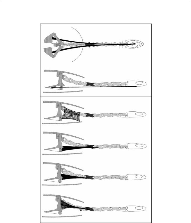
Figure 7.2 Chelodina longicollis. Terminology of the most important structural elements of a cervical vertebra (C7). (A) Lateral view, (B) posterior view, and (C) anterior view (terminology after Romer & Parsons, 1977).
Cervical Anatomy and Function in Turtles |
167 |
Lateral view
Dorsal view
Frontal view
Caudal view
C1 |
C2 |
C3 |
C4 |
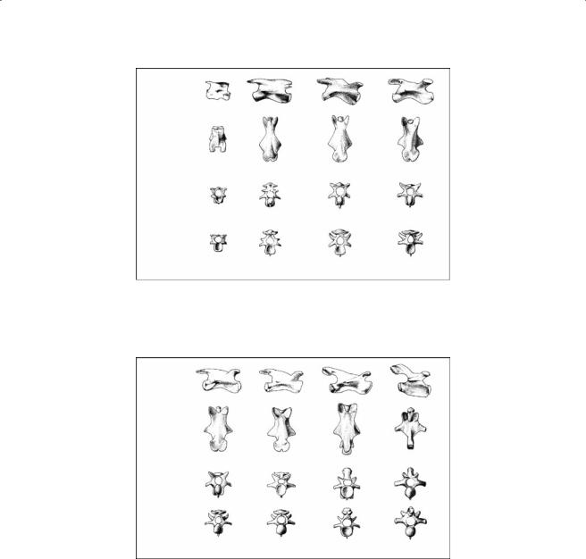
Figure 7.3 Lateral, dorsal, frontal, and caudal views of the first four cervical (C1-C4) vertebrae in
C. longicollis.
Lateral view
Dorsal view
Frontal view
Caudal view
C5 |
C6 |
C7 |
C8 |
Figure 7.4 Lateral, dorsal, frontal, and caudal views of the last four cervical (C5-C8) vertebrae in C. longicollis.
Figure 7.6). The orientation of the zygapophyses is variable throughout the neck. Because of the absence of the iliocostalis and logissimus groups (discussed later), the transverse processes are only poorly developed (Figure 7.5 and Figure 7.6). Because of the strong divergence of the postzygapophyses and the absence of a well-developed neural spine, a hiatus intervertebralis (Bojanus, 1821) is created. In Apalone, this hiatus is covered by a thick membrane (Ogushi, 1913).
7.3.2Cervical Joints
Joint S(kull)-C1 (occipito-cervical joint). This procoelous ball-and-socket type joint allows the skull to move independently from the neck. The occipital is spherical. The ridges of the congruent cotyle on C1 are well developed. C1 lacks anterior zygapophyses. The connection between the
168 |
Biology of Turtles |
Lateral view
Dorsal view
Frontal view
Caudal view
C1 |
C1´ |
C2 |
C3 |
C4 |
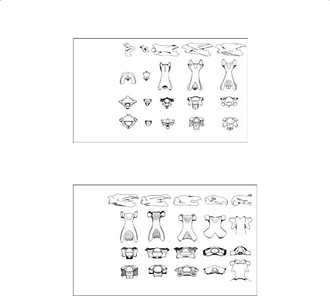
Figure 7.5 Lateral, dorsal, frontal, and caudal views of the first four cervical (C1-C4) vertebrae in A. spinifera. C1′ is the intercentrum of the atlas.
Lateral view
Dorsal view
Frontal view
Caudal view
C5 |
C6 |
C7 |
C8 |
D1 |
Figure 7.6 Lateral, dorsal, frontal, and caudal views of the last four cervical (C5-C8) and the first dorsal vertebra (D1) in A. spinifera.
neural arch of C1 and the base of the skull is ligamentous. The joint is similar in both Apalone and
Chelodina.
Joints C2-1, C3-2, C4-3, C5-4 (anterior part of the neck). The articulation facets of vertebrae composing these joints are very similar. The vertebral centra are all opisthocoelous and have an anterior elliptical cotyle, of which the long axis is oriented vertically in Chelodina and horizontally in Apalone, and a posterior condyle with enlarged lateral ridges. The articular facets of the zygapophyses of the most cranial joint of this group (C2-1) are less strongly developed. The elongated neural spine of C2 extends anteriorly beyond the articular facets of the posterior zygapophyses of C1 in both groups. The posterior zygapophyses of C1 are clearly separated. In the more caudal joints, the zygapophyses are elongated and slightly elevated (Figure 7.3). The articular facets of the anterior zygapophyses are slightly concave and their outline is kidney-shaped. They are oriented in a dorso-medial direction. The posterior zygapophyseal facets are ventrolaterally inclined. Lateral mobility is strongly reduced in Apalone compared to Chelodina because of the paired post-zygapophyses.

Cervical Anatomy and Function in Turtles |
169 |
Joints C6-5 and C7-6. The central joints C6-5 and C7-6 are procoelous in Chelodina but opisthocoelous in Apalone. In posterior view, the posterior condyle of C5 in Chelodina has the shape of a raindrop. Alternatively, in Chelodina the condyle of C6 is almost spherical. The congruent cotyle on the anterior side of C6 and C7 has pronounced lateral and ventral ridges. The articular facets of the posterior zygapophysis are well developed and also have a kidney-shaped outline. The bilateral posterior zygapophyseal facets contact each other medially, forming a semicircular or horseshoeshaped articular surface. The articular facets of the anterior zygapophyses are more oval (almost circular) as compared to the more distal joints. In Apalone, the articulatory facets of the post-zyg- apophyses are positioned horizontally. The post-zygapophyses themselves are nearly in line with the long axis of the vertebrae (less than 6°).
Joint C8-7. C8-7 is opisthocoelous and the shape of its condyle and cotyle resemble previously described ophisthocoelous joints in both groups. The cotyle has enlarged lateral ridges in Chelodina and is fully split in Apalone. The posterior zygapophysis of C7 is elongated, inclined in dorsal direction in both groups (Figure 7.4). The articular facets are clearly distinguishable from those of the remainder of the zygapophyses and are laterally oriented in Chelodina. The rather short but strongly elevated anterior zygapophyses of C8 tend to incline dorsomedially in Chelodina but more laterally in Apalone.
Joint D1-C8 (cervico-dorsal joint). D1-C8 is procoelous in Chelodina. In Apalone, this joint lacks a central articulation. In Chelodina, the rather flattened condyle on the posterior part of C8 is almost circular. The articular facet faces caudo-ventrally. The circular congruent cavity on the anterior part of D1 is shallower than those on the more anterior joints. The elongate posterior zygapophysis in Chelodina is more elevated in the dorsal direction than that of joint C8-7 (Figure 7.4). The articular facets of the zygapophyses are more inclined than those in the previously described joints. On C8, the articular facets of both sides fuse and are clearly distinguishable from the posterior zygapophysis. Their appearance is that of a vertically positioned cylinder (approximating a vertical hinge joint). The anterior zygapophyses on D1 are rather short and present an increased dorsomedial inclination of the articular facets. In Apalone, the post-zygapophyses of C8 are extremely well developed and wider than those of the more anteriorly situated vertebrae (Figure 7.6). The articulatory facets are strongly concave. The pre-zygapophyses of D1 have been rearranged to horizontally oriented cylinders. Only ventroflexion is possible at this joint.
7.3.3Musculature
The following short description of the cervical musculature is based on the terminology of Shah (1963) for Chelodina and that of Ogushi (1913) for Apalone. Other morphological accounts on the cervical musculature in turtles (Bojanus, 1819; George & Shah, 1954, 1955; Scanlon, 1982) were consulted to clarify the position of certain muscles where needed.
7.3.3.1 Chelodina
m. constrictor colli. This is a thin sheet-like muscle which covers the first two thirds of the neck. The muscle has a dual origin: some fibers originate on the squamosal and the dorsal connective tissue associated with the occipital spine and others originate on the neural spines of C2-C6. The fibers run ventrally and insert midventrally on a central connective tissue sheet (median raphe).
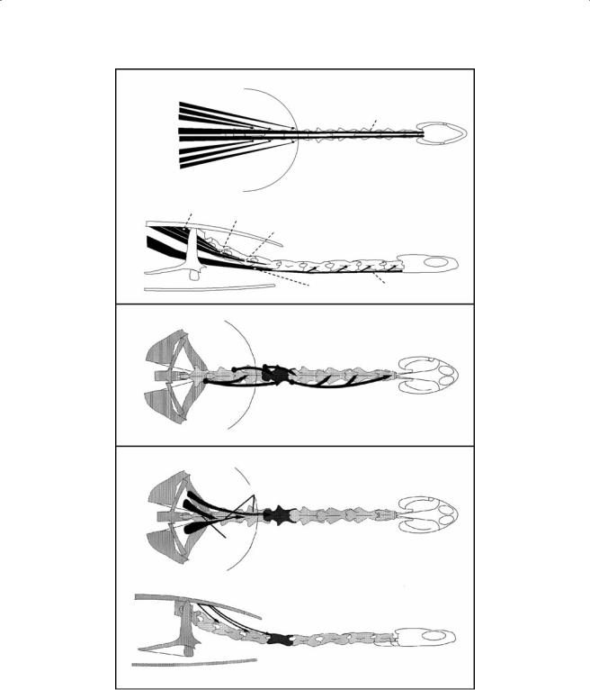
m. retrahens capitis et collique (Figure 7.7). This muscle consists of a number of well-developed and individualized bundles that connect the carapace to the neck and head. The first bundle is the longest and originates at the lateral aspect of D8 and the adjacent part of the carapace. The muscle runs cranially and inserts by means of a long tendon at the basioccipital. A number of fibers from the first bundle insert on the lateral aspect of C1-C5. The second bundle originates at the level of D5. The muscle runs cranially and inserts tendinously at the lateral aspect of C5. The third bundle
170 |
Biology of Turtles |
|
|
Carapace |
m. retrahens capitis collique |
|
|
|
|
Bundle 2 * |
|
|
Tendon |
Bundle 3 * |
|
|
|
Bundle 4 * |
|
|
|
Bundle 1 * |
|
|
Skull |
Bundle 1 * |
|
|
|
Bundle 4 * |
|
|
|
Bundle 3 * |
|
|
|
Bundle 2 * |
|
|
|
Bundle 4 |
Bundle 3 |
|
|
|
Bundle 2 |
|
|
* |
|
|
|
|
|
|
|
Pectoral girdle |
|
Bundle 1 |
Tendon |
|
|
||
|
|
|
m. longissimus cervicis |
|
Bundle 6 |
Bundle 5 |
|
|
Bundle 7 |
Bundle 4 |
|
m. longissimus thoracis
Lateral bundles
Medial
bundle
Figure 7.7 Schematic representation of the m. retrahens capitis collique, the m. longissimus cervicis, and the m. longissimus thoracis in C. longicollis.
Cervical Anatomy and Function in Turtles |
171 |
m. semispinalis
m. testocapitis
Lateral bundle
Medial bundle
m. spinalis cervico-capitis
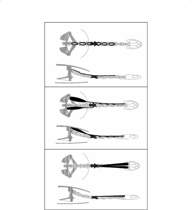
Figure 7.8 Schematic representation of the m. semispinalis, the m. testocapitis, and the m. spinalis cer- vico-capitis in C. longicollis.
172 |
Biology of Turtles |
m. rectus cervicis
m. scalenus complex
m. scalenus I
m.scalenus II
m. scalenus III
m.scalenus IV
Tendon of m. retrahens capitis collique
Figure 7.9 Schematic representation of the m. rectus cervicis and the m. scalenus complex in C. longicollis.

Cervical Anatomy and Function in Turtles |
173 |
m. constrictor hyoideus
|
|
|
mm. intertransversarii colli |
b8 |
b6 |
b4 |
b2 |
|
b7 |
b5 |
b3 |
m. longus colli
6
5 |
4 |
3 |
2 |
1 |
Figure 7.10 Schematic representation of the m. constrictor hyoideus, the mm. intertransversarii colli, and the m. longus colli in C. longicollis.

174 |
Biology of Turtles |
originates at D4 and inserts at the lateral aspect of C7. The fourth bundle originates at D3 and inserts at the transverse process of C8.
m.longissimus cervicis (LC, Figure 7.7). This is a segmentally arranged muscle with a more complex arrangement. The LC of C7 originates on the pre-zygapophysis of C8. The muscle belly runs anteriorly and divides into a more medial and a lateral bundle. The lateral bundle inserts on the post-zygapophysis of C6 and the medial on the lateral aspect of C5. The LC 6 originates on the pre-zygapophysis of C7 and inserts on the post-zygapophysis of C5. The LC 5 originates on the pre-zygapophysis of C6 and inserts on the post-zygapophysis of C4. The LC 4 originates on the prezygapophysis of C5 and inserts mainly on the neural arch of C1. During the course of the muscle, fibers diverge and insert on the post-zygapophyses of C3 and C2 as well.
m. longissimus thoracis (Figure 7.7). This muscle is responsible for lateral flexion of the posterior part of the neck and consists of one medial and two lateral bundles. All bundles originate at the ventral aspect of the carapace, cranial to the origin of the m. testocapitis. The medial bundle inserts at the lateral aspect of C8. The first lateral bundle inserts by means of a tendon on C6 at the level of the post-zygapophysis. The fibers of the lateral most bundle run initially ventral to the previous bundle but run more laterally at the level of C7. The muscle bundle inserts tendinously at the level of the post-zygapophysis of C5.
mm. semispinalis (Figure 7.8). This is a series of short serially arranged muscles that span adjacent vertebrae and run from C2 to C8. The muscles originate at the neural spine of the more posterior vertebra and run anterolaterally to insert on the post-zygapophysis of the anterior vertebra.
m. testocapitis (Figure 7.8). This is a very long muscle that consists of two distinct muscle bundles that connect the carapace to the head and neck. Both bundles originate at the ventral aspect of the carapace, lateral to the scapula. The fibers of the medial bundle converge onto a strong tendon that inserts at the lateral aspect of C5. The lateral bundle inserts through a complex arrangement of tendons in the anterior part of the neck at the lateral aspect of C4-C2.
m. spinalis cervico-capitis (Figure 7.8). This is superficial dorsal cervical muscle bundle. The muscle originates at the neural spines of C3-C5 and runs obliquely anteriorly. The more medially positioned fibers insert on the lateral aspect of the occipital spine and the more lateral fibers on the dorsal aponeurosis covering the m. adductor mandibulae.
m. rectus cervicis (Figure 7.9). This is a very long and ventrally positioned muscle that connects the pectoral girdle with the ventral aspect of the hyobranchium. The muscle originates at the mid-dorsal aspect of the coracoid. The fibers run ventrally and join the fibers of the m. constrictor hyoideus of the same side. At the level of the hyobranchium, the two muscles separate again and the fibers of the m. rectus cervicis insert partially on the second ceratobranchial and partially on the basibranchium.
m. scalenus complex (Figure 7.9). This is a series of four muscles that connect the pectoral girdle with the lateral aspect of the posterior neck. The most superficial bundle is very thin and runs between the acromion, the dorsal aspect of the plastron, and the posterior most cervical vertebrae (C6-C8). The second and ventral most bundle of the scalenus complex originates on the acromion but distally to the first bundle. The fibers insert at the ventrolateral aspect of C4 and C5. The third bundle originates partly on the scapula and partly on the acromion. The fibers converge on a flat tendon that inserts at the ventrolateral aspect of C5. The fourth part is the shortest and originates across the entire width of the scapula. The muscle inserts on the proximal aspect of the tendinous insertion of the second bundle of the m. retrahens capitis et collique.
m. constrictor hyoideus (Figure 7.10). This muscle originates at the lateral aspect of C6, runs cranially and inserts at the posterior aspect of the first ceratobranchial. Although its primary function is presumably related to the abduction and retraction of the ceratobranchial, it presumably also plays a role during lateral bending of the neck.
mm. intertransversarii colli (Figure 7.10). This is another segmentally arranged muscle that has a dual origin. The lateral bundle originates at the ventral aspect and the medial bundle at the
Cervical Anatomy and Function in Turtles |
175 |
diapophysis of the cervical vertebra. Both bundles join and insert at the posterior aspect of the vertebral centrum of the more cranially positioned vertebra.
m. longus colli (Figure 7.10). This muscle consists of a series of segmentally arranged small muscle bundles that run in close association to the m. retrahens capitis et collique. The longus colli bundles of the first five vertebrae originate at the ventral aspect of the respective vertebrae. Insertion takes place by means of the tendinous insertion of the second bundle of the m. retrahens capitis et collique on the lateral aspect of C4-C1. The muscles in the more posterior part of the neck are somewhat more complex. The m. longus colli 8 originates on the ventral side of C8 and inserts on the tendon of the second bundle of the m. retrahens capitis et collique. The longus colli 7 originates at the ventral aspect of C7 and inserts by means of the insertion of the tendon of the second bundle of the m. retrahens capitis et collique at the lateral aspect of C5. The longus colli 6 originates at the ventral aspect of C6 and inserts at the lateral aspect of C5.
7.3.3.2 Apalone
Notable in Apalone is the strong reduction of the epaxial musculature (Hofstetter & Gasc, 1969), the absence of the longissimus system, and the absence of the iliocostalis system that is characteristic for all turtles.
m. constrictor colli (m. sphincter colli, Ogushi, 1913). This muscle is poorly developed in Apalone and consists of two discrete parts. The anterior part covers the lateral and ventral aspect of the anterior neck region. It originates on the dorsal connective tissue associated with the occipital spine and runs ventrally to insert on the median raphe. The posterior part is often difficult to discern and originates more posteriorly in the neck on the nuchal connective tissue associated with the neural spines and inserts ventrally on the median raphe.
m. rectus cervicis (m. coraco-hyoideus, Ogushi, 1913). This is the ventral-most muscle in the cervical region. The muscle has a dual origin—one part originates on the dorsal aspect of the proximal part of the procoracoid, and the other part originates at the dorsal aspect of the epicoracoid. The muscle runs anteriorly and inserts at the posterior aspect of the basibranchium.
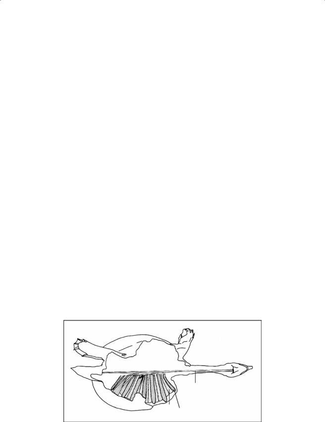
m. carapaco-basioccipitis (Figure 7.11). This is a very long muscle that runs from the pelvic girdle to the cranium. Three distinct origins on the crista mediana ventralis of the first, second, and third caudal vertebrae are present. In addition, three accessory sites of origin can be discerned: on the proximal aspect of the fifth rib, across the entire length of the sixth costal plate, and along the entire seventh rib and associated costal plate. The fibers of this last bundle initially run at an angle of at most 90° to the long axis of the remainder of the muscle.
m. carapaco-basioccipitis
Costal Rib
plate
Figure 7.11 Schematic representation of the m. caracapo-basioccipitis in A. spinifera.