

Budras_Анатомия лошади
.pdfNerve |
Innervation |
Comments |
Ventral branches |
Muscles of hindlimb |
•Lateral branches
•Medial branches
V. Caudal (coccygeal) nerves (Cd1–Cd5) |
|
Dorsal branches |
Dorsal sacrocaudal mm. and intertransversarii; |
|
skin on dorsal surface of tail |
Ventral branches |
Ventral sacrocaudal mm. and intertransversarii; |
|
skin on ventral surface of tail |
Leave vertebral canal through ventral sacral foramina
Form roots of sacral plexus (see further on)
Form cauda equina
Form dorsal sacral plexus
Form ventral sacral plexus
Brachial plexus (pp. 6/7; 8/9; 10/11)
Network of ventral branches of spinal nerves C6, 7, and 8, and T1 and 2, which form the roots of the plexus that perforate the scalenus muscle to approach the forelimb craniomedial to the shoulder joint.
Suprascapular nerve (7.9) |
Supraand infraspinatus |
From C6 and 7; crosses cranial border pf scapula |
Subscapular nerve (7.3) |
Subscapularis |
Chiefly from C6 and 7 |
Axillary nerve (7.14) |
|
Chiefly from C7 and 8; most of the nerve passes |
|
|
between subscapularis and teres major to lateral |
|
|
surface of limb |
• Muscular branches |
Caudal part of subscapularis, teres major and |
|
|
minor, deltoideus, and articularis humeri |
|
•Craniolateral cutaneous branch of arm
•Cranial cutaneous antebrachial nerve (7.26; 9.24)
Skin lateral to prox. part of humerus Skin on cranial surface of forearm
Musculocutaneous nerve (7.7) |
|
Chiefly from C7, but also from C6 and C8; forms |
|
|
ansa axillaris with median n. |
• Proximal muscular branch (7.a) |
Coracobrachialis and biceps |
Crosses lateral surface of coracobrachialis to reach |
|
|
biceps |
•Distal muscular branch (7.b)
•Medial cutaneous antebrachial nerve (7.30; 9.29)
Radial nerve (7.11)
•Muscular branches
•Caudolateral cutaneous branch of arm
•Deep branch (7.18)
•• Muscular branches
• Superficial branch (7.27)
•• Lateral cutaneous antebrachial nerve (7.29; 9.27)
Median nerve (7.8; 9.11)
•Muscular branches
•Medial palmar nerve (7.37)
N. digitalis palmaris communis II (9.16)
•••Communicating branch
•••Medial (palmar) digital nerve
••••Dorsal branch (11.9)
••••Branch to digital cushion (11.10)
• Lateral palmar nerve (7.39; 9.15)
N. digitalis palmaris communis III (9.15)
••••Deep branch
••••Lateral and medial palmar metacarpal nerves (7.n, m)
Brachialis
Skin on medial surface of forearm, carpus, and into metacarpus
Triceps, tensor fasciae antebrachii, anconeus
Skin lateral to distal part of humerus
Extensor(s) carpi radialis, carpi ulnaris, digitalis communis, digitalis lateralis, carpi obliquus
Skin on lateral surface of forearm
Flexor carpi radialis, humeral and radial heads of deep digital flexor
In metacarpus connects to lateral palmar n. which descends on lateral aspect of flexor tendons
Hoof dermis, hoof cartilage, coffin joint, navicular bone and bursa
Skin on dorsomedial surface of fetlock and digit, dorsal part of pastern joint, coronary dermis Digital cushion
From C7, 8, and T1; passes between teres major and long head of triceps to lateral surface of limb, then crosses lateral supracondylar crest (see also Radial Paralysis)
Does not proceed to metacarpus and digit as in other domestic mammals
Chiefly from C8 and T1, contributions also from C7 and T2; accompanies brachial a.; in forearm accompanies median a. between deep digital flexor and flexor carpi radialis
Passes carpus in carpal canal accompanying supf. and deep flexor tendons, continues to fetlock joint on medial aspect of these tendons
Continues medial palmar n. into digit; runs palmar to medial digital a.; crosses deep surface of ligament of ergot
May be double
Above carpus receives palmar br. of ulnar n., passes carpus in substance of flexor retinaculum, then lies on lateral aspect of flexor tendons where it receives communicating br. from medial palmar n.
Interosseus |
|
Fetlock joint; skin on dorsolateral and dorso- |
Deeply embedded on axial surface of splint bones, |
medial aspect of digit |
then subcutaneous across fetlock joint |
105

Nerve |
Innervation |
Comments |
••• Lateral (palmar) digital nerve |
Same as medial digital n. (see above) |
Continues lateral palmar n. into digit |
(9.17) |
|
|
•••• Dorsal branch |
|
May be double |
•••• Branch to digital cushion |
|
|
Ulnar nerve (7.12) |
|
Chiefly from C8 and T1, occasionally T2; lies caudal |
|
|
to brachial vessels and crosses the caudal (extensor) |
|
|
aspect of elbow joint |
• Caudal cutaneous antebrachial nerve (7.23) |
Skin on caudal aspect of forearm |
|
• Muscular branches |
Flexor(s) carpi ulnaris, digitalis supf., digitalis |
|
|
profundus (ulnar head) |
|
• Dorsal branch (7.43; 9.14) |
Dorsolateral skin of carpus and metacarpus |
Emerges just prox. to accessory carpal bone and |
|
|
turns dorsodistally |
• Palmar branch (7.40; 11.d) |
|
Joins lateral palmar n. at the carpus (see above) |
Cranial and caudal pectoral nerves |
Pectoral muscles and subclavius |
|
(9.e; 9.c) |
|
|
Long thoracic nerve (59.r) |
Thoracic part of serratus ventralis |
Chiefly from C7 and C8 |
Lateral thoracic nerve (59.s) |
Cutaneus trunci and ventral skin on thorax and |
Chiefly from C8 and T1 (see also intercostobrachial |
Thoracodorsal nerve (7.2) |
abdomen |
n., 59.g) |
Latissimus dorsi |
Chiefly from C8 |
Lumbosacral plexus
LUMBAR PLEXUS (pp. 18/19; 20/21; 76/77; 80/81)
Ventral branch of L1 (iliohypogastric n.) (21.2)
Ventral branches of L2–6 form the roots of the lumbar plexus
No communication with neighboring ventral brr., therefore not part of lumbar plexus
• Lateral branch |
Abdominal mm. except rectus; skin of flank |
Penetrates oblique abd. mm. and continues as lateral |
|
|
cutaneous br. |
• Medial branch |
Caudal parts of abd. mm.; skin on ventral abd. |
Ventral branch of L2 (ilioinguinal n.) |
wall; udder and prepuce |
|
|
(21.3; 75.2; 77.18) |
|
• Lateral branch |
Abdominal mm. except rectus; skin of flank |
• Medial branch |
Caudal parts of abd. mm.; peritoneum in vicinity |
|
of inguinal canal |
Genitofemoral nerve |
|
(21.5; 73.11; 75.3; 77.20) |
|
• Genital branch |
Internal abdominal oblique m. and cremaster; |
|
vaginal tunic; skin of prepuce, scrotum, and |
|
udder |
• Femoral branch |
Medial skin of thigh |
Lateral cutaneous femoral nerve (21.6; 77.10; |
Psoas major; skin on cranial part of thigh and |
65.11) |
stifle |
Femoral nerve |
|
(19.12) |
|
• Muscular branches |
Sartorius, quadriceps, pectineus, and part of |
|
adductor |
Passes subperitoneally to vicinity of deep inguinal ring
From L2 and L3
Penetrates oblique abd. mm. and continues as lateral cutaneous br.
Crosses bifurcation of deep circumflex iliac vessels, passes through inguinal canal
From L2–L4; crosses deep circumflex iliac vessels; passes through inguinal canal
Passes through vascular lacuna
From L3 and L4
From L3 to L6; passes between psoas minor and iliopsoas to muscular lacuna where it detaches saphenous n.
• Saphenous nerve (19.25; 73.9) |
Sensory brr. to stifle joint; skin on medial |
Accompanies femoral vessels until it becomes |
|
surface of thigh, leg, and metatarsus |
subcutaneous |
Obturator nerve |
Ext. obturator, pectineus, gracilis, and adductor |
From L4 to L6, also S1; passes through obturator |
(19.5) |
|
foramen |
UQL|XbVxUmAVqC+HnIZ42uaraA==|1288009611
106

Nerve |
Innervation |
Comments |
Sacral plexus (pp 18/19; 20/21; 80/81) |
Roots from S1 to S5 |
|
Cranial gluteal nerve |
Gluteal mm. and tensor fasciae latae |
From L6 to S2; emerges from greater sciatic foramen |
(19.8) |
|
|
Caudal gluteal nerve (19.2)
Caudal cutaneous femoral nerve (21.17)
Caudal clunial nerves
Ischiatic nerve (19.4)
•Muscular branches
•Common peroneal nerve (19.17)
•Lateral cutaneous sural nerve (19.24)
•• Superficial peroneal nerve (19.39)
Vertebral heads of biceps, semitendinosus, and semimembranosus; also supf. gluteal m.
Skin on caudal part of thigh
Skin on caudal part of thigh
Small muscles in vicinity of hip joint; semimembranosus and pelvic heads of biceps and semitendinosus
Skin lateral and distal to stifle joint Perforates distal part of biceps
From L6 to S2; emerges from greater sciatic foramen
From S1 and S2; lies on lateral surface of sacrosciatic ligament
From L5 to S2; through greater sciatic foramen to lie on lateral surface of sacrosciatic lig., then turns ventrally caudal to hip joint
(see Table p. 90)
Crosses lateral surface of gastrocnemius and is under the skin at the stifle joint where it divides into supf. and deep peroneal nn.
Arises opposite stifle joint space and passes distally between lateral and long digital extensor muscles
••• |
Muscular branches |
Lateral digital extensor |
|
••• Dorsal and lateral cutaneous branches |
Skin dorsolateral to hock joint, metatarsus, and |
|
|
|
|
digit |
|
• Deep peroneal nerve (19.32) |
|
Passes deeply between lateral and long dig. exten- |
|
|
|
|
sors, then distally on tibialis cranialis to dorsal sur- |
|
|
|
face of hock joint and under extensor retinacula to |
|
|
|
dorsal surface of metatarsus and digit |
•• |
Muscular branches |
Tibialis cranialis, lateral and long dig. extensors, |
|
|
|
extensor brevis, peroneus tertius |
|
•• |
Medial dorsal metatarsal nerve (23.8) |
Skin on dorsomedial aspect of digit |
Descends on medial surface of large metatarsal bone |
•• |
Lateral dorsal metatarsal nerve (23.9) |
Skin on dorsolateral aspect of digit |
Descends dorsal to lateral splint bone |
• Tibial nerve (19.13) |
|
Descends between the two heads of gastrocnemius |
|
|
|
|
and divides prox. to hock joint into med. and lat. |
|
|
|
plantar nn. |
•• |
Caudal cutaneous sural nerve (19.30; 23.6) |
Skin on caudal aspect of leg |
Accompanies lateral saphenous v.; passes distally on |
|
|
|
common calcanean tendon across lateral surface of |
|
|
|
hock to fetlock joint |
•• |
Muscular branches |
Popliteus, extensors of hock and flexors of digit |
|
•• |
Medial plantar nerve (19.38; 21.26) |
|
Crosses plantar aspect of hock and in metatarsus lies |
|
|
|
on medial aspect of flexor tendons |
•••N. digitalis plantaris communis II (21.27; 23.10)
•••• Communicating branch (23.12) |
In metatarsus connects to lateral plantar n. on |
|
lateral aspect of flexor tendons |
•••• Medial (plantar) digital nerve (23.15)
• Lateral plantar nerve (19.35; 21.26)
The plantar digital nerves have a similar distribution as the palmar digital nerves (see above)
Deep branch |
Interosseus |
•••Lateral and medial plantar metatarsal nerves (23.13; 23.14)
Deeply embedded on axial surface of splint bones; then subcutaneous, but only the medial nerve reaches the hoof dermis
•• Lateral (plantar) digital nerve (23.16)
The plantar digital nerves have a similar distribution as the palmar digital nerves (see above)
Pudendal nerve (19.f) |
Rectum, reproductive organs |
From S2 to S4; crosses ischial arch with int. puden- |
• Superficial perineal nerves |
Skin of perineal region, cranially to scrotum and |
dal a. |
|
udder |
|
• Deep perineal nerve |
Ischiocavernosus, bulbocavernosus, urethralis, |
|
|
retractor penis (clitoridis) |
|
• Dorsal nerve of penis (clitoris) |
Penis (clitoris) |
|
Caudal rectal nerve (19.c) |
|
From S4 and S5 |
• Muscular branches |
Anal sphincters, coccygeus, levator ani |
|
• Cutaneous branches |
Skin around anus |
|
107

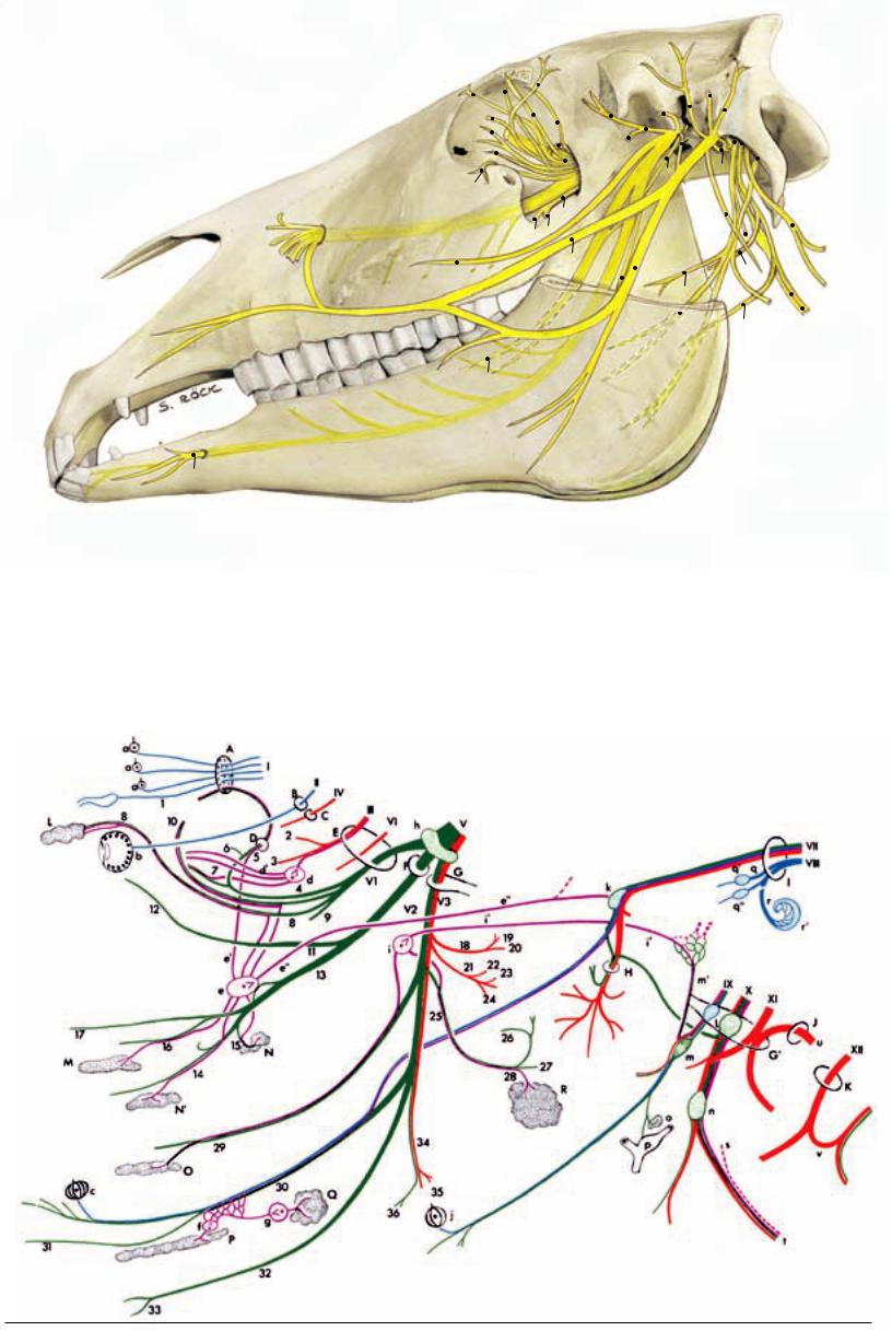
4. Cranial Nerves
Nerv |
Fig. |
Name (fiber type) |
Innervation |
Comments |
I |
|
Olfactory n. (special sense: olfaction) |
Olfactory region in nasal cavity |
Orgin: olfactory bulb; leaves skull by cribri- |
|
|
|
|
form plate |
(1) |
|
Vomeronasal n. (special sense: |
Vomeronasal organ |
|
|
olfaction) |
|
II |
|
Optic n. (special sense: sight) |
Optic part of retina |
III |
41.17 |
Oculomotor n. (s, m, psy) |
|
(2) |
|
Dorsal branch (m) |
Dorsal rectus, levator palp. sup., retractor |
|
|
|
bulbi muscles |
(3) |
|
Ventral branch ( m, psy) |
Medial and ventral recti, ventral oblique |
|
|
|
muscles |
IV |
41.11 |
Trochlear n. (m) |
Dorsal oblique muscle |
V |
|
Trigeminal n. |
|
VI |
|
Ophthalmic n. (s) |
Skin surrounding eyes, ethmoid bone, |
|
|
|
lacrimal gl., upper eyelid |
(4) |
41.21 |
Nasociliary n. (s) |
|
(5) |
41.20 |
Ethmoidal n. (s) |
Olfactory area of nasal cavity |
(6) |
41.19 |
Infratrochlear n. (s) |
Conjunctiva, 3rd eyelid, lacrimal caruncle |
|
|
|
and skin near medial angle of eye |
(7) |
41.22 |
Long cilicary nn. (s, psy) |
Iris, cornea, ciliary m. |
(8) |
41.12 |
Lacrimal n. (s, psy, sy) |
Lacrimal gl., skin and conjunctiva near lat. |
|
|
|
angle of eye |
(9) |
41.9 |
Frontal n. (s) |
Skin of forehead and upper eyelid; br. to |
|
|
|
frontal sinus |
(10) |
41.10 |
Zygomaticotemporal br. |
Skin of temporal region, lacrimal gl. |
|
|
(s, psy) |
|
V2 |
39.27 |
Maxillary n. (s) |
|
(11) |
41.27 |
Zygomatic n. |
|
(12) |
41.16 |
Zygomaticofacial br. (s) |
Lower eyelid, tarsal gll. |
(13)Pterygopalatine n. (s, psy)
(14) |
41.d |
Greater palatine n. (s, psy) |
Mucosa and gll. of hard palate |
(15) |
41.e |
Lesser palatine n. (s, psy) |
Mucosa and gll. of soft palate |
(16) |
41.c |
Caudal nasal n. (s) |
Mucosa of nasal cavity and gums |
(17) |
39.27' |
Infraorbital n. (s) |
Skin of nose, nostrils and upper lip; also |
|
41.b |
|
via alveolar brr. upper teeth and maxillary |
|
|
|
sinuses |
Developmentally an evagination of the diencephalon; leaves skull by optic foramen Origin: mesencephalon; leaves skull by orbital fissure
Origin: mesencephalon; leaves skull by trochlear foramen
Origin: rhombencephalon; nerve of 1st pharyngeal arch
Leaves skull by orbital fissure
Detaches a br. to frontal sinus
Leaves orbit by trochlear notch; brr. to frontal sinus
Receive psy.* fibers from ciliary gangl.
Usually double; receives psy. fibers from pterygopalatine gangl. and sy. fibers from int. carotid plexus
Perforates periorbita and leaves orbit through supraorb. for. where it becomes supraorbital n. (41.8)
Receives psy. fibers from pterygopalatine gangl.
Leaves skull by round foramen
Leave orbit near lat. angle of eye Receives psy. fibers from pterygopalatine gangl.
Passes through gr. palatine canal
Passes through sphenopalatine for. Leaves skull by infraorbital for.
V3 |
39.23 |
Mandibular n. (s, m) |
(18)Masticatory n. (m)
(19) |
|
Deep temporal nn. (m) |
Temporalis muscle |
(20) |
39.7 |
Masseteric n. (m) |
Masseter muscle |
(21) |
|
Medial pterygoid n. (m) |
Med. pterygoid muscle |
(22) |
|
Tensor tympani n. (m) |
Tensor tympani muscle |
(23) |
|
Tensor veli palatini n. (m) |
Tensor veli palatini |
(24) |
|
Lateral pterygoid n. (m) |
Lat. pterygoid muscle |
(25) |
39.21 |
Auriculotemporal n. (s, sy, psy) |
|
(26) |
37.26 |
Transverse facial br. (s) |
Skin of masseteric region and over tem- |
|
|
|
poromandibular joint |
(27) |
|
Communicating br. (s) |
Carries sensory fibers to dorsal and ventral |
|
|
|
buccal brr. of facial n (VII) |
(28) |
|
Parotid brr. (s, psy) |
Parotid gl. |
(29) |
|
Buccal n. (s, psy) |
Buccal gll. and mucosa of buccal part of |
|
|
|
oral cavity |
(30) |
39.13 |
Lingual n. (s, psy, and gustatory) |
Mucosa of apex and body of tongue |
(31) |
|
Sublingual n. (s) |
Mucosa of prefrenular part of oral cavity |
(32) |
39.25 |
Inferior alveolar n. (s) |
Lower teeth and gums |
(33) |
37.i |
Mental n. (s) |
Skin of lower lip and chin |
(34) |
39.15 |
Mylohyoid n. (m) |
Mylohyoideus muscle |
(35) |
|
Rostral digastric br. (m) |
Rostral belly of digastricus |
(36) |
|
Submental brr. (s) |
Skin of rostral part of intermandibular |
|
|
|
space |
Leaves skull by oval notch of for. lacerum
Passes laterally through mandibular notch At its origin lies the small otic gangl.
Receives psy. fibers from otic gangl Detaches brr. to temporomandibular joint
The sensory fibers are thus distributed to the side of the face
Bring psy. fibers to the parotid gl. Receives psy. fibers from otic gangl.
Receives psy. and gustatory fibers via chorda tympani (VII)
Enters mandible by mandibular for. Leaves mandible by mental for.
108
Fiber quality: m = motor; s = sensory; sy = sympathetic; psy = parasympathetic
Cranial Nerves
|
6 |
|
|
|
|
|
|
|
|
|
|
|
|
|
|
|
|
|
|
|
|
|
|
|
|
|
|
|
40 |
|
|
|
|
|
|
|
|
|
|
|
|
|
|
|
|
|||
|
|
|
|
|
|
|
|
|
|
10 |
|
|
|
|
|
|
|
18 |
|
|
|
|
|
|
|
|
|
|
41 |
|
|
|
|
|
|
|
|
|
||||||||||
|
|
|
|
|
|
|
|
|
|
|
|
|
|
|
|
|
|
|
|
|
|
|
|
|
|
|
|
|
|
|
||||||||||||||||||
|
|
|
|
|
|
|
|
|
|
|
9 |
|
|
|
|
|
|
|
|
|
|
|
|
|
|
|
|
|
|
|
|
|
|
|
|
|
|
|
|
|
||||||||
|
|
|
|
|
|
|
|
|
|
|
|
|
|
|
|
|
|
|
|
|
|
|
|
|
|
|
|
|
|
|
|
|
|
|
|
|
|
|
|
|||||||||
|
|
|
|
|
|
|
|
|
|
|
|
|
|
|
|
|
|
|
|
|
|
|
|
|
|
|
|
|
|
|
|
|
|
|
|
|
|
|
||||||||||
|
3 |
|
|
|
|
|
|
|
|
|
|
|
|
|
|
|
|
|
|
|
|
|
|
|
|
|
|
IX |
||||||||||||||||||||
|
|
|
|
|
|
|
|
|
|
|
20 |
|
|
|
|
|
|
|
V3 27 |
VII |
||||||||||||||||||||||||||||
|
|
|
|
|
|
|
|
|
|
|||||||||||||||||||||||||||||||||||||||
|
|
|
|
|
|
|
|
|
|
|
|
|
|
|
|
|
|
|
|
|
|
|
||||||||||||||||||||||||||
|
|
|
|
|
|
|
|
|
|
|
|
|
|
5 |
|
|
|
|
|
|
|
|
|
|
|
|
X |
|||||||||||||||||||||
|
|
VI |
|
|
|
|
|
|
|
|
|
|
|
|
|
|
|
|
|
|
|
|
|
|
|
|
||||||||||||||||||||||
|
|
|
|
|
|
|
|
|
|
|
|
|
|
|
|
|
|
|
|
|
|
|
|
|
|
|
|
|
||||||||||||||||||||
|
|
|
|
|
|
|
|
|
|
|
|
|
|
|
4 |
|
|
|
|
39 |
|
|
|
|
|
|
|
|
|
|
|
|
|
XII |
||||||||||||||
|
|
|
|
|
|
|
|
|
|
|
|
|
|
|
|
|
|
|
|
|
|
|
|
|
|
|
||||||||||||||||||||||
|
|
|
|
|
|
|
|
|
|
|
|
|
|
|
|
|
|
|
|
|
|
|
|
|||||||||||||||||||||||||
|
|
|
|
|
|
|
|
|
|
|
|
|
|
V2 |
|
|
|
|
|
|
|
|
|
|
|
|
|
|
|
|
|
|
|
|
|
|
|
|
||||||||||
|
12 |
|
|
|
|
|
|
|
|
|
|
|
|
|
|
|
|
|
|
|
|
|
|
|
|
|
|
|
|
|
|
|
|
|
|
|
|
|
|
|
||||||||
|
|
|
|
|
|
|
|
|
|
|
|
|
|
|
|
|
|
|
|
|
|
|
|
|
|
|
|
|
|
|
|
|
|
|
|
|
|
|
|
|
|
|
|
|
||||
|
|
|
|
|
|
13 |
|
|
|
|
|
|
|
|
|
|
|
|
|
|
|
|
|
|
|
|
|
|
|
|
|
|
|
|
|
|
|
XI |
||||||||||
|
|
|
|
|
|
|
|
|
|
|
|
|
|
|
|
|
|
|
|
|
|
|
|
|
|
|
|
|
|
|
|
|
|
|||||||||||||||
17 |
29 |
|
|
|
15 |
|
|
|
|
|
|
|
|
|
|
|
|
|
|
|
|
|
|
49 |
|
|
|
|
|
|
|
|
|
|
|
|
||||||||||||
|
|
|
|
|
|
|
|
|
|
|
|
|
|
|
|
|
|
|
|
|
|
|
|
|
|
|
|
|
|
|
|
|
||||||||||||||||
|
|
|
|
|
|
|
|
|
|
|
|
|
|
|
|
|
|
|
|
|
|
|
|
|
|
|
|
|
|
|
|
|
|
|
||||||||||||||
|
|
|
|
|
|
|
|
|
|
|
|
|
|
|
|
|
|
|
|
|
|
|
|
|
|
|
|
|
|
|
|
|
|
|
|
|
|
|
|
|
|
|
|
|
|
|
||
|
|
|
|
|
|
30 |
32 |
|
|
|
|
|
44 |
|
|
|
|
|
|
|
|
|
|
|
|
|
|
|
|
|
|
|
|
|||||||||||||||
|
|
|
|
|
|
|
|
|
|
|
|
|
|
|
|
|
|
|
|
|
|
|
|
|
|
|
|
|
|
|
||||||||||||||||||
|
|
|
|
|
|
|
|
|
|
|
|
|
|
|
|
|
|
|
|
|
|
|
|
|
|
|
|
|
||||||||||||||||||||
|
|
|
|
|
|
|
|
34 |
|
|
|
|
|
|
|
|
|
|
|
|
|
|
|
|
|
|
|
|
|
|||||||||||||||||||
|
|
|
|
|
|
|
|
|
|
|
|
|
|
|
|
|
|
|
|
|
|
|
|
|
|
|
|
|
|
|
|
|
|
|
|
|
|
s |
|
|
|
|
|
|
||||
|
|
|
|
|
|
|
|
|
|
|
|
|
|
|
|
|
|
|
|
|
|
|
48 |
|
|
|
|
|
|
|
|
|
|
|
|
|
|
|
|
t |
||||||||
|
|
|
|
|
|
|
|
|
|
|
|
|
|
|
|
|
|
|
|
|
|
|
|
|
|
|
|
|||||||||||||||||||||
|
|
|
|
|
|
|
|
|
|
|
|
|
|
|
|
|
|
|
|
|
|
|
|
|
|
|
|
|
|
|
|
|
|
|
|
|
|
|
|
|
|
|
|
|
|
|
|
|
|
|
|
|
|
|
|
|
|
|
|
|
|
|
|
|
|
|
|
|
|
|
|
|
|
|
|
|
|
|
|
|
|
|
|
|
|
|
|
|
|
|
|
|
|
|
|
|
|
 53 50
53 50
31
33
A |
Cribriform plate |
K |
Hypoglossal canal |
a |
Olfactory region |
j |
Vallate and foliate papillae |
s |
trunk |
B |
Optic canal |
L |
Lacrimal gland |
b |
Retina |
k |
Geniculate ganglion |
s' |
cervical ganglion |
C |
Trochlear foramen |
M |
Nasal glands |
c |
Fungiform papillae |
l |
Proximal ganglia |
t |
trunk |
D |
Ethmoidal foramen |
N Palatine glands in soft |
d |
Ciliary ganglion |
m |
Distal (petrosal) ganglion |
u |
root of accessory nerve |
|
E |
Orbital fissure |
|
palate |
d' |
Short ciliary nerves |
m' Tympanic nerve |
v |
cervicalis |
|
F |
Round foramen |
N' Palatine glands in hard |
e |
Pterygopalatine ganglion |
n |
Distal (nodose) ganglion |
|
|
|
G |
Oval notch of foramen lacerum |
|
palate |
e' |
Orbital branches |
o |
Carotid body |
|
|
G’ Jugular foramen |
O |
Buccal glands |
e'' Nerve of pterygoid canal |
p |
Carotid sinus |
● |
of special senses |
||
H |
Stylomastoid foramen |
P |
Sublingual gland |
f |
Sublingual ganglion |
q |
Vestibular nerve |
● |
neuron |
I |
Internal acoustic meatus |
Q |
Mandibular gland |
g |
Mandibular ganglion |
q' |
Superior vestibular ganglion |
● |
neuron |
J |
Foramen magnum |
R |
Parotid gland |
h |
Trigeminal ganglion |
q'' Inferior vestibular ganglion |
● |
neuron |
|
|
|
|
|
i |
Otic ganglion |
r |
Cochlear nerve |
● |
neuron |
|
|
|
|
i' |
Minor petrosal nerve |
r' |
Spiral ganglion |
|
|
109

Cranial Nerves
Nerv |
Fig. |
Name (fiber type) |
Innervation |
Comments |
VI |
41.28 |
Abducent n. (m) |
Lateral rectus, lateral portion of retractor |
Origin: rhombencephalon; leaves skull by |
|
|
|
bulbi |
orbital fissure |
VII |
37.18 |
Facial n. (m, s, psy, gustatory) |
|
Origin: medulla oblongata; has facial and in- |
|
|
|
|
termediate components; enters int. acoustic |
|
|
|
|
meatus, bears geniculate gangl. in facial |
|
|
|
|
canal; leaves skull by stylomastoid for.; n. of |
|
|
|
|
2nd pharyngeal arch |
(37) |
|
Greater petrosal n. (psy) |
Gll. of nasal cavity and palate |
Runs in petrosal canal; joins deep petrosal n. |
|
|
|
|
(sy.) to form n. of petrosal canal which goes |
(38) |
|
Stapedial n. (m) |
Stapedius muscle |
to pterygopalatine gangl. |
|
|
|||
(39) |
|
Chorda tympani (gustatory, psy) |
Salivary gll. |
Leaves petrous temporal bone by petrotym- |
|
|
|
|
panic fissure and joins lingual n. (V3) |
(40) |
37.27 |
Internal auricular br. (s) |
Skin on int. surface of external ear |
Is joined by auricular br. of vagus |
(41) |
37.30 |
Caudal auricular n. (m) |
Muscles of external ear |
Is joined by dorsal brr. of first two cervical |
|
|
|
|
nn. |
(42) |
|
Auriculopalpebral n. (m) |
Orbicularis oculi, interscutularis, zygoma- |
Joins brr.of auriculotemporal n. (V3) |
|
|
|
ticus, levator nasolabialis, mm. of external |
|
|
|
|
ear |
|
(43) |
37.12 |
Dorsal buccal br. (m) |
Muscles of upper lip and nose |
Receives communic. br. from auriculotempo- |
|
|
|
|
ral n. (V3); n. injured in facial paralysis |
(44) |
37.13 |
Ventral buccal br. (m) |
Muscles of cheek and lower lip |
(45) |
|
Cervical br. (m) |
Parotidoauricularis, cut. mm. of head and |
|
|
|
neck |
(46) |
|
Digastric br. (m) |
Caudal belly of digastricus |
(47) |
|
Stylohyoideus brr. (m) |
Stylohyoideus |
VIII |
|
Vestibulocochlear n. (special sense: |
Macula of utriculus and sacculus, am- |
|
|
balance and hearing) |
pullary crests, spiral gangl. of cochlea |
IX |
47.28 |
Glossopharyngeal n. |
|
|
51.3 |
(m, s, psy, gustatory) |
|
(48) |
51.4 |
Pharyngeal br. (s, m) |
Pharyngeal mucosa, stylopharyngeus |
(49) |
51.c |
Carotid sinus br. (s) |
Baroreceptors in wall of carotid sinus, |
|
|
|
chemoreceptors in carotid body |
(50) |
51.5 |
Lingual brr. (s, gustatory) |
Mucosa of root of tongue and pharynx |
X |
47.26 |
Vagus (s, m, psy) |
|
(51) |
|
Auricular br. (s) |
Skin of external acoustic meatus |
(52) |
51.d |
Pharyngeal br. (s, m) |
Pharyngeal mm. except stylopharyngeus; |
|
|
|
mucosa of pharynx and larynx |
(53)Cranial laryngeal n. (s, m)
(54) |
49.a |
• Internal br. (s) |
Laryngeal mucosa rostral to glottis |
(55) |
49.b |
• External br. (m) |
Cricothyroideus |
|
|
Cardiac brr. (psy) |
Heart |
|
|
Recurrent (caudal) laryngeal n. |
Brr. to trachea and esophagus; laryngeal |
|
|
(m, s, psy) |
mm. except cricothyroideus; laryngeal |
|
|
|
mucosa caudal to glottis |
Unites with ventral brr. of cervical nn.
Origin: rhombencephalon; into petrous temporal bone through int. acoustic meatus
Origin: medulla oblongata; leaves skull by jugular for.; n. of 3rd pharyngeal arch; lies in a fold of guttural pouch
Takes part in forming pharyngeal plexus
Communicates with lingual n. (V3)
Origin: medulla oblongata; leaves skull by jugular for.; n. of 4th pharyngeal arch; in a fold of guttural pouch
Communicates with int. auricular br. (VII) Takes part in forming pharyngeal plexus
Arises from distal gangl.
Passes into larynx through thyroid for.
Leaves vagus in thorax; passes up the neck; thought to be involved in “roaring”
XI |
47.23 |
Accessory n. (m) |
Origin of cranial root: medulla oblongata, |
|
51.15 |
• Cranial root |
spinal root: cervical spinal cord; leaves skull |
|
|
• Spinal root |
by jugular for.; lies against guttural pouch |
|
(56) |
Internal br. (m) |
Connects with vagus |
(57)External br. (m)
|
51.r |
• Dorsal br. (m) |
Trapezius |
|
|
51.q |
• Ventral br. (m) |
Sternocephalicus |
|
XII |
47.29 |
Hypoglossal n. (m) |
Intrinsic tongue musculature; genio-, |
Origin: medulla oblongata; leaves skull by |
|
51.14 |
|
stylo-, and hyoglossi |
hypoglossal canal; forms ansa cervicalis with |
|
|
|
|
ventral br. of C |
110
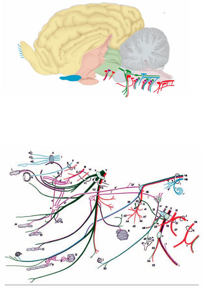
Cranial Nerves
Brain:
 Telencephalon
 Diencephalon
 Mesencephalon
 Metencephalon
Myelencephalon
I
|
|
|
II |
|
|
|
|
|
|
|
|
|
|
|
|
|
III |
IV |
|
|
|
|
|
|
|
|
|
|
|
VI |
|
|
|
|
|
|
|
|
|
|
|
|
|
VIII |
|
|
|
|
|
|
|
|
|
V1 |
|
VII |
IX |
XII |
|
|
|
|
|
|
|
V2 |
|
X XI |
|
|
|
|
|
|
|
|
|
V3 |
|
|
|
||
|
|
|
|
|
|
|
|
|
|
|
|
A |
Cribriform plate |
K |
Hypoglossal canal |
a |
Olfactory region |
|
j |
Vallate and foliate papillae |
s |
Sympathetic trunk |
|
B |
Optic canal |
L |
Lacrimal gland |
b |
Retina |
|
k |
Geniculate ganglion |
s' |
Cranial cervical ganglion |
|
C |
Trochlear foramen |
M |
Nasal glands |
c |
Fungiform papillae |
l |
Proximal ganglia |
t |
Vagosympathetic trunk |
||
D |
Ethmoidal foramen |
N Palatine glands in soft |
d |
Ciliary ganglion |
|
m |
Distal (petrosal) ganglion |
u |
Spinal root of accessory nerve |
||
E |
Orbital fissure |
|
palate |
d' |
Short ciliary nerves |
m' Tympanic nerve |
v |
Ansa cervicalis |
|||
F |
Round foramen |
N' Palatine glands in hard |
e |
Pterygopalatine ganglion |
n |
Distal (nodose) ganglion |
|
|
|||
G |
Oval notch of foramen lacerum |
|
palate |
e' |
Orbital branches |
|
o |
Carotid body |
|
|
|
G’ Jugular foramen |
O |
Buccal glands |
e'' Nerve of pterygoid canal |
p |
Carotid sinus |
|
● Neuron of special senses |
||||
H |
Stylomastoid foramen |
P |
Sublingual gland |
f |
Sublingual ganglion |
q |
Vestibular nerve |
● Sensory neuron |
|||
I |
Internal acoustic meatus |
Q |
Mandibular gland |
g |
Mandibular ganglion |
q' |
Superior vestibular ganglion |
● Parasympathetic neuron |
|||
J |
Foramen magnum |
R |
Parotid gland |
h |
Trigeminal ganglion |
q'' Inferior vestibular ganglion |
● Sympathetic neuron |
||||
|
|
|
|
i |
Otic ganglion |
|
r |
Cochlear nerve |
● Motor neuron |
||
|
|
|
|
i' |
Minor petrosal nerve |
r' |
Spiral ganglion |
|
|
||
111

Contributions to Clinical-Functional Anatomy
Arthroscopy and Endoscopy in Veterinary
Medicine |
ASTRID B. M. RIJKENHUIZEN |
General Remarks. Veterinary medicine today without arthroscopy (Gr. arthros = joint; scopein = to examine) in the treatment of joint diseases is something that cannot be imagined. This so-called keyhole surgery, or also minimally invasive surgery, is realized by application of the arthroscope, an optical system of lenses. It is one of the greatest advances of the last decade in the recognition and treatment of joint diseases and has almost completely replaced the conventional surgery of joints. In contrast to conventional surgery with a wide access, this minimally invasive procedure has several advantages. These are less stress for the horse, better demonstration of the anatomical structures of the joint, the minimizing of soft tissue trauma, shorter operation times for many manipulations, less postoperative pain, extension of therapeutic possibilities, shorter healing time, a better cosmetic result, and a quicker recovery of efficiency. A substantial advantage of this technically high quality method is improved diagnostics should the clinical and radiographic as well as the sonographic examination fail to clarify the problem. Endoscopy also serves for diagnostic examination and minimally invasive manipulations at the internal surfaces of the body cavities and hollow organs, for example the gastrointestinal tract. The procedure is similar to arthroscopy with application of similar methods and instruments.
Instruments and Objectives of Arthroscopy. Arthroscopy is utilized predominantly for endoscopic examination of joints but also for other synovial structures such as synovial sheaths (tendovaginoscopy) and synovial bursae (bursascopy). In doing this, the arthroscope is introduced through a small incision of the skin and soft tissues into joint cavities or synovial sheaths or bursae. The visual impression is transmitted by a camera to a monitor to facilitate orientation to the surgeon. In this way the internal structures of the joint, synovial sheath or bursa can be directly observed. The surgeon sees these internal structures only by way of the monitor. Installation of the monitor is at shoulder-level of the surgeon and faces the surgeon. Additional mechanical or surgical instruments can be introduced to permit minimally invasive treatments; for example, the removal of bone or bone-fragments.
What Possibilities of Evaluation are Provided by Arthroscopy? Cartilage, bone, the joint capsule with synovial and fibrous layers, menisci, collateral ligaments, cruciate ligaments, and tendons associated with the joints as, for example, the long digital extensor, will be assessed.
•Degenerative changes (among other things, frayed ligaments, formation of fissures, traces of where the bones have rubbed against and damaged one another, partial losses of bone, rarefaction or thickening or detachment of cartilage, arthrotic alterations such as marginal swellings, cysts of articular cartilage).
•Inflammatory changes of the joint capsule (among other things, number, size and shape of synovial villi), hyperemic changes, adhesions as well as partial or complete rupture).
•Traumatic changes (fragments, etc.).
Possible Applications of Arthroscopy
a.Diagnosis: When clinical or diagnostic imaging examination does not result in a certain diagnosis, direct arthroscopic visualization of the intraarticular structures may deliver additional information. Also a possibility exists to obtain biopsy material of altered structures directly viewed.
b.Instituting therapy at the same time that the diagnosis is made.
• Removal of osseous or cartilaginous free bodies (corpora libera) that are occurring freely in synovia;
• In joint inflammation, synoviectomy for bacterial examination and culture (partial removal of the altered stratum synoviale);
• Tapping of synovia for bacterial examination;
• Arthroscopic procedures of osteosynthesis; for example, fractures of the third carpal bone (os carpale III);
• Synoviectomy (partial removal of the ulcerated synovial membrane) in cases of arthritis;
• Removal of osteochondromas (tumors of osseous and cartilaginous tissue);
• Smoothing and suturing of fissures in tendons or menisci;
• Flushing of the joint cavity to remove articular detritus and infectious material, for example, during treatment of articular hemorrhage and septic arthritis; this can be done with visual monitoring of the procedure:
• Removal of cysts and necrotic tissue;
• Smoothing of marginal excrescences of joints;
• Reconstruction of cruciate ligament in case of rupture.
Arthroscopy is most suited for treatment of osteochondrosis dissecans.
Which joints and synovial structures are treated arthroscopically?
Almost every joint is suitable for arthroscopy. Nowadays, it is applied in the examination and treatment of the hock joint, fetlock joint, femoropatellar joint, femorotibial joint, radiocarpal an intercarpal joints. But also the coffin joint, pastern joint, hip joint, shoulder, elbow, and temporomandibular joints have possible application. For tendovaginoscopy in the horse, the flexor aspect of the fetlock region, carpal region, and the tendon sheaths of the flexor aspect of the hock joint are suitable. For bursoscopy, the navicular bursa, intertubercular bursa and the subtendinous and calcaneal bursae and also acquired (subcutaneous) bursae can be examined.
Virtually any structure that is filled with synovial fluid can be examined endoscopically.
What instruments will be used?
The main instrument that is used is a rigid arthroscope (4 mm diameter) with a viewing angle of 0° to 30° or 45° or 70°. It contains optical device that is connected by a light-conducting cable to a cold light source (150–300 watt). Smaller optical instruments with a diameter of 2.7 mm can be used for smaller joints, for example, the coffin joint. Usually longer optical instruments are employed for the femoropatellar joint in order to assess the articular surfaces distal to the patella. The angle of view can be altered simply by turning the optical device without moving the entire instrument. In this way, the operator is able to hold the optical device away from other instruments and is thus able to protect it from damage during manipulation. The optical device is connected to a video camera that is connected to a monitor and digital image processor for recording.
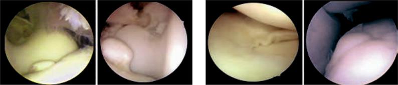
Fig. 1. Arthroscopy of the tarsal tendon sheath. (Courtesy of Prof. Dr. Rijken- 112 huizen)
Fig. 2. Tendon and tendon sheath, opened wedge-like. The inner lining of the tendon sheath (peritendineum) is formed as a double-layered mesotendineum that surrounds the tendon as an epitendineum. The connective tissue that conducts the vessels can, as a vinculum tendineum, pass from the tendon to the underlying bone. (Courtesy of Institut f. Veterinär-Anatomie, Berlin)
A pump is used to expand the joint cavity with fluid or gas. This maintains a constant pressure in the joint during manipulations.
This expansion is necessary to provide space within the joint for a far reaching inspection and surgical manipulations. Trocars are used to introduce the instrument, which is pushed through the trocar. The passage within the trocar is sealed by valves. In this way, neither gas nor fluid escapes from the joint during the changing of instruments. The passage of the optical instrument through the different compartments of the joint space is essential for examination of the joint. The operator is able to evaluate the individual structures one after the other. As mentioned, arthroscopy is used not only for diagnosis but at the same time for minimally invasive manipulations. Special instruments have been developed for this purpose. They are also introduced by special access to the joint. Among these are hand instruments such as punch, pliers, scissors, knives, curettes and rasps and, in addition, motor driven instruments for smoothing articular cartilage and menisci. Laser and thermocautery instruments are also used.
How is the arthroscope used?
The positioning of the patient in lateral or dorsal recumbency depends on the manipulation to be performed. This should be done in such a way that movement, manipulation, of the joint to be examined is possible. As in conventional operations the surgery is realized under aseptic conditions. First, a cannula is introduced into the joint and by this means the joint is filled with physiological saline or gas (carbon dioxide or air) and the joint cavity expanded. After that, a small incision is made in the skin and, using a blunt or sharp trocar, the arthroscope is introduced into the joint space after which the trocar is replaced by the optical instrument. One or two additional joint openings are made for the introduction of special surgical instruments. Certain manipulations such as the extirpation of an osteochondroma, are carried out after the ligation of blood vessels. For minimally invasive procedures, the principle of the socalled triangular technique is employed. This means that the surgical instruments and the optical instrument are introduced at the points of a triangle. The triangle technique provides optimal viewing with the necessary freedom of movement and, besides that, additional protection for the sensitive optical instrument. Later, sutures close the small openings in the joint needed for the operation. If possible, a sterile dressing is applied.
Follow-up treatment in arthroscopic manipulations
Depending on the indication and seriousness of the operation, the limbs and joints are fully weight-bearing following the procedure.
If fragments of bone and cartilage were removed, a follow-up control radiograph is appropriate.
Complications of arthroscopy
During the procedure, technical problems may occur that impair visibility. Damage to blood vessels may lead to hemorrhage and reduce visibility. This impairment of visibility can be removed by
increasing fluid-flow or by irrigation. Iatrogenic complications as, for example, injuries to cartilage, blood vessels, and nerves that are caused by the treating veterinarian must be considered. Synovial fluid may leak out at the injection site. There may be herniation at the site of incision. Occasionally, there may be observed an increase in secondary periosteal bone or ossifications within the joint. Infections are dreaded complications that, in the extreme, may result in ankylosis.
Special preconditions for successful endoscopy
To carry out a procedure like this in which direct observation of the field of operation is lacking, orientation is exclusively by monitor, and there is a loss of the three dimensional sense that requires a systematic training. Solid anatomical knowledge and a rich experience are essential preconditions for successful minimally invasive manipulations. A well-adjusted team of surgeons, a trained staff and a well equipped operating room are additional requirements for successful surgery.
Endoscopy of the hock joint (talocrural joint, Fig. 3)
Arthroscopy of the talocrural joint is most common because therapeutic options are favorable here and a good overview can be obtained by expanding the joint space. The dorsal and plantar parts of this compound joint are most valuable. Positioning: dorsal or lateral recumbency. Access: Standard access is dorsomedial, as far distal as possible. Indications: osteochondrosis dissecans (OCD) and traumata of the articular ridges of the trochlea of the talus, of the tibial cochlea as well as the lateral and medial malleoli.
Arthroscopy of the stifle joint (Fig. 4)
Arthroscopy of the stifle joint is often done. The stifle or genual joint is subdivided into a femoropatellar joint with its own joint capsule and the femorotibial joint with medial and lateral joint capsules. Orientation within the femoropatellar joint is excellent. In the femorotibial joint only the cranial and caudal parts are visible. Positioning: dorsal recumbency or, in rare cases, lateral recumbency. Access: Standard access for the femoropatellar joint is craniomedial, between the medial and intermediate patellar ligaments. But access can also be achieved by splitting the intermediate patellar ligament or entering between the lateral and intermediate patellar ligaments. Starting from the femoropatellar joint access can be gained to the femorotibial joint. The medial joint can be separately examined by positioning the arthroscope between the medial and intermediate patellar ligaments, directing the instrument caudoproximally and somewhat axially. The lateral joint cavity can also be reached from this access.
•Indications ([1], [2]):
•Osteochondrosis dissecans (OCD) of the medial ridge of the trochlea
•Cysts of the medial condyle
•Cruciate ligament repair following rupture of the ligament
•Repair of the meniscus following meniscal tears
Fig. 3. Arthroscopy of the tarsocrural joint. Photographs of an OCD of the sagittal |
Fig. 4. Arthroscopy of the stifle joint. Left: Meniscus of the lateral femorotibial joint. |
|
ridge of the tibia. (Courtesy of Prof. Dr. Rijkenhuizen) |
Right: OCD of the lateral ridge of the femur (femoropatellar joint). (Courtesy of |
|
|
Prof. Dr. Rijkenhuizen) |
|
|
|
113 |
Thoracic Limb |
BIANCA PATAN |
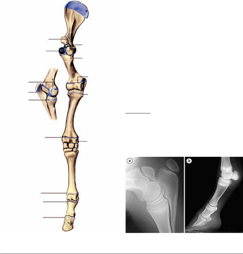
4.1. Ossification of the individual thoracic limb bones takes place predominantly from several ossification centers, which are separated from each other by cartilaginous growth zones (growth plates or physes). Some of these apophyses and epiphyseal growh plates close only after birth and for this reason, for a certain time, are still demonstrable by radiograph (Figs. 4.1.1, 4.1.2). [3] Knowledge of their localization and of the time of closure is of paramount clinical importance as the growth plates may feign fracture lines in young animals. Moreover, they represent a certain locus minoris resistentiae (weak point) and fractures of the growth plates (so-called Salter-Harris fractures) are not rare. Depending on the configuration of the fracture line or the involvement of the epiphysis and/or metaphysis, 6 types can be differentiated. [5] On the thoracic limb, the proximal growth plates of the ulna and radius as well as the distal plates of the humerus, scapula, radius and Mc III (cannon bone) are most frequently encountered. [4] Treatment and prognosis depend on the kind of fracture. In most cases, a surgical approach (internal fixation of the fracture fragments by osteosynthesis) is indicated. [6] [5] If the fracture is stable and there is no dislocation of the bone fragments and no joint involvement, a conservative treatment is possible. [6]
12–24 months
ca. 5 months
approx. 3–4 months
24–36 months
11–24 months
11–24 months
24–36 months
11–24 months
11–24 months
(medial view)
20–24 months
up to 12 months
UQL|XbVxUmAVqC+HnIZ42uaraA==|1288009770 |
approx. 6 months |
|
|
|
approx. 12 months |
|
8–12 months |
Fig. 4.1.1. Apophyseal and epiphyseal lines of fusion of the thoracic limb of the horse. (Closure of the growth plates after Butler et al., 2000). (Courtesy of Institut
114 f. Veterinär-Anatomie, Berlin)
Unequal growth in the area of the growth plate can result in angular limb deformities. In such cases, an axial deviation of the distal limb in a lateral direction is called a valgus position; an axial deviation in the medial direction, a varus position. [7] [8] The distal epiphysis of the radius is most often affected, in which, because of a greater growth potential on the medial aspect, a carpus valgus results. [7] In addition to the asymmetrical growth of the growth plate cartilage, an incomplete ossification and deformation of the carpal bones may be the cause of the axial deviation. [8] [9] Moreover, very often a low-grade carpus valgus may develop in newborn foals as a result of slack periarticular ligaments. This usually corrects itself during the first postnatal weeks. [8] [9] On the basis of radiographs in which lines through the longitudinal axis of the radius and metacarpal bone III are drawn, the origin and degree of severity of the axial deviation (intersection or angular deviation of the lines) can be determined (Fig. 4.4). [10] A special form of axial deviation in the carpal region (offset knee or bench knee) consists of a valgus deviation at the distal end of the radius and a varus deviation at the proximal metacarpal bone III. [7] For the correction of axial deviations in the area of the growth plate, also surgical manipulations, which have an impact on bone growth in the growth plate, are in use besides conservative methods of treatment. In this way, the growth of the bone can be inhibited on the convex aspect, actually the growth plate can be overbridged temporarily by suitable osteosynthetic procedures. [8] [9] Alternatively or in combination with this, bone growth on the concave aspect can be stimulated by an incision and lifting of the periosteum. [7] [8] [9] The usefulness of the foregoing is, to be sure, controverted. [11] In both surgical procedures, the time of growth plate closure must be taken into account. [7] [8]
4.2.Scapula. Fractures are relatively rare. Most often, the supraglenoid tubercle is affected in which case the fracture-line usually reaches the shoulder joint. Owing to the pull of the attaching muscles (biceps brachii and coracobrachialis muscles), the fragments are as a rule displaced cranially and distally. [12] [13]
4.3.Fractures of the humerus occur often as oblique or spiral fractures of the metaphysis or of the diaphysis. [14] Avulsions in the area of the deltoid tuberosity or of the tubercles can also occasionally be observed. [13] In addition, incomplete fractures due to fatigue or stress fractures (fissures) in young thoroughbreds in training are not rare. In the proximal part of the bone, the caudolateral cortex is predominantly affected; distally, the fractures are often located craniomedially or caudally. For diagnosis, scintigraphya is the method of choice as the fracture lines are sometimes not recognizable radiographically. [15] [16].
aImage-producing method with the use of a radioactive substance (as a rule, technetium 99m) that, following intravenous injection, is selectively concentrated in a specific tissue. For scintigraphy of bone, the radioactive isotope is coupled to a bone marker (polyphosphonate) that, with active bone formation, is accumulated in hydroxyapatite crystals. The radiation given off with radioactive breakdown can be recorded with the help of a so-called gamma-camera in which case there is a locally elevated radiation intensity, which points to an increased metabolic activity in a particular bone.
Fig. 4.1.2. Growth plates in the region of the elbow (a) and on the distal limb (b) of a 2.5 months old warmblood filly-foal. (Courtesy of Klinik für Orthopädie bei Huf- u. Klauentieren, Veterinärmed. Univ. Wien)