книга / MasterPass _ Pharmacology in 7 Days for Medical Students
.pdf
PHARMACOLOGY IN 7 DAYS FOR MEDICAL STUDENTS
channels in outer membrane of bacteria. It is believed that beta-lactam ring (6-amino- penicillanic acid nucleus) of the bacteria is a structural analogue of D-alanyl-D-alanine. During transpeptidation in the presence of penicillins, beta-lactam ring is incorporated in the cross links rather than D-alanyl-D-alanine and this renders that cross links between peptidoglycan chains weak.
Cephalosporins
1They bind to specific penicillin biding protein (PBP)-receptors on bacteria causing inhibition of cell wall synthesis by blocking transpeptidation of peptidoglycans.
2They also cause activation of autolytic enzymes in the cell wall causing bacterial death.
Sulfonamides
They are bacteriostatic and inhibit protein synthesis. Sulfonamide susceptible organisms, unlike mammals, cannot use exogenous folate but must synthesise it from PABA.
This pathway is essential for the synthesis of purines and nucleic acids. Since sulfonamides are structural analogues of PABA, they inhibit dihydropteroate synthase and thus folate production. Sulfonamides inhibit both gram positive and negative bacteria. Rickettsiae are not inhibited, rather stimulated by sulfonamides. Combination of a sulfonamide with an inhibitor of dihydrofolate reductase (like trimethoprim) provides synergistic activity because of sequential inhibition of folate synthesis.
Tetracyclines
Tetracyclines are bacteriostatic and act by inhibiting bacterial protein synthesis at ribosomal level. They reach their site of action (30S ribosomal subunit of susceptible bacteria) through cell membrane ‘porin’ channels by simple diffusion or by active energy-dependent transport process. They bind reversibly to 30S ribosomal subunit and block biding of amino-acyl tRNA to the receptor site on mRNA-ribosomal complex. Addition of amino acids to the growing peptide chain is prevented thus inhibiting protein synthesis producing bacteriostatic effect.
Tetracyclines also interfere with oxidative phosphorylation, glucose oxidation, bacteria respiration and cell permeability.
Quinolones
They are bactericidal and block bacterial DNA synthesis by inhibiting the following enzymes:
1DNA gyrase (topoisomerase-II).
2Topoisomerase-IV.
Chloramphenicol
It is bacteriostatic for all susceptible microorganisms by protein synthesis inhibition at ribosomal level by binding reversibly to 50S ribosomal subunit near the binding site for macrolides and clindamycin (thus these drugs can interfere with each others actions). Chloramphenicol inhibits binding of amino acids containing aminoacyl tRNA to the acceptor site of the mRNA-ribosomal complex. Interaction between peptidyl transferase and its amino acid substrate is prevented. Amino acids are not added to the growing peptide chain so protein synthesis is inhibited.
74

MECHANISMS OF ACTION
Chloramphenicol may be bactericidal for Haemophilus influenzae, Nisseria meningitidis and some strains of bacteroides.
Amoebicides
Amoebicides irreversibly inhibit protein synthesis by blocking the movement of ribosomes along mRNA. They act only against amoebic trophozoites.
Aminoglycosides
They are bactericidal against aerobic, Gram –ve bacteria and some Gram +ve cocci. Aminoglycosides are irreversible inhibitors of protein synthesis. The initial event is passive diffusion via poring channels across the out membrane. Drug is then actively transported across the inner membrane into the cytoplasm by an oxygen-dependent process. The transmembrane electrochemical gradient supplies the energy for this process and transport is coupled to a proton pump. Low extracellular pH and anaerobic conditions inhibit the transport by reducing the gradient. Transport may be enhanced by drugs which inhibit cell wall synthesis such as β-lactam penicillins or vancomycin. This enhancement may be the basis of synergism of these antibiotics with aminoglycosides. Once inside the cytoplasm, aminoglycosides bind to specific 30S ribosomal subunit proteins (S12 in case of streptomycin). Protein synthesis is inhibited in at least three ways:
1Aminoglycosides distort the structure of 30S ribosomal subunit, thus interfering with the initiation of protein synthesis.
2Misreading of mRNA, which causes incorporation of incorrect amino acids into the peptide resulting in the formation of nonfunctional or toxic proteins.
3Break up of polysomes into nonfunctional monosomes.
These activities occur more or less simultaneously and the overall effect is irreversible and lethal for the cell.
Aminoglycosides also damage inner cytoplasmic membrane by incorporating defective proteins in the cytoplasmic bacterial membrane. The membrane loses its permeability throwing small and large molecules (structural and functional proteins) leading to bacterial cell death. This is called energy-dependent phase-II transport.
Macrolides
At usual doses, macrolides are bacteriostatic; at higher doses, however, they become bactericidal. Their binding site is either the same or in close proximity to that of clindamycin and chloramphenicol. These drugs bind with 50S subunit of the bacterial ribosome thus inhibiting the translocation step of protein synthesis. Additionally, they may also interfere with the transpeptidation step.
Metronidazole
This drug is particularly effective against anaerobic protozoan parasites (including entamoeba histolytica, etc.). These organisms possess electron-transport proteins that participate in electron removal reactions. The nitro group of metronidazole acts as an ‘electron acceptor’ resulting in formation of cytotoxic compounds that bind to proteins and DNA rendering them nonfunctional. This leads to cell death.
75

PHARMACOLOGY IN 7 DAYS FOR MEDICAL STUDENTS
Clonidine
Clonidine, being lipid soluble, crosses the BBB by simple diffusion and is taken up by the adrenergic neurons and the vasomotor centre of the brain. It is an active drug unlike α-methyl dopa, which is a pro-drug. Clonidine acts as an agonist on α2-receptors situated post-synaptically on the neurons. These α-receptors are α2A in nature. When stimulated, they cause increase in the negative feedback on the release of noradrenaline in brain stem resulting in decreased release from centre to periphery mainly in the heart. This causes fall in blood pressure especially in supine and upright positions by causing decrease in the force of contraction and heart rate leading to decrease in cardiac output. Bradycardia is more marked due to stimulation of parasympathetic system by the drug. Fall in blood pressure is accompanied by decrease in the concentration of noradrenaline and renin in the plasma. Postural hypotension is very mild and is seen in hypovolemic patients only. This is because of agonist effect on the peripheral α2B and α1 adrenergic receptors. In smooth muscles of blood vessels, it acts as an agonist causing vasoconstriction.
Alpha-methyl dopa
It is a structural analogue of levodopa. It is usually given orally. Its absorption through GIT is incomplete. The drug crosses the BBB by an active transport process responsible for the transport of aromatic amino acids. After having reached the brain, it is taken up by adrenergic neurons in the brain stem area where it is converted into α-methyl nor-dopamine, which in turn is converted into α-methyl nor-epinephrine. This process of conversion goes on side by side both in brain and in peripheral sympathetic adrenergic neurons. So it is concentrated in the intraneuronal granules in brain and in periphery and is stored there displacing noradrenaline from the storage vesicles. It is released as α-methyl epinephrine both in centre and in periphery. In periphery, it acts as α1 post-synaptic receptor agonist leading to vasoconstriction and increase in blood pressure. It does not act as antihypertensive peripherally. In the brain, α-methyl nor-epinephrine acts as agonist at α2 post-synaptic receptors causing increase in the negative feedback on release of noradrenaline. So less noradrenaline is released resulting in decreased sympathetic flow from centre to periphery. Methyl dopa mainly produces its antihypertensive effects by causing vasodilatation and decreasing total peripheral resistance by causing variably reduction in heart rate and blood pressure.
β2 selective adrenoceptor agonist drugs
These drugs are given by inhalational, oral and parenteral routes. They are β2-selective adrenoceptor agonists and cause bronchodilatation.
Methylxanthines
In high concentrations in vitro, they inhibit several members of phosphodiesterase (PDE) enzyme family. Since phosphodiesterases hydrolyse cyclic nucleotides, this inhibition results in higher concentration of intracellular cAMP and in some tissues cGMP. cAMP is responsible for a myriad of cellular functions including:
1Stimulation of cardiac functions.
2Relaxation of smooth muscles.
3Reduction in the immune and inflammatory activity of specific cells.
In the PDE enzyme family, amongst the various isoforms of phosphodiesterases,
76

MECHANISMS OF ACTION
PDE4 appears to be the most directly involved in the actions of methylxanthines on airway smooth muscle and on inflammatory cells. The inhibition of PDE4 in inflammatory cells reduces their release of cytokines and chemokines, which in turn results in a decrease in immune cell migration and activation.
Another proposed mechanism is inhibition of cell surface receptors for adenosine. These receptors modulate adenylyl cyclase activity, and adenosine has been shown to provoke contraction of isolated airway smooth muscle and histamine release from airway mast cells.
Corticosteroids (as antiasthmatics)
At biochemical level: Corticosteroids decrease the production of pro-inflammatory mediators (like cytokines, leukotrienes, prostaglandins, platelet-activating factor, etc.).
At cellular level: Corticosteroids inhibit the proliferation of T-lymphocytes and are cytotoxic to certain subsets of T-cells (→ ↓ cellular immunity). They also increase the catabolism of IgG (→ ↓ humoral immunity).
In asthmatics, corticosteroids do not directly cause bronchodilatation. In fact, by decreasing the production and release of pro-inflammatory mediators they reduce bronchial reactivity to different allergens and thus cause a reduction in the frequency of asthma exacerbations.
Besides decreasing the production and release of pro-inflammatory mediators, corticosteroids also cause contraction of engorged vessels in the bronchial mucosa. They also potentiate the effects of β- agonists on the airways.
Mast cell stabilisers
Mast cell stabilisers cause alteration in the function of delayed Cl– channels in the cell membrane inhibiting cell activation. This action on airway nerves is thought to be responsible for nedocromil’s inhibition of cough on:
AMast cells for inhibition of early response to antigen challenge.
BEosinophils for inhibition of inflammatory response to inhalation of allergens.
The inhibitory effect on mast cells appears to be specific for cell type since cromolyn has little inhibitory effect on mediator release from human basophils. It may also be specific for different organs, since cromolyn inhibits mast cells degranulation in human and primate lungs but in the skin. This in turn may reflect known differences in mast cells found at different sites as in their neutral protease content.
Leukotriene pathway inhibitors
They either cause inhibition of 5-lipoxygenase thereby preventing leukotriene synthesis or cause inhibition of binding of LTD4 to its receptor on target tissues, thereby preventing its action.
Adrenocorticosteroids
They enter the cell and bind to cytosolic receptors that transport the steroid into the nucleus. The steroid-receptor complex alters gene expression by binding to glucocorticoid response elements (GREs) or mineralocorticoid-specific elements. Tissue specific responses to steroids are made possible by the presence in each tissue of different protein regulators that control the interaction between the hormone receptor complex and particular response element.
77

PHARMACOLOGY IN 7 DAYS FOR MEDICAL STUDENTS
Heparin
Heparin is a heterogeneous mixture of sulfated mucopolysaccharides. It binds to antithrombin-III (ATIII) forming a heparin-ATIII complex. This complex binds and irreversibly inactivates thrombin (activated factor II), factors IXa, Xa, XIa, XIIa, and XIIIa. In the presence of heparin, ATIII proteolyses clotting factors 1000-fold faster than in its absence.
Warfarin
Warfarin inhibits Vit-K dependent synthesis of factors X, VII, IX and X in the liver by inhibiting the enzyme Vit-K epoxide reductase.
Hormonal contraception (oral/parenteral/implanted)
Combination pills: The combinations of oestrogens and progestins exert their contraceptive effect largely through selective inhibition of pituitary functions that results in inhibition of ovulation. They also produce a change in the cervical mucus, uterine endometrium, motility and secretion in fallopian tubes. All these changes decrease the likelihood of conception and implantation.
Progestins alone pills: Continuous use of progestins alone does not always inhibit ovulation. The other factors mentioned therefore play a major role in the prevention of pregnancy when these agents are used.
Postcoital pills: The exact mechanism of action is not well understood. However, it appears that they inhibit LH surge thus inhibiting ovulation. They probably also produce the same changes in the cervical mucus, uterine endometrium and fallopian tubes as being produced by the combination pills.
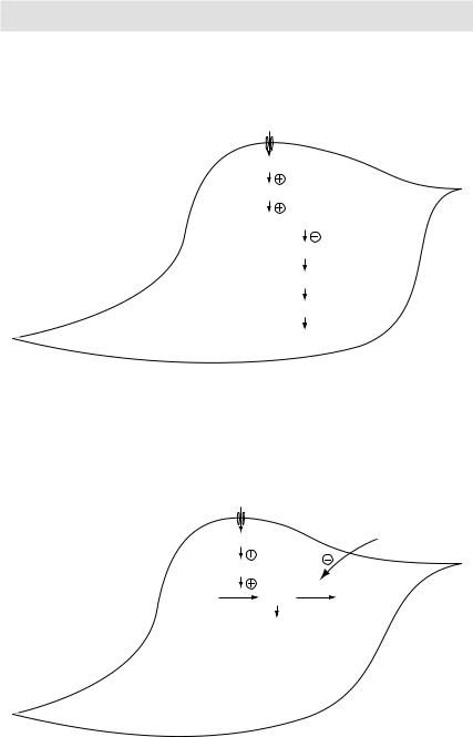
Heparin AT-III
Heparin AT-III
Active Heparin AT-III clotting factor
Inactive Heparin AT-III clotting factor
Figure 3.14 Mechanism of action of heparin and antithrombin-III (AT-III)
78

MECHANISMS OF ACTION
Organic nitrates and nitrites
1Biochemical: When these drugs are taken orally, they are denitrated in the body (especially in the vascular and liver bed of endothelium) in a stepwise fashion. Initially one radical is removed, then another, by glutathione iso-reductase
enzyme. Hydrolysis takes place and metabolites are released.
Release of nitric oxide radical → activation of guanylyl (guanylate) cyclase → accumulation of cGMP → activation of cGMP dependent kinases → dephosphorylation of myosin light chain → vasodilatation (venular and arteriolar).
2Hemodynamic: venodilatation → decreased preload → arteriolar dilatation → decreased.
Phosphodiesterase (PDE) inhibitors
Phosphodiesterase (PDE) inhibitors (Sildenafil, tadalafil, vardenafil) fill the penis with blood and are thus used in men with erectile dysfunction. Physiologically, NO released from penile nerve terminals stimulates guanylyl cyclase in the smooth muscle of corpus cavernosum → ↑ intracellular cGMP → smooth muscle relaxation → ↑ inflow of blood → erection. It is pertinent to mention here that if we artificially increase the in vivo NO level by coadministering nitrates and PDE inhibitors, generalised vasodilatation and profound hypotension may result. Thus prescribing PDE inhibitors in patients who are taking nitrates (say due to unstable angina) is contraindicated.
Radioactive iodine I131 therapy
I131 is administered orally in solution form as sodium I131. It is absorbed rapidly, after which it is avidly taken up by the thyroid tissue and incorporated in the storage follicles. I131 destroys thyroidal tissue by the emission of β-rays, which are cytotoxic, ionising and have got short-range (so destroy only follicle cells, not surrounding structures). They have an effective t½ of 5 days and a penetration range of 400–2000μm.
Unlike thioamides and iodine salts, which decrease thyroid hormone synthesis and/or release only transiently, I131 causes complete destruction of thyroid tissue within a few weeks producing permanent hypothyroidism and a cure to thyrotoxicosis without surgery.
Being radioactive, I131 therapy is contraindicated in pregnant ladies and lactating mothers.
Indications of I131 therapy
Thyrotoxicosis due to:
1Graves’ disease.
2Toxic multi-nodular goitre.
3Toxic nodule.
I131 is the only isotope used for the treatment of thyrotoxicosis; others are used in diagnosis. It is the drug of choice for the treatment of Graves’ disease in patients <35yrs of age (except in pregnant/lactating women, in whom it is contraindicated).
Advantages of I131 therapy
1Because of once daily dosage, I131 has good compliance.
2Being odourless, it is not unpleasant to take it.
79

PHARMACOLOGY IN 7 DAYS FOR MEDICAL STUDENTS
Mechanism of vascular smooth muscle relaxation: Nitrates are chemically reduced to release nitric oxide (NO) leading to vasodilatation
Nitric oxide
Nitric oxide
Guanylyl cyclase
GTP  cGMP
cGMP
Myosin phosphatase enzyme
Myosin dephosphorylation
Actin-Myosin crossbridges disengage
Myocyte |
Relaxation |
Figure 3.15 Mechanism of vascular smooth muscle relaxation
Nitric oxide |
|
|
Nitric oxide |
Sildenafil |
|
Guanylyl cyclase |
|
|
GTP |
cGMP PDE |
GMP |
|
Relaxation |
(inactive) |
|
|
|
•PDE degrades cGMP to inactive GMP
•Inhibitors of PDE→↑cGMP→relaxation
Myocyte
Figure 3.16 Mechanism of action of phosphodiesterase (PDE) inhibitors
80

MECHANISMS OF ACTION
3It is cost-effective.
4It is safe (mortality is rare). Fears of radiation-induced genetic damage, leukaemia or neoplasia have not been realised after >50yrs of clinical experience with radioiodine therapy.
5It is a painless procedure, done in an OPD. No hospitalisation of patient is required and thus no work loss is incurred.
6Patient is saved from the hazards of surgery.
Thyroid hormone synthesis and transport
Synthesis of both triiodothyronine (T3) and thyroxine (T4) requires iodine, which is derived from food and iodine supplements (iodine-fortified salt, etc.). Iodine is taken up by the thyroid gland by an active process. Once inside the thyroid gland, the iodide ion combines with tyrosine to form monoiodotyrosine (MIT) and diiodotyrosine (DIT). Next, two molecules of DIT combine to form T4, and one molecule each of MIT and DIT combine to form T3. Once synthesised, these hormones are stored in the thyroid gland. On their release, both T4 and T3 circulate in the blood combined with a protein called thyroxine-binding globulin (synthesised by the liver). In peripheral tissues, some of the T4 is converted to T3 by a process of deiodination. Although T3 is released as such by the thyroid gland also, most of the circulating T3 comes by this process of deiodination of T4 to T3. T3 is about 10 times more potent than T4. It is basically T3 that exerts most of the physiological effects of thyroid hormones at the target organs.
Thyroid-stimulating hormone (TSH) released by the pituitary gland stimulates T4 and T3 synthesis by the thyroid gland. Higher levels of T4 and T3 in turn inhibit pituitary release of TSH thus providing an effective negative feedback control mechanism.
Antithyroid drugs: thioamides
Drugs included: propylthiouracil; carbimazole.
These drugs inhibit the synthesis (not release) of thyroid hormones. They block the process of T4 and T3 synthesis at multiple points:
1They inhibit iodination of the tyrosine residues.
2They block the coupling of MIT and DIT.
3Propylthiouracil alone, when given in high doses, also blocks the peripheral conversion of T4 to T3.
Since these drugs only inhibit the synthesis of thyroid hormones (and not release), their pharmacological effects starts only when the already synthesised and stored thyroid hormone molecules are depleted. This takes about 3–4 weeks.
Iodide salts and iodine
These drugs decrease both the synthesis (by inhibiting the iodination of tyrosine) and release of thyroid hormones. Because of the latter effect, unlike thioamides, these drugs have a rapid onset of action (within 2–7 days). Additionally, these drugs also decrease the size and vascularity of the enlarged hyperplastic thyroid gland. They are thus most beneficial when given in the pre-op period when, by decreasing the vascularity of the thyroid gland, they decrease the chances of per-op and post-op haemorrhage from the thyroid tissue.
The effects of iodide salts and iodine are transient. After a few weeks, the thyroid gland stops responding and escapes from the iodide block rendering these drugs ineffective.
81

PHARMACOLOGY IN 7 DAYS FOR MEDICAL STUDENTS
I– Na+
I– Na+
Thyroid |
T |
TG |
T |
follicular |
|
|
|
cell |
T |
T |
T |
Thyroid peroxidase |
(organification) |
T |
DOT |
|
TG |
MIT MIT MIT |
|
Colloid space
Thyroid peroxidase |
(coupling) |
Phagolysosome
MIT |
|
T4 |
T3 |
TG |
DOT |
|
T |
T |
MIT |
|
T4 |
T3 |
TG |
DOT |
|
T |
T |
Lysosome
I–
 T4, T3, MIT, DIT Proteolysis of
thyroglobulin
Plasma
 Deiodinase
  T4
T4  T3
T3
Peripheral conversion
Figure 3.17 Thyroid hormones synthesis, storage and release
82

MECHANISMS OF ACTION
These drugs are usually given in the form of Lugol’s solution (iodine and potassium iodide).
Aspirin and NSAIDs
As we know that arachidonic acid (derived from the cell membrane lipids) metabolism follows two pathways – lipoxygenase and cyclooxygenase. The latter pathway leads to the formation of three eicosanoids, i.e. prostacyclin, prostaglandins (PGE and PGF) and thromboxane.
Cyclooxygenase (COX) enzyme exists in 2 isoforms – COX-1 and COX-2. COX-1 is found in many body tissues, primarily in the non-inflammatory cells; COX-2, however, is found primarily in the inflammatory cells (polymorphonuclear cells, lymphocytes, etc.).
Aspirin and other non-selective NSAIDs: inhibit both COX-1 and COX-2 in turn inhibiting the synthesis of eicosanoids throughout the body. Eicosanoids are important mediators of inflammation. For example, they are involved in increasing or decreasing vascular and bronchial tone, leukocyte chemotaxis, platelet aggregation, etc. By inhibiting eicosanoids synthesis, these drugs exert their anti-inflammatory effect. For example, PG synthesis in CNS (stimulated by pyrogens) leads to fever. NSAIDs by suppressing PG synthesis, thus produce an antipyretic effect. PGs produced in the injured tissues activate the nociceptors. NSAIDs exert an analgesic effect by suppressing PG synthesis in the injured tissues. PG synthesis in the stomach protects the gastric lining from the cytotoxic effects of HCl. This cytoprotection is lost when PG synthesis is inhibited by the NSAIDs leading to gastritis and peptic ulceration.
Difference between aspirin and other non-selective NSAIDs: Unlike other non-selective NSAIDs, aspirin (but not its active metabolite, salicylate) acetylates and thus irreversibly inhibits COX-1 and COX-2 enzymes. This leads to a longer duration of action (especially the antiplatelet effect).
COX-2 selective NSAIDs (celecoxib and rofecoxib): only inhibit the COX-2 enzyme primarily found in the inflammatory cells. These agents thus do not inhibit eicosanoids synthesis throughout the body. Thus, at least theoretically, these agents should have lesser GI side effects (gastritis and peptic ulceration) – something not uncommonly seen with protracted therapy with aspirin and other non-selective NSAIDs.
GPIIb-IIIa receptor antagonists
Fibrinogen strands bind with GPIIb-IIIa receptors present on platelet membranes leading to platelet aggregation. GPIIb-IIIa receptor antagonists (like tirofiban and abciximab) by inhibiting GPIIb-IIIa receptors inhibit platelet aggregation. Use of these potent antiplatelet drugs is reserved in selected cases of unstable angina and non-ST elevation myocardial infarction (NTEMI) and those undergoing percutaneous coronary intervention.
Clopidogrel, ticlopidine and dipyridamole
cAMP is one of the ‘natural antiplatelets’ found in human bodies. An increase in the intracellular concentration of cAMP in platelets leads to decrease platelet aggregation and vice versa (the exact mechanism not clearly understood). Physiologically, ADP receptor activation causes inactivation of adenylyl cyclase enzyme leading to a fall in cAMP levels. Drugs like clopidogrel and ticlopidine irreversibly inhibit the ADP
83
